Research Article | | Peer-Reviewed

Isolation and Probiotic Characterization of Lactic Acid Bacteria from Kindirmou and Pendidam in Adamawa Region (Cameroon)

Received: 8 March 2024    Accepted: 20 March 2024    Published: 12 April 2024
Views:       Downloads:
Abstract

The microorganisms intended for use as probiotics in food formulation should exert health benefit effects and be regarded as safe for animals and humans uses. The aim of this study was to evaluate the probiotic potential of lactic acid bacteria (LAB) isolated from pendidam and kindirmou, two traditional fermented milks (TFM) produced in the Adamawa region (Cameroon). Twenty-five samples (pendidam: 13 and kindirmou: 12) were randomly collected in five markets of Ngaoundere (n = 17 samples) and Meiganga (n = 8 samples). These samples were screened for their antimicrobial activity, and nine TFMs were retained. Lactic acid bacteria were isolated from these samples and their antimicrobial activity was already evaluated. Based on the inhibition zone, twenty-two LABs were retained and examined in vitro for potential probiotic properties based on their low pH tolerance, resistance to bile salts, tolerance to simulated gastrointestinal juices, hydrophobicity, autoaggregation, gelatinase and hemolytic activities. The outcome of these parameters studied was used as input data for a principal component analysis (PCA) to select the most promising isolate, and the six potential probiotic isolates were characterized through a biochemical profile. The characterized isolates have been identified as Lactiplantibacillus plantarum, Lacticaseibacillus casei, and Lactococcus lactis. Traditional fermented milks contain LAB with important properties that can be utilized in the formulation of functional foods.

Published in Advances in Biochemistry (Volume 12, Issue 2)
DOI 10.11648/j.ab.20241202.11
Page(s) 35-48
Creative Commons

This is an Open Access article, distributed under the terms of the Creative Commons Attribution 4.0 International License (http://creativecommons.org/licenses/by/4.0/), which permits unrestricted use, distribution and reproduction in any medium or format, provided the original work is properly cited.

Copyright

Copyright © The Author(s), 2024. Published by Science Publishing Group

Keywords

Probiotic, Lactic Acid Bacteria, Pendidam, Kindirmou, Cameroon

1. Introduction
Probiotics are microorganisms that provide health benefits to humans or animals. FAO/WHO defines probiotics as “live microorganisms that, when administered in adequate amounts, confer benefits to the host”. Consumption of probiotics has been shown to be helpful in overcoming various clinical conditions ranging from infantile diarrhoea, antibiotic-associated diarrhoea, relapsing Clostridium difficile colitis, Helicobacter pylori infection, inflammatory disease to cancer and female uro-genital infections . They also contribute by improving lactose intolerance, lowering serum cholesterol levels, and increasing the utilization of nutrients . In general, 6 to 7 log10 of probiotic bacteria per mL or g of food has been recommended for health benefits . The criteria for being considered as probiotic bacteria are numerous and strict . To be considered as a probiotic, a microorganism must fulfill criteria such as non-pathogenic or without an antibiotic resistance profile ; must overcome physical and chemical barriers such as acid and bile in the gastro intestinal tract ; must be able to adhere to intestinal surfaces and to inhibit pathogens microorganism .
Actually, lactic acid bacteria (LAB) together with bifidobacteria are the most investigated probiotics in recent decades because of their health benefit . Fermented foods include desirable edible microorganisms that are good for human health . It is possible and important to conduct research on the strains of traditional fermented food products that have fascinating probiotic potential as a source of novel candidates . Traditional fermentation methods can be a valuable source of endogenous LAB since they are spontaneous and unrestrained . Traditional fermented milk (TFM) is the main source of isolated active strains with significant biological activity, according to a number of researchers .
In Cameroon, particularly among pastoral communities in the Adamawa region of the country such as Foulbe and Bororo, large amounts of TFM products such as kindirmou (fermented milk) and pendidam (skimmed fermented milk) have been consumed for centuries because of their health benefit effect . They are commonly made from raw cow milk using spontaneous fermentation or backslopping techniques . The microbiota of the two products has been studied, and the authors reported the presence of Lactobacillus delbrueckii N2, Limosilactobacillus fermentum TM1, Lactiplantibacillus plantarum G88, and Lacticaseibacillus paracasei subsp. tolerans N2-produicing biosurfactant with good antioxidant properties and antimicrobial activity from pendidam . However, there is a lack of information on the preliminary probiotic characteristics of isolated lactic acid bacteria. The aim of this study is to assess the probiotic potential of LAB isolated from traditional fermented milks (pendidam and kindirmou) in the Adamawa region of Cameroon.
2. Materials and Methods

2.1. Culture Media and Reagents

Tryptic Soy Agar and broth (TSA and TSB respectively), Mueller Hinton agar (MHA) and De Mann Rogosa and Sharpe (MRS), culture media, were obtained from Biolife (Biolife Italiana, Milano, Italy). The porcine bile salt, pepsin, and pancreatin were purchased from Sigma-Aldrich (Sigma Chemical Co., St-Louis, United States). The antibiotics: Amoxicillin + Clavulanic acid, Penicillin G, Cefixime, Chloramphenicol, Tetracycline, Streptomycin, Erythromycin, Sulfonamide and Oxolinic acid were purchased from Sigma-Aldrich (Bio-Rad, Boulevard Raymond Poincare, France).

2.2. Microbial Strains

The microbial strains Candida albicans, Rhodotorula mucilaginosa LMSA 2.08, Candida parasilopsis LMSA 2.09, Kluveyromyces marxianus CLIB 282, Debaryomyces hansenii CLIB 197, Saccharomyces cereviseae CLIB 227, Morganella morganii, Klebsiella pneumoniae, Proteus mirabilis, Bacillus cereus ATCC 19615, Listeria monocytogenes ATCC 19115 were obtained from the UBOCC at the LUBEM (Laboratoire Universitaire de Biodiversité et Ecologie Microbienne, Brest, France) and were used for antimicrobial tests (as indicator strains).

2.3. Sampling

In the Adamawa region, Ngaoundere and Meiganga are the main towns for large-scale dairy production. As for the sampling technique, all markets in the different towns were selected, and samples were collected in a guided, random procedure from the main vendors in each market. An explanation of the study and authorization were first obtained from each vendor before the samples were collected. A total of 25 samples (pendidam: 13 and kindirmou: 12) were randomly collected in five markets of Ngaoundere (n = 17 samples) and Meiganga (n = 8 samples) cities using guidelines described by the Codex Alimentarus (1999) . That included: (i) Grand-Marché (7º19’ 15.81” N; 13º35’ 06.46” E; n = 4); (ii) Marché Bantai (7º19’ 11.55” N; 13º35’ 27.14” E; n = 4); (iii) Petit-Marché (7º19’ 39.10” N; 13º35’ 00.25” E; n = 4); (iv) Marché de Dang (7º43’60.45” N; 13º55’ 74.71” E; n = 6) at Ngaoundere and Marché de Meiganga (6º31’ 08.65” N; 14º17’ 26.33” E; n = 8) at Meiganga.

2.4. pH and Titratable Acidity Determination of Traditional Fermented Milk

Titratable acidity was determined by titration of 10 mL of sample with 0.1 N NaOH using alcoholic phenolphthalein (0.5%; m/v) as an indicator and the pH measured with a pH meter (pH 201 Microprocessor, Hanna Instruments Srl, Ronchi di Villafranca, Italy).

2.5. Antimicrobial Activity of Traditional Fermented Milk Assay

The antimicrobial potential of TFMs was determined as well as described by Mbawala et al. . Briefly, 25 µL of each TFM was introduced into 6 mm diameter wells made of MHA medium containing microorganism test (~ 106 CFU/mL for both yeast and bacteria). Subsequently, the Petri dishes were kept for 30 min at room temperature (20 – 25°C) and then incubated at 37°C / 24 h for the bacteria and 25°C / 48 h for yeast. The diameters of inhibition were measured, and the samples with the highest values were retained for LABs isolation and screening .

2.6. Isolation and Screening of Lactic Acid Bacteria for Antimicrobial Activity

The isolation of LAB from different samples were done using de Man Rogosa Sharpe (MRS) agar following the method described by de Man et al. . Serial dilution (10-1 to 10-7) of 10 mL of each sample were done in 90 mL of physiological saline. One hundred microliter aliquots of the appropriate dilutions were surface-plated on MRS agar, incubated at 37°C for 48 – 72 h. All Gram-positive, catalase-negative, whitish colonies and oxidase-negative bacteria were selected for the assessment of their antimicrobial potential. The antimicrobial activity of isolated LAB was conducted using the method described by Fleming et al. . Briefly, 15 µL (~ 108 CFU/mL) of cultures (16 ± 2 h) were spotted onto MRS agar and then incubated for 18 h at 30°C. Thereafter, each culture was overlaid with 7 mL of Mueller Hinton soft agar (0.7% agar) inoculated with 100 µL (106 – 108 CFU/mL) of an overnight culture of the indicator bacteria and yeast. The plates were incubated for 24 h and 48 h for bacteria and yeast. Inhibition zone around each spot was measured and the isolates with the greatest values were retained for probiotic characterization tests.

2.7. Probiotic Characterization of Isolates

2.7.1. Resistance to Acidic Conditions

The resistance of the LABs to acidic conditions was assessed as described by Sieladie et al. . For each LAB, 100 µL of microbial suspension (~ 108 CFU/mL) were cultured in MRS broth adjusted to pH 2.0, 3.0, and 6.5 using 1 M HCl. Bacterial tolerance was evaluated by determining the optical density at 620 nm after 6 and 24 h of incubation at 37°C. Acid tolerance was classified according to the methods used by Sieladie et al. .
Survival (%)=100*ODpH6.5-ODpH2 or 3ODpH6.5 

2.7.2. In vitro Evaluation of Resistance to Human Digestive System

The resistance of LAB to gastrointestinal conditions was evaluated using the method described by Guo et al. . One milliliter of bacteria suspension (~108 bacteria) was mixed with 9 mL of simulated gastric juice [3 g/L pepsin (Sigma-Aldrich, USA) in sterile saline water (NaCl, 0.85%; w/v) solution adjusted to pH 2.0 using 1 M HCl)]; simulated pancreatic juice [pancreatin (Sigma-Aldrich, USA) 1 g/L in sterile saline water adjusted to pH 8.0 using 0.1 M NaOH] respectively. After 1, 90 (for simulated pancreatic juice) and 180 (for simulated gastric juice) minutes of incubation at 37°C, the tolerance of bacteria was evaluated by plating on MRS agar. For the evaluation of LAB resistance to simulated intestinal juice, 1 mL of bacteria suspension was mixed with 9 mL of pancreatic juice supplemented with 0.45% bile salts (w/v), incubated for 1 and 240 min at 37°C, enumerated by plating on MRS agar and then incubated at 37°C for 48 h. The resistance of LAB to bile salts was tested according to the method of Guo et al. . Briefly, 1 mL of LAB culture of 18 h (108 – 1010 CFU/mL) were inoculated into tubes containing 9 mL of solution of bile salts at 0.2 and 0.45% (w/v) and incubated at 37°C for 24 h. After incubation, 1 mL of the previously obtained suspension was diluted, cultured on MRS agar, and then incubated at 37°C for 48 h.
The rate of survival was calculated as follows:
Survival (%)=100*N1N0
N1: Total viable count of LAB after incubation in SGJ or SPJ or SIJ; N0: Total viable count of LAB strains before incubation;

2.7.3. Autoaggregation Test

Autoaggregation test was carried out according to the method of Kos et al. . Bacterial cells were cultured in MRS broth, incubated at 37°C for 18 h, and then young cells were collected by centrifugation (5000 g at 4°C / 15 min). Four milliliters of microbial suspension (~ 108 CFU / mL) were prepared in phosphate buffered saline (PBS) using the 2McFarland standard and then incubated for 5 h at room temperature (28 ± 2°C). After each 1 h interval, 100 µL of suspension was mixed with 3.9 mL of PBS and absorbance (A) was measured at 600 nm. The percentage of autoaggregation was expressed as follows:
Autoaggregation (%)=1-AtA0*100
Where At is the absorbance at time t = 1, 3, and 5 h and A0 is the absorbance at t = 0 h.

2.7.4. Microbial Adhesion to Solvents

The adhesion of bacteria to organic solvent (MATS) was evaluated according to the method described by Kos et al. . In this study, two solvents were tested including xylene (Baker Chemicals, Deventer, Holland), and chloroform (Scharlau Chloroform, Spain). The young culture (16 ± 2 h at 37°C) was harvested by centrifugation (5000 g at 4°C for 15 min), washed twice using PBS and the bottom was used for the microbial suspension (approximately 108 CFU/mL) preparation using potassium nitrate solution (KNO3: 0.1 mol/L at pH = 6.2). The absorbance of the cell suspension was measured at 600 nm (A0). One milliliter of each solvent was added to 3 mL of microbial suspension and thoroughly homogenized using a vortex (for 2 min). The aqueous phase was collected after 20 minutes, and its absorbance at 600 nm (A1) was measured. The percentage of bacterial adhesion in the solvent was calculated as follows:
Bacterial adhesion (%)=1-A1A0*100

2.7.5. Susceptibility of LAB to Antibiotics

The susceptibility of LABs to antibiotics was performed using the disc diffusion method as previously described . Cells from 16 ± 2 h old cultures were prepared in sterile saline water (0.85% NaCl, m/v) using the 0.5McFarland standard. The suspension was diluted (1:100), cultured on Muller-Hinton agar surface, dried at room temperature (28 ± 2°C) and then antibiotics were applied. Antibiotics used were selected according to their site of action, frequency of utilization and include (i) inhibitors of cell wall synthesis: Amoxicillin + Clavulanic acid, 20/10 µg; Penicillin G, 6 µg; Cefixime, 10 µg; (ii) inhibitor of protein synthesis: Chloramphenicol, 30 µg; Tetracycline, 30 µg; Streptomycin, 500 µg; Erythromycin, 15 µg; (iii) inhibitors of the synthesis of folic acid/nucleic bases: Sulfonamide, 200 µg; and (iv) inhibitor of DNA gyrase enzyme: Oxolinic acid, 10 µg. After incubation, inhibition diameters were recorded and the susceptibility of the isolated was evaluated as described by Sieladie et al. .

2.7.6. Gelatinase and Hemolysis Activities

The production of gelatinase and the hemolysis activity of LAB strains were determined using the method of Eaton and Gasson . The cultures of 16 ± 2 h old was plated by streaking on MRS agar supplemented with 3% (w/v) gelatin and incubated at 37°C for 24 h and then incubated at 4°C for 5 h. Colonies surrounded by clear zones were considered to be gelatinase producers. The hemolysis activity of LAB was evaluated by streaking 12 h old bacteria on 5% sheep blood agar (Biomerieux, France). After incubation at 37°C for 48 h, the colony surrounded by a clear zone was considered as bacteria with beta-hemolysis activity. Staphylococcus aureus ATCC 25923 was used as a positive control.
The isolates showing interesting probiotic properties have been subjected to biochemical characterization using API 50 CHL kitTM and results were recorded after 24 and 48 h at 37°C. Species were determined using Apident 2.0 database (BioMérieux, France) and confirmed using online API web services (https://apiweb.biomerieux.com). The specificity of these galleries was at least 92%.

2.8. Statistical Analysis

The analysis was carried out in triplicate, results were expressed as means ± standard deviation. Data were analyzed by analysis of variance (ANOVA); differences between means were tested using the Duncan Multiple Ranking test in Statgraphic® Centurion XVII software (Statpoint Technology, Inc. USA). The probiotic characteristics (hydrophobicity, autoaggregation, gastric, pancreatic, intestinal simulated juices tolerance, and of bile salts tolerance) of the LAB were subjected to the Principal Component Analysis (PCA) to discriminate LAB isolates using Statistical XLStat 2017 software.
3. Results and Discussion

3.1. pH, Titratable Acidity, and Screening of Fermented Milks

The pH values of the pendidam vary from 3.38 to 3.65, and for kindirmou samples, they vary from 3.63 to 4.11. The titratable acidity of the pendidam varies from 111.9 to 345°D and from 67.5 to 167.7°D for kindirmou. According to the origin, the lowest pH values and the highest titratable acidity values were obtained in Ngaoundere, while the highest values of pH and lowest values of titratable acidity were obtained in Meiganga. ANOVA shows a significant difference between samples (P ≤ 0.05). Jiwoua and Milière explained that the low value of pH and the high value of titratable acidity compared to raw milk are related to the presence of organic acids, mainly lactic acid produced during fermentation. Mbawala et al. explain that variation observed either pH or titratable acidity could be due to climatic variations, the sampling site, and the manufacturing process.
Concerning the antimicrobial activity of TFMs against test strains, some samples were active with inhibition diameter ranges from 1.3 to 9.6 mm. The inhibition zone obtained could be explained by the organic acids produced and released in the samples. Mbawala et al. also reported the similar observations and explained that by the presence of molecules such as hydrogen peroxide, bacteriocins, and biosurfactants produced by microorganisms, including lactic acid bacteria.

3.2. Antimicrobial Activity of Lactic Acid Bacteria

Based on the antimicrobial activities of TFMs described previously, nine of them were selected. Forty LABs were isolated and twenty-two showed antimicrobial activity against at least one indicator microorganism (Table 1). The inhibition diameters were isolate dependent with the highest value of 72 mm. The most sensitive strains were L. monocytogenes and B. cereus, while S. cereviseae, C. parapsolosis and D. hansenii were more resistant. No inhibition activity was observed against S. cereviseae.
The inhibitory effect obtained in this study could be attributed to antimicrobial compounds produced and released by the isolates such as organic acids, hydrogen peroxide, biosurfactants, diacetyl, bacteriocin or the synergy between some of them . In the present study, Gram-positive bacteria were more sensitive to the antimicrobial compounds produced. This is based on the difference in cell envelope composition between the two groups of bacteria. Researchers reported that bacteriocins were most active in Gram-positive pathogen bacteria (i.e., L. monocytogenes, B. subtilis) and added that bacteriocins act by forming pores in the cytoplasmic membrane leading to the disruptions in cell function . In contrast, Song and Richard reported the resistance of L. monocytogenes and explained that resistance to pore formation through a membrane change in composition and properties, a decrease in surface hydrophobicity and a lower affinity of the bacterial surface for antimicrobial compounds. The resistance observed in Gram-negative bacteria is due to the outer membrane acting as an efficient permeability barrier against macromolecules and hydrophobic substances. In the present study, weak activities were observed on the yeasts particularly on S. cereviseae, C. parapsolosis and D. hansenii. Voulgari et al. showed that LAB isolated from fermented products produced protein-like compounds with antifungal activity against selected strains except S. cereviseae. In addition, low activities of LAB against D. hansenii, K. marxianus, and S. cerevisiae have been reported . Other compounds with antifungal activity have always been documented, such as cyclic dipeptides, hydroxylated fatty acids, phenyllactic acid, and substances assimilated to bacteriocins . Based on these results, 22 isolates with the predominance of lactobacilli were retained. The occurrence of lactobacilli with a high antagonistic activity of fermented products has been documented .
Figure 1. Inhibition percentage of LAB under acidic condition.
Table 1. Antimicrobial activity of lactic acid bacteria isolated from traditional fermented milks.

LAB

Diameter of inhibition (mm)

M. morganii

P. mirabilis

L. monocytogenes

B. cereus

K. pneumoniae

PD1

48.0 ± 1.4k

41.2 ± 0.3i

65.5 ± 0.4jk

57.5 ± 0.7hi

50.5 ± 0.7m

PD2

48.5 ± 0.7k

34.3 ± 0.4h

72.5 ± 0.6m

64.6 ± 0.8e

25.0 ± 0.6b

PD3

32.0 ± 1.4fg

31.8 ± 0.2fgh

51.0 ± 1.0c

45.5 ± 0.5d

44.4 ± 0.5k

PD4

35.5 ± 0.7i

21.5 ± 0.3bc

66.5 ± 0.6kl

59.7 ± 0.3jk

19.6 ± 0.8a

PD5

24.5 ± 0.3b

14.5 ± 0.7a

53.5 ± 0.4d

57.5 ± 0.6hi

30.2 ± 0.3e

PD6

33.7 ± 0.3ghi

39.7 ± 1.0i

70.5 ± 0.7m

60.4 ± 0.5k

34.4 ± 0.5h

PD8

29.0 ± 0.2e

29.6 ± 0.8def

56.0 ± 0.8e

57.5 ± 0.7hi

30.5 ± 0.7ef

PB1

31.7 ± 0.4fg

21.3 ± 0.4bc

57.0 ± 1.0ef

52.7 ± 0.5f

31.6 ± 0.4fg

PB2

24.2 ± 0.3b

32.4 ± 0.5gh

57.5 ± 0.5ef

56.5 ± 0.4d

28.2 ± 0.2d

PB3

32.0 ± 1.2bc

41.7 ± 0.8i

63.4 ± 0.7hij

57.5 ± 1.0hi

34.5 ± 0.7h

PB4

34.0 ± 0.8ghi

31.0 ± 1.0efg

64.5 ± 0.7ijk

54.6 ± 0.2h

39.3 ± 0.4i

PD9

21.0 ± 1.0a

34.0 ± 0.6h

38.7 ± 0.3b

30.7 ± 0.8a

24.5 ± 0.2b

PD10

35.0 ± 0.7hi

27.1 ± 1.0d

61.2 ± 1.0gh

54.6 ± 0.6g

23.7 ± 0.3b

PD11

32.7 ± 1.0fgh

29.5 ± 0.7def

62.5 ± 0.8ghi

58.4 ± 0.5ij

26.4 ± 0.5c

PD12

28.4 ± 0.5de

27.2 ± 1.2d

56.0 ± 0.6e

40.5 ± 0.3c

34.5 ± 0.2h

PG1

26.6 ± 0.8cd

22.2 ± 0.3bc

61.7 ± 0.3gh

58.4 ± 0.5ij

26.5 ± 0.8c

PG2

24.7 ± 0.8bc

20.0 ± 1.0b

58.5 ± 0.6f

50.6 ± 0.8

34.3 ± 0.4h

PG3

39.6 ± 0.8j

31.0 ± 0.6efg

67.7 ± 0.2l

61.2 ± 0.2k

42.1 ± 0.2j

PG4

33.9 ± 0.1ghi

29.5 ± 0.7def

68.0 ± 0.2l

58.2 ± 0.3ij

44.1 ± 0.2k

PG5

39.6 ± 0.5j

30.0 ± 1.0efg

27.0 ± 1.0a

34.5 ± 0.7b

32.3 ± 0.4g

KM1

30.5 ± 0.7ef

28.7 ± 1.0de

60.8 ± 1.0g

57.3 ± 0.4hi

24.3 ± 0.3b

KB3

31.2 ± 1.0f

23.5 ± 0.7c

62.2 ± 0.3gh

56.4 ± 0.5h

48.2 ± 0.4l

Table 1. Continued.

LAB

Diameter of inhibition (mm)

C. parapsilosis

S. cerevisiae

C. albicans

R. mucilaginosa

D. hansenii

K. marxianus

PD1

16.5 ± 0.7a

0a

16.2 ± 0.3b

14.5 ± 0.7b

26.1 ± 0.1i

26.2 ± 0.4k

PD2

30.3 ± 0.2h

0a

24.4 ± 0.5e

18.4 ± 0.6c

20.5 ± 0.7g

14.5 ± 0.7c

PD3

32.5 ± 0.6i

0a

29.5 ± 0.7hi

14.6 ± 0.8b

18.1 ± 0.1ef

20.2 ± 0.4fg

PD4

22.4 ± 0.5e

0a

24.4 ± 0.6e

18.4 ± 0.6c

14.2 ± 0.3d

22.7 ± 1.0k

PD5

16.1 ± 0.1a

0a

26.3 ± 0.4f

22.1 ± 0.1f

10.5 ± 0.7c

12.5 ± 0.2b

PD6

21.5 ± 0.5cde

0a

14.8 ± 0.1b

14.4 ± 0.6b

24.0 ± 0.0h

18.5 ± 0.7e

PD8

25.0 ± 0.4f

0a

24.6 ± 0.8e

22.5 ± 0.7f

8.4 ± 0.6b

21.5 ± 0.3ghi

PB1

22.2 ± 0.8de

0a

20.4 ± 0.5c

10.4 ± 0.6a

8.5 ± 0.7b

18.3 ± 0.4e

PB2

16.4 ± 0.6a

0a

24.3 ± 0.4e

20.3 ± 0.4d

26.2 ± 0.4i

21.8 ± 0.3hi

PB3

20.2 ± 0.2bc

0a

34.5 ± 0.7j

21.7 ± 0.4ef

17.6 ± 3.3ef

12.5 ± 0.7b

PB4

20.5 ± 0.2bc

0a

36.2 ± 0.3i

28.1 ± 0.1i

16.2 ± 0.3e

10.7 ± 1.1a

PD9

30.4 ± 0.4h

0a

30.4 ± 0.5k

10.3 ± 0.5a

19.0 ± 0.0fg

16.2 ± 0.4d

PD10

28.5 ± 0.3g

0a

26.6 ± 0.8f

14.2 ± 0.4b

6.5 ± 0.7a

12.5 ± 0.7b

PD11

28.2 ± 0.5g

0a

22.3 ± 0.4d

20.8 ± 0.3de

20.2 ± 0.3g

18.4 ± 0.6e

PD12

29.4 ± 0.1gh

0a

39.5 ± 0.7l

24.2 ± 0.3g

22.5 ± 0.7h

21.0 ± 1.4gh

PG1

26.1 ± 1.0f

11.0 ± 0.8e

26.8 ± 1.0fg

25.3 ± 0.4h

17.6 ± 0.8ef

26.5 ± 0.6k

PG2

28.8 ± 0.8gh

9.2 ± 0.4d

24.5 ± 0.7e

18.2 ± 0.4c

23.2 ± 0.3h

19.3 ± 0.4ef

PG3

20.6 ± 0.6bcd

3.4 ± 0.5b

34.4 ± 0.5j

22.1 ± 0.1f

20.3 ± 0.4g

20.2 ± 0.3fg

PG4

22.2 ± 0.3de

6.7 ± 0.1c

34.5 ± 0.7j

18.3 ± 0.4c

23.4 ± 0.6h

24.4 ± 0.6k

PG5

19.5 ± 0.7b

0a

10.9 ± 0.2a

24.2 ± 0.3g

16.2 ± 0.4

19.2 ± 0.4ef

KM1

23.0 ± 1.0e

0a

20.2 ± 0.6c

28.4 ± 0.6i

20.5 ± 0.7g

26.2 ± 0.3k

KB3

22.7 ± 1.0e

0a

28.2 ± 0.1gh

24.3 ± 0.4g

29.6 ± 0.8j

30.5 ± 0.7l

PB: Bantaille pendidam; PD: Dang pendidam; PG: Grand- marché pendidam; KM: Meiganga kindirmou; KB: Bantaille kindirmou; Means followed by distinct letters in the same column are different by the Duncan test (P < 0.05).

3.3. Resistance to Acidic Conditions

In this study, the inhibition percentage at pH 3 and 2 during 24 h has been done and the data are illustrated in Figure 1. Tolerance under acidic conditions is dependent on time and isolate. Indeed, we noted that more than 56% of the strains survived after 3 h at pH 3, but after 6 h, these percentages decreased to thresholds below 50% (Figure 1). Variation in LAB viability with incubation time has been showed by several authors . For example, Velez et al. working with 17 strains obtained that 11 declined their viability to undetectable levels after 30 min of exposure while only L. paracasei A-1, Leuconostoc mesenteroides B-1 and Lacticaseibacillus rhamnosus GG were able to survive over 90 min of exposure. Kalui et al. working with 18 strains of L. plantarum obtained 100% survival at pH 2.5 after 3 h of exposure, while 10% tolerated a pH of 2. Sieladie et al. had a survival percentage of less than 50% for all strains after 6 h exposure at pH 2. Similarly, Liong and Shah. , Kalui et al. observed that resistance to acidic conditions of L. casei and L. plantarum strains decreased during the first three hours. The observed resistance variation could be explained by bacterial diversity, the ability of the isolates to develop resistance mechanisms such as the use of proton pump, the decarboxylation of amino acids and the progressive expression of regulators that promote changes in the parietal structure of the cell encounter .
Table 2. Microbial load (log CFU/mL) of lactic acid bacteria isolates under stress conditions.

Isolates

Initial count

Bile salts (%)

SGJ

SPJ

SIJ

0.2

0.45

180 min

90 min

240 min

KB3

7.76

6.75 ± 0.21ef

6.21 ± 0.11efg

7.59 ± 0.20abc

7.61 ± 0.34bcde

7.67 ± 0.02fg

KM1

7.66

6.18 ± 0.30d

5.86 ± 0.15de

7.57 ± 0.09abc

7.44 ± 0.22bc

7.50 ± 0.20efg

PB1

7.68

6.27 ± 0.07de

5.99 ± 0.21ef

7.76 ± 0.12bcde

9.05 ± 0.41f

7.38 ± 0.04ef

PB2

9.11

8.21 ± 0.26h

7.21 ± 0.10jkl

9.26 ± 0.08fgh

8.79 ± 0.19f

8.9 ± 0.01ij

PB3

7.77

6.25 ± 0.15de

6.12 ± 0.13ef

7.79 ± 0.02bcde

7.49 ± 0.26bcd

5.57 ± 0.22c

PB4

7.79

4.75 ± 0.21b

4.83 ± 0.11b

7.81 ± 0.14bcde

7.88 ± 0.24cde

5.61 ± 0.19c

PD1

9.11

5.38 ± 0.02c

5.39 ± 0.10c

9.38 ± 0.12gh

8.84 ± 0.13f

4.44 ± 0.07b

PD10

7.91

7.52 ± 0.23g

6.88 ± 0.22ij

7.72 ± 0.07abcd

8.07 ± 0.23e

6.60 ± 0.09d

PD11

7.72

7.33 ± 0.42g

6.52 ± 0.05ghi

7.54 ± 0.07ab

7.57 ± 0.11bcde

7.35 ± 0.09e

PD12

7.65

5.47 ± 0.48c

4.85 ± 0.16b

8.09 ± 0.09de

7.22 ± 0.04ab

6.47 ± 0.12d

PD2

9.18

7.48 ± 0.05g

7.25 ± 0.12kl

8.85 ± 0.31f

9.8 ± 0.35h

9.14 ± 0.21j

PD3

9.08

3.25 ± 0.23a

2.70 ± 0.14a

9.43 ± 0.24h

8.91 ± 0.27f

8.64 ± 0.08i

PD4

7.89

6.24 ± 0.23de

5.64 ± 0.05cd

8.14 ± 0.13e

8.87 ± 0.27f

3.50 ± 0.13a

PD5

9.11

8.07 ± 0.19h

7.56 ± 0.07lm

9.00 ± 0.22fg

9.19 ± 0.24fg

8.09 ± 0.01h

PD6

9.15

7.84 ± 0.08gh

7.55 ± 0.19lm

9.19 ± 0.43fgh

9.61 ± 0.12gh

8.71 ± 0.14i

PD8

7.83

6.73 ± 0.03ef

6.67 ± 0.16i

7.99 ± 0.14cde

8.03 ± 0.43de

7.68 ± 0.04g

PD9

9.03

7.47 ± 0.36g

6.63 ± 0.06hi

9.19 ± 0.22fgh

9.06 ± 0.29f

8.74 ± 0.16i

PG1

7.96

7.48 ± 0.07g

7.12 ± 0.16jk

7.95 ± 0.01bcde

6.85 ± 0.23a

7.51 ± 0.03efg

PG2

9.04

8.29 ± 0.08h

8.23 ± 0.12n

8.94 ± 0.11f

8.92 ± 0.21f

8.88 ± 0.14ij

PG3

7.69

6.57 ± 0.16def

6.30 ± 0.21fgh

7.64 ± 0.14abc

7.56 ± 0.02bcde

7.58 ± 0.09efg

PG4

7.45

6.84 ± 0.19f

6.26 ± 0.21fg

7.33 ± 0.06a

7.81 ± 0.08cde

7.33 ± 0.02e

PG5

9.03

8.18 ± 0.01h

7.76 ± 0.31m

8.85 ± 0.13f

8.78 ± 0.04f

8.83 ± 0.18i

PB: Bantaille pendidam; PD: Dang pendidam; PG: Grand- marché pendidam; KM: Meiganga kindirmou; KB: Bantaille kindirmou; Means followed by distinct letters in the same column are different by the Duncan test (P < 0.05). SGJ, SPJ, and SIJ: simulated gastric, pancreatic and intestinal juices respectively.

3.4. Effect of Simulated Gastric Juices and Bile Salts on the Viability of Strains

The effect of simulated gastric (SGJ), pancreatic (SPJ), intestinal juices (SIJ), and bile salts (BS) on the 22 bacterial viability has been assessed and the data are illustrated in Table 2. We noted that all selected isolates could grow in all simulated gastric juices with a survival rates upper than 50%. Therefore, more than 68% of the strains survived after 180 min in SGJ at pH 2.0 and more than 90% after 90 min of incubation in SPJ at pH 8.0. There is a significant decrease (P < 0.05) of survival rate at 0.45% bile salt.
Several authors reported a higher survival rate of LAB under SGJ, SPJ, SIJ, and bile salts stress conditions. Futhermore, the effect of pepsin and pancreatin present in simulated juices on the survival of isolates has also been reported. Charteris et al. working with SGJ in the same conditions observed that L. fermentum KLD exhibited a higher survival rate (70% of the initial count) and were considered intrinsically tolerant to gastric transit. De Sant’anna et al. observed that among 24 strains, 8 showed growth inhibition lower than 60% in the SGJ (at pH 2). Maragkoudakis et al. working with 29 strains (after 3 h in the presence of pepsin) obtained L. rhamnosus ACA-DC 112 and L. paracasei subsp. paracasei ACA-DC 130 as the best survival while, 14 strains displayed loss of viability of upper 3 log UFC and 13 strains were completely inhibited. Guo et al. observed for all strains that pepsin improved their survival under acidic conditions when compared to MRS acid medium without pepsin. Concerning pancreatin, their presence reduced to less than 1 log UFC or no loss after 4 h of exposure in simulated pancreatic juices .
In the present study, bile salt tolerance was also found to be isolate-dependent. Researchers reported that probiotic bacteria such as L. acidophilus were found to excrete Bile Salt Hydrolases (BSH) that catalyze the hydrolysis of glycine- and taurine-conjugated bile salts into free bile salts and amino acid residues, thereby reducing the toxicity of the bile salts . However, the direct relationship between the tolerance of bile salts and the production and activity of BSH it is not well established. For example, a high resistance rate has been reported under 1% bile salt with nine strains of lactobacilli and among them, only one exhibited important BSH activity . In addition, Minelli et al. observed that among 4 strains of L. casei growing in MRS supplemented with 1% (w/v) Oxgall, no BSH activity was observed.

3.5. Hemolysis and Gelatinase Activity

None of the isolates was found to be positive for gelatinase and hemolysis activities compared to S. aureus used as a control. The absence of hemolytic and gelatinase activities is one of the safety criterion for the selection for probiotic strains . Siladie et al. showed that all strains of L. plantarum isolated from raw cow's milk in the western part of Cameroon were negative for hemolysis and gelatinase activity. The GelE gene is one of the genes responsible for gelatinase activity, but Eaton and Gasson. reported that gelE expression is highly influenced by some factors such as culture conditions and manipulation techniques. The species belonging to Enterococcus genus such as E. faecium, E. avium, E. maldoratus, and E. raffinosus are most prevalent and responsible of several public health concerns due to its ability to transfer genes (cylL L and cylL S) of virulence factors .

3.6. Susceptibility to Antibiotics

Other safety criteria for the selection of strains for the probiotics purpose in food formulation is their susceptibility to antibiotics. Data from this study revealed that more than 50% of the isolates were resistant to penicillin, sulfonamide, oxolinic acid, streptomycin, and cefixime. About 86% were sensitive to amoxicillin/clavulanic acid, tetracycline, erythromycin and chloramphenicol (Table 3). Cocci were resistant to streptomycin while lactobacilli were resistant to lactams, quinolones, aminoglycosides, sulfonamides, and sensitive to cyclins, macrolides and phenicols. Antibiotic resistance depends on the isolates tested and these are in agreement with those of literature. Muñoz-Atienza et al. obtained an antibiotic resistance rate of 33, 11 and 0% for strains belonging to the Lactobacillus, Enterococcus and Lactococcus genera. Similarly, Birri et al. working on 80 strains of 09 different species reported a total susceptibility to chloramphenicol, erythromycin, tetracycline and a resistance to aminoglycosides and glycopeptides. Several studies reported that Lactobacillus spp. are generally susceptible to antibiotics that inhibit protein synthesis, such as chloramphenicol, erythromycin, clindamycin and tetracycline . De Sant’anna et al. reported the sensitivity to clindamycin and erythromycin that all LAB, while 87.5%, showed moderate sensitivity to tetracycline. The European Food Safety Authority (EFSA) requires that the bacteria used for the food formulation be free of acquired antibiotic resistance genes to limit the transfer of these genes . The multi-drug resistance of lactic acid bacteria have been reported by several authors . For example, Singh et al. working on nine strains of Lactobacillus reuteri found that several of them were resistant to polymyxin B, gentamycine, cefazolin, ampicillin, vancomycin, cephalothin and cefuroxime.
According to the resistance of cocci to streptomycin, Ammor et al. reported that Pediococcus are intrinsically resistant to vancomycin, streptomycin, ciprofloxacin and trimethoprim-sulphamethoxazole. Natural resistance to multiple classes of antibiotics is probably due to cell wall structure and membrane permeability, complemented in some cases by the efflux mechanism. However, the differences in resistance between some strains could also be explained by some non-specific mechanisms, such as multidrug transporters or defective cell wall autolysis .
PB: Bantaille pendidam; PD: Dang pendidam; PG: Grand- marché pendidam; KM: Meiganga kindirmou; KB: Bantaille kindirmou; (S): sensible (Diameter of inhibition>15 mm); (MS): Moderately sensible (Diameter of inhibition 10-15 mm); (R): resistant (Diameter of inhibition <10 mm);

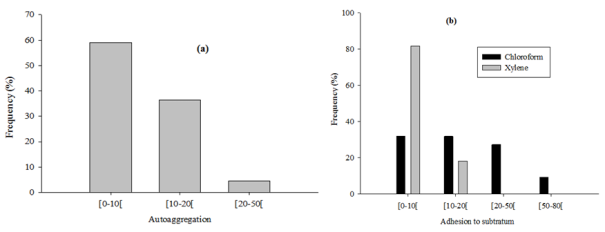
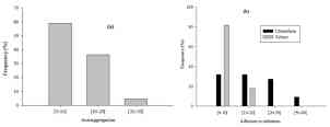
3.7. Autoaggregation and Hydrophobicity Properties of Isolates

The autoaggregation values ranged from 7.8 to 32.8% after 5 h of incubation (Figure 2a). Concerning hydrophobicity using xylene, cell surface hydrophobicity depended on the isolates with a maximum value of 16.4% (Figure 2b). For the adhesion to chloroform, values ranged from 39 and 77% with the maximum values of 39.1% (PD8), 42% (PD10), 60.3% (PB1) and 76.6% (PD12). The PD8, PD10, PB1, and PD12 isolates showed more hydrophilic cell surface properties with strong affinity for chloroform, which means they are strong electron donors.
Figure 2. Adhesion to the substratum (a) and autoaggregation (b) of LAB.
Autoaggregation and hydrophobicity are known as adhesive properties of strains to epithelial cells and mucosal surfaces. It has been suggested to be an important property of many bacterial strains used as probiotics . Cell-forming aggregates can potentially decrease pathogenic bacteria adhesion to the intestinal mucosa, preventing colonization and improving elimination from the intestinal environment . Similarly, García-Cayuela et al. working on one hundred twenty-six L. plantarum strains showed that aggregated communities survive and proliferate under conditions that reduce the prevalence of single non-aggregated cells like S. aureus, L. monocytogenes and E. coli. In contrast, Collado et al. obtained lower autoaggregation percentage values with L. acidophilus NCFM (12.6%), L. casei Shirota, L. rhamnosus LC-705 (18.2%) and L. salivarius Ls-33 (15.2%). The results obtained with different techniques may not be comparable, and that could explain the variability observed. Kos et al. showed with L. acidophilus M92 that the culture method could affect bacterial aggregation and observed that cells obtained after culture in broth medium and suspension in PBS buffer (pH 7.2) had greater characteristics. Janković et al. observed the similarly results using L. plantarum B.
Concerning hydrophobicity or adhesion to hydrocarbons, it is likely affected by environmental factors such as chemical, physical, and enzymatic treatments and can result in a modification of the physiochemical properties of cell surfaces, which later affects bacterial hydrophobicity . Tuomola et al. working on four Lactobacillus strains reported that the adhesion of L. acidophilus 1 and L. rhamnosus GG to human intestinal mucus glycoproteins was due to their proteinaceous structures. Greene and Klaenhammer. proposed that S-layer proteins and other cell-associated proteins are involved in cell protection, surface recognition, and adherence of L. acidophilus BG2FO4. Kos et al. suggested that only pronase- and pepsin sensitive proteins of the surface layer are responsible for the hydrophobicity of the cell surface in bacteria. Frece et al. obtained a better adhesion of L. acidophilus M92 in comparison to L. plantarum L4 and E. faecium L3 examined and explained by the presence of S-layer proteins on the surface of L. acidophilus M92. Therefore, several authors reported that the presence of proteinaceous structures on the cell surface results in higher hydrophobicity, while hydrophilic surfaces are associated with the presence of polysaccharides . On the other hand, Granato et al. reported that lipoteichoic acids are one of the factors responsible for the adhesion of L. johnsonii LaI. According to Sorongon et al. , bacterial hydrophobicity could also be affected by the culture medium used, thermal treatment, and bacterial age.

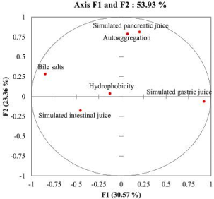
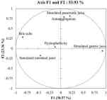
3.8. Correlation Analysis

The discrimination of lactic acid bacteria using all probiotic characteristics tested and isolates was carried out through principal component analysis and the results obtained show that the 06 variables could be organized into two components F1 and F2 expressing 53.93% of the variations (Figure 3). Three components have eigenvalues greater than 1. The contribution of variables to factors (Table 4) showed that F1 (30.57%) was related to the tolerance of simulated gastric juice and bile salts tolerance, F2 (23.36%) was related to the tolerance of simulated pancreatic juice and the autoaggregation property and F3 (18.60%) to the hydrophobicity property and to the tolerance of simulated intestinal juice.
Figure 3. Biplot shows the projection of the isolates onto the plane formed by F1 and F2.
About 50% of the strains have some similar characteristics, while the correlation between autoaggregation property, simulated gastric juice survival and bile salts survival was observed. PD6 presented the highest autoaggregation value inversely to strain PB1.
Furthermore, there was a negative correlation between hydrophobicity and the simulated gastric juice that could be explained by the physicochemical composition of the microbial cell surface structure. Bile salts and simulated intestinal juice tolerance were positively correlated due to analytical methods that included the use of bile acids in both cases. We noted a positive and significant correlation between simulated pancreatic juice and autoaggregation, demonstrating that the physicochemical composition of the surface structure of the bacterial cell was not affected by the presence of pancreatin. Kos et al. found similar results and reported that Lactobacillus autoaggregation was not influenced by treatment using proteolytic enzymes.
Table 3. Contribution of variables to the factors in the PCA based on correlations.

Variables

F1

F2

F3

Hydrophobicity

-0.126

0.038

0.795

Autoaggregation

0.07

0.79

-0.235

Simulated gastric juice

0.922

-0.061

-0.149

Simulated pancreatic juice

0.203

0.813

0.026

Simulated intestinal juice

-0.455

-0.176

-0.635

Bile salts

-0.846

0.284

0.047

Values shown in bold are significant with α = 0.05
Based on the principal component analysis and the probiotic properties of the 22 isolates tested, six, including PD2, PD6, PB3, PB4, PD9, and KB3 exhibited interesting probiotic aptitudes. Comparison of biochemical profile on the API online portal allowed us to identify isolates PD2, PD6, PB3, PD9 as L. casei (identification percentage: 99.8 – 100%), PB4 as L. plantarum (identification percentage = 99.9%) and KB3 as L. Lactis (identification percentage = 98.9%).
4. Conclusion
In summary, L. casei, L. plantarum, and L. lactis isolated from traditional fermented cow’s milks of Cameroon showed interesting probiotic properties in vitro. As a preliminary test, in vitro tests have shown definite probiotic potential for the selected isolates. They are an interesting candidate for functional food formulations. However, the final selection would also depend on their probiotic and functional characteristics in vivo.
Abbreviations
LAB: Lactic acid bacteria
MATS: Microbial adhesion to solvents
TFM: traditional fermented milks
ANOVA: Analysis of variance
TSA: Tryptic Soy Agar
TSB: Tryptic Soy broth
MHA: Mueller Hinton agar
MRS: De Mann Rogosa and Sharpe
SGJ: Simulated gastric juice
SPJ: Simulated pancreatic juice
SIJ: Simulated intestinal juice
BS: Bile salts
BSH: Bile salt hydrolases
Author Contributions
Nodem Sohanang Francky Steve: Conceptualization, Resources, Software, Formal Analysis, Methodology, Writing – original draft, Writing – review & editing.
Mohammadou Bouba Adji: Supervision, Validation, Investigation, Visualization, Methodology, Project administration.
Sokamte Tegang Alphonse: Software, Methodology, Writing – original draft, Writing – review & editing.
Mbawala Augustin: Supervision, Funding acquisition, Validation, Investigation, Visualization, Project administration, Writing – review & editing.
Tatsadjieu Ngoune Leopold: Supervision, Validation, Investigation Project administration, Writing – review & editing.
Funding
The authors declare that they have not received any internal or external funding.
Data Availability Statement
Data supporting the findings of this study are available from the corresponding author, upon reasonable request.
Conflicts of Interest
The authors declare no conflicts of interest.
References
[1] FAO/WHO, “FAO/WHO expert consultation on evaluation of health and nutritional properties of probiotics in food including powder milk with live lactic acid bacteria,” 2002.
[2] K. Angmo, A. Kumari, Savitri, and T. C. Bhalla, “Probiotic characterization of lactic acid bacteria isolated from fermented foods and beverage of Ladakh,” LWT - Food Sci. Technol., vol. 66, no. October, pp. 428–435, 2016,
[3] A. Abushelaibi, S. Al-Mahadin, K. El-Tarabily, N. P. Shah, and M. Ayyash, “Characterization of potential probiotic lactic acid bacteria isolated from camel milk,” LWT - Food Sci. Technol., vol. 79, pp. 316–325, 2017,
[4] Z. Guo, J. Wang, L. Yan, W. Chen, X. ming Liu, and H. ping Zhang, “In vitro comparison of probiotic properties of Lactobacillus casei Zhang, a potential new probiotic, with selected probiotic strains,” LWT - Food Sci. Technol., vol. 42, no. 10, pp. 1640–1646, 2009,
[5] Y. Moreno, M. C. Collado, M. A. Ferrús, J. M. Cobo, E. Hernández, and M. Hernández, “Viability assessment of lactic acid bacteria in commercial dairy products stored at 4°C using LIVE/DEAD® BacLightTM staining and conventional plate counts,” Int. J. Food Sci. Technol., vol. 41, no. 3, pp. 275–280, 2006,
[6] T. Faye, A. Tamburello, G. E. Vegarud, and S. Skeie, “Survival of lactic acid bacteria from fermented milks in an in vitro digestion model exploiting sequential incubation in human gastric and duodenum juice,” J. Dairy Sci., vol. 95, no. 2, pp. 558–566, 2012,
[7] F. M. de Sant’Anna et al., “Assessment of the probiotic potential of lactic acid bacteria isolated from Minas artisanal cheese produced in the Campo das Vertentes region, Brazil,” Int. J. Dairy Technol., vol. 70, no. 4, pp. 592–601, 2017,
[8] L. De Vuyst, M. R. Foulquié Moreno, and H. Revets, “Screening for enterocins and detection of hemolysin and vancomycin resistance in enterococci of different origins,” Int. J. Food Microbiol., vol. 84, no. 3, pp. 299–318, 2003,
[9] M. Sidira et al., “Effect of probiotic-Fermented milk administration on gastrointestinal survival of Lactobacillus casei ATCC 393 and modulation of intestinal microbial flora,” J. Mol. Microbiol. Biotechnol., vol. 19, no. 4, pp. 224–230, 2011,
[10] D. V. Sieladie, F. N. Zambou, P. M. Kaktcham, A. Cresci, and F. Fonteh, “Probiotic properties of lactobacilli strains isolated from raw cow milk in the western highlands of Cameroon,” Innov. Rom. Food Biotechnol., vol. 9, no. August 2015, pp. 12–28, 2011.
[11] M. P. Vélez, K. Hermans, T. L. A. Verhoeven, S. E. Lebeer, J. Vanderleyden, and S. C. J. De Keersmaecker, “Identification and characterization of starter lactic acid bacteria and probiotics from Columbian dairy products,” J. Appl. Microbiol., vol. 103, no. 3, pp. 666–674, 2007,
[12] M. Iranmanesh, H. Ezzatpanah, and N. Mojgani, “Antibacterial activity and cholesterol assimilation of lactic acid bacteria isolated from traditional Iranian dairy products,” LWT - Food Sci. Technol., vol. 58, no. 2, pp. 355–359, 2014,
[13] J. M. Mathara et al., “Functional characteristics of Lactobacillus spp. from traditional Maasai fermented milk products in Kenya,” Int. J. Food Microbiol., vol. 126, no. 1–2, pp. 57–64, 2008,
[14] M. Abdelbasset and K. Djamila, “Antimicrobial activity of autochthonous lactic acid bacteria isolated from Algerian traditional fermented milk Raib,” African J. Biotechnol., vol. 7, no. 16, pp. 2908–2914, 2008,
[15] D. Guilhem, “Le lait de vache dans les sociétés peules Pratiques alimentaires et symbolisme d ’ un critère identitaire,” pp. 1–8, 2006.
[16] N. M. M. Tchamba et al., “Assessment of Probiotic Potential of Lactic Acid Bacteria Isolated from Bottle Gourds (Calabash) of Milk Fermentation of Mbéré, Cameroon,” J. Pharm. Res. Int., vol. 33, pp. 356–369, 2021,
[17] W. Liu et al., “Isolation and identification of lactic acid bacteria from Tarag in Eastern Inner Mongolia of China by 16S rRNA sequences and DGGE analysis,” Microbiol. Res., vol. 167, no. 2, pp. 110–115, 2012,
[18] D. Libouga, J. Essia Ngang, and H. Halilou, “Qualité de quelques laits fermentés camerounais,” Sci. Aliments, vol. 25, no. 1, pp. 53–66, Feb. 2005,
[19] J. Maïworé, L. Tatsadjieu, and I. Piro-metayer, “Identification of yeasts present in artisanal yoghurt and traditionally fermented milks consumed in the northern part of Cameroon,” vol. 6, pp. 0–8, 2019,
[20] A. Mbawala, M. M. Pahane, and H. T. Mouafo, “Effect of manufacturing practices on the microbiological quality of fermented milk (Pendidam) of some localities of Ngaoundere (Cameroon),” Int. J. Curr. Microbiol. Appl. Sci., vol. 3, no. 11, pp. 71–81, 2014.
[21] T. H. Mouafo, A. Mbawala, and R. Ndjouenkeu, “Effect of different carbon sources on biosurfactants’ production by three strains of Lactobacillus spp.,” Biomed Res. Int., vol. 2018, 2018,
[22] M. T. Hippolyte, M. Augustin, T. M. Hervé, N. Robert, and S. Devappa, “Application of response surface methodology to improve the production of antimicrobial biosurfactants by Lactobacillus paracasei subsp. tolerans N2 using sugar cane molasses as substrate,” Bioresour. Bioprocess., vol. 5, no. 1, 2018,
[23] Codex Alimentarus, “Méthodes d’analyse et d’échantillonnage recommandées, Première partie, Méthodes d’analyse et d’échantillonnage par ordre alphabétique des catégories et noms de produit,” pp. 18–24, 1999.
[24] A. Mbawala, P. Y. Mahbou, H. T. Mouafo, and L. N. Tatsadjieu, “Antibacterial activity of some lactic acid bacteria isolated from a local fermented milk product (Pendidam) in Ngaoundere, Cameroon,” J. Anim. Plant Sci., vol. 23, no. 1, pp. 157–166, 2013.
[25] J. C. de Man, M. Rogosa, and M. E. Sharpe, “A medium used for the cultivation of Lactobacilli,” J Appl Bacteriol, vol. 23, pp. 130–135, 1960.
[26] H. P. Fleming, J. L. Etchells, and R. N. Costilow, “Microbial inhibition by an isolate of Pediococcus from cucumber brines.,” Appl. Microbiol., vol. 30, no. 6, pp. 1040–2, 1975.
[27] B. Kos, J. Šušković, S. Vuković, M. Sǐmpraga, J. Frece, and S. Matošić, “Adhesion and aggregation ability of probiotic strain Lactobacillus acidophilus M92,” J. Appl. Microbiol., vol. 94, no. 6, pp. 981–987, 2003,
[28] CASFM/EUCAST, “Comité de l’Antibiogramme de la Société Française de Microbiologie,” Eur. Soc. Clin. Microbiol. Infect. Dis., vol. V1.0 2019, pp. S3–S4, 2019,
[29] T. J. Eaton and M. J. Gasson, “Molecular Screening of Enterococcus Virulence Determinants and Potential for Genetic Exchange between Food and Medical Isolates” Appl. Environ. Microbiol., vol. 67, no. 4, pp. 1628–1635, 2001,
[30] C. Jiwoua and J. B. Millière, “Flore lactique et entérocoques du lait caillé (Pindidam) produit dans l’Adamaoua (Cameroun),” Lait, vol. 70, no. 5–6, pp. 475–486, 1990.
[31] K. Voulgari, M. Hatzikamari, A. Delepoglou, P. Georgakopoulos, E. Litopoulou-Tzanetaki, and N. Tzanetakis, “Antifungal activity of non-starter lactic acid bacteria isolates from dairy products,” Food Control, vol. 21, no. 2, pp. 136–142, 2010,
[32] H. J. Song and J. Richard, “Antilisterial activity of three bacteriocins used at sub minimal inhibitory concentrations and cross-resistance of the survivors,” Int. J. Food Microbiol., vol. 36, no. 2–3, pp. 155–161, 1997,
[33] J. Magnusson, K. Ström, S. Roos, J. Sjögren, and J. Schnürer, “Broad and complex antifungal activity among environmental isolates of lactic acid bacteria,” FEMS Microbiol. Lett., vol. 219, no. 1, pp. 129–135, 2003,
[34] J. Magnusson and J. Schnürer, “Lactobacillus coryniformis subsp. coryniformis Strain Si3 Produces a Broad-Spectrum Proteinaceous Antifungal Compound,” Appl. Environ. Microbiol., vol. 67, no. 1, pp. 1–5, 2001,
[35] J. Schnürer and J. Magnusson, “Antifungal lactic acid bacteria as biopreservatives,” Trends Food Sci. Technol., vol. 16, no. 1–3, pp. 70–78, 2005,
[36] D. K. D. Dalié, A. M. Deschamps, and F. Richard-Forget, “Lactic acid bacteria - Potential for control of mould growth and mycotoxins: A review,” Food Control, vol. 21, no. 4, pp. 370–380, 2010,
[37] A. Mechai, M. Debabza, and D. Kirane, “Screening of technological and probiotic properties of lactic acid bacteria isolated from Algerian traditional fermented milk products,” Int. Food Res. J., vol. 21, no. 6, pp. 2451–2457, 2014.
[38] T. P. Singh, G. Kaur, R. K. Malik, U. Schillinger, C. Guigas, and S. Kapila, “Characterization of Intestinal Lactobacillus reuteri Strains as Potential Probiotics,” Probiotics Antimicrob. Proteins, vol. 4, no. 1, pp. 47–58, Mar. 2012,
[39] M. Guetouache and B. Guessas, “Characterization and identification of lactic acid bacteria isolated from traditional cheese (Klila) prepared from cows milk,” African J. Microbiol. Res., vol. 9, no. 2, pp. 71–77, 2015,
[40] C. M. Kalui, J. M. Mathara, P. M. Kutima, C. Kiiyukia, and L. E. Wongo, “Functional characteristics of Lactobacillus plantarum and Lactobacillus rhamnosus from ikii, a Kenyan traditional fermented maize porridge,” African J. Biotechnol., vol. 8, no. 18, pp. 4363–4373, 2009,
[41] M. T. Liong and N. P. Shah, “Acid and bile tolerance and cholesterol removal ability of lactobacilli strains,” J. Dairy Sci., vol. 88, no. 1, pp. 55–66, 2005,
[42] P. D. Cotter and C. Hill, “Surviving the Acid Test: Responses of Gram-Positive Bacteria to Low pH,” Microbiol. Mol. Biol. Rev., vol. 67, no. 3, pp. 429–453, 2003,
[43] W. P. Charteris, P. M. Kelly, L. Morelli, and J. K. Collins, “Antibiotic susceptibility of potentially probiotic Bifidobacterium isolates from the human gastrointestinal tract,” Lett. Appl. Microbiol., vol. 26, no. 5, pp. 333–337, 1998,
[44] P. A. Maragkoudakis, G. Zoumpopoulou, C. Miaris, G. Kalantzopoulos, B. Pot, and E. Tsakalidou, “Probiotic potential of Lactobacillus strains isolated from dairy products,” Int. Dairy J., vol. 16, no. 3, pp. 189–199, 2006,
[45] B. E. Minelli et al., “Assessment of novel probiotic Lactobacillus casei strains for the production of functional dairy foods,” Int. Dairy J., vol. 14, no. 8, pp. 723–736, 2004,
[46] S. Salminen et al., “Demonstration of safety of probiotics - A review,” Int. J. Food Microbiol., vol. 44, no. 1–2, pp. 93–106, 1998,
[47] D. Donohue and S. Salminen, “Safety of probiotic bacteria,” Asian Pacific J. Clin. Nutr., vol. 5, pp. 25–28, 1996.
[48] E. Muñoz-Atienza et al., “Antimicrobial activity, antibiotic susceptibility and virulence factors of Lactic Acid Bacteria of aquatic origin intended for use as probiotics in aquaculture,” BMC Microbiol., vol. 13, no. 1, 2013,
[49] D. J. Birri, D. A. Brede, G. T. Tessema, and I. F. Nes, “Bacteriocin Production, Antibiotic Susceptibility and Prevalence of Haemolytic and Gelatinase Activity in Faecal Lactic Acid Bacteria Isolated from Healthy Ethiopian Infants,” Microb. Ecol., vol. 65, no. 2, pp. 504–516, 2013,
[50] A. Hummel, W. H. Holzapfel, and C. M. A. P. Franz, “Characterisation and transfer of antibiotic resistance genes from enterococci isolated from food,” Syst. Appl. Microbiol., vol. 30, no. 1, pp. 1–7, 2007,
[51] A. S. Hummel, C. Hertel, W. H. Holzapfel, and C. M. A. P. Franz, “Antibiotic resistances of starter and probiotic strains of lactic acid bacteria,” Appl. Environ. Microbiol., vol. 73, no. 3, pp. 730–739, 2007,
[52] M. S. Ammor, A. Belén Flórez, and B. Mayo, “Antibiotic resistance in non-enterococcal lactic acid bacteria and bifidobacteria,” Food Microbiol., vol. 24, no. 6, pp. 559–570, 2007,
[53] M. C. Collado, J. Meriluoto, and S. Salminen, “Adhesion and aggregation properties of probiotic and pathogen strains,” Eur. Food Res. Technol., vol. 226, no. 5, pp. 1065–1073, 2008,
[54] Y. Bao et al., “Screening of potential probiotic properties of Lactobacillus fermentum isolated from traditional dairy products,” Food Control, vol. 21, no. 5, pp. 695–701, 2010,
[55] T. García-Cayuela et al., “Adhesion abilities of dairy Lactobacillus plantarum strains showing an aggregation phenotype,” Food Res. Int., vol. 57, pp. 44–50, 2014,
[56] T. Janković, J. Frece, M. Abram, and I. Gobin, “Aggregation ability of potential probiotic Lactobacillus plantarum strains,” Int. J. Sanit. Eng. Res., vol. 6, no. 1, pp. 19–24, 2012.
[57] T. Wadstroum, K. Andersson, M. Sydow, L. Axelsson, S. Lindgren, and B. Gullmar, “Surface properties of lactobacilli isolated from the small intestine of pigs,” J. Appl. Bacteriol., vol. 62, no. 6, pp. 513–520, 1987,
[58] Q. Li, X. Liu, and J. Zhou, “Aggregation and adhesion abilities of 18 lactic acid bacteria strains isolated from traditional fermented food,” Int. J. Agric. Policy Res., vol. 3, no. February, pp. 84–92, 2015.
[59] J. D. Greene and T. R. Klaenhammer, “Factors involved in adherence of lactobacilli to human Caco-2 cells,” Appl. Environ. Microbiol., vol. 60, no. 12, pp. 4487–4494, 1994.
[60] E. M. Tuomola, A. C. Ouwehand, and S. J. Salminen, “Chemical, physical and enzymatic pre-treatments of probiotic lactobacilli alter their adhesion to human intestinal mucus glycoproteins,” Int. J. Food Microbiol., vol. 60, no. 1, pp. 75–81, 2000,
[61] C. Pelletier, C. Bouley, C. Cayuela, S. Bouttier, P. Bourlioux, and M. N. Bellon-Fontaine, “Cell surface characteristics of Lactobacillus casei subsp. casei, Lactobacillus paracasei subsp. paracasei, and Lactobacillus rhamnosus strains,” Appl. Environ. Microbiol., vol. 63, no. 5, pp. 1725–1731, 1997.
[62] D. Granato et al., “Cell surface-associated lipoteichoic acid acts as an adhesion factor for attachment of Lactobacillus johnsonii La1 to human enterocyte-like Caco-2 cells,” Appl. Environ. Microbiol., vol. 65, no. 3, pp. 1071–1077, 1999.
[63] J. Frece, B. Kos, I. K. Svetec, Z. Zgaga, V. Mrša, and J. Šušković, “Importance of S-layer proteins in probiotic activity of Lactobacillus acidophilus M92,” J. Appl. Microbiol., vol. 98, no. 2, pp. 285–292, 2005,
[64] M. L. Sorongon, R. A. Bloodgood, and R. P. Burchard, “Hydrophobicity, Adhesion, and Surface-Exposed Proteins of Gliding Bacteria,” Appl. Environ. Microbiol., vol. 57, no. 11, pp. 3193–3199, 1991.
Cite This Article
  • APA Style

    Steve, N. S. F., Adji, M. B., Alphonse, S. T., Augustin, M., Leopold, T. N. (2024). Isolation and Probiotic Characterization of Lactic Acid Bacteria from Kindirmou and Pendidam in Adamawa Region (Cameroon). Advances in Biochemistry, 12(2), 35-48. https://doi.org/10.11648/j.ab.20241202.11

    Copy | Download

    ACS Style

    Steve, N. S. F.; Adji, M. B.; Alphonse, S. T.; Augustin, M.; Leopold, T. N. Isolation and Probiotic Characterization of Lactic Acid Bacteria from Kindirmou and Pendidam in Adamawa Region (Cameroon). Adv. Biochem. 2024, 12(2), 35-48. doi: 10.11648/j.ab.20241202.11

    Copy | Download

    AMA Style

    Steve NSF, Adji MB, Alphonse ST, Augustin M, Leopold TN. Isolation and Probiotic Characterization of Lactic Acid Bacteria from Kindirmou and Pendidam in Adamawa Region (Cameroon). Adv Biochem. 2024;12(2):35-48. doi: 10.11648/j.ab.20241202.11

    Copy | Download

  • @article{10.11648/j.ab.20241202.11,
      author = {Nodem Sohanang Francky Steve and Mohammadou Bouba Adji and Sokamte Tegang Alphonse and Mbawala Augustin and Tatsadjieu Ngoune Leopold},
      title = {Isolation and Probiotic Characterization of Lactic Acid Bacteria from Kindirmou and Pendidam in Adamawa Region (Cameroon)},
      journal = {Advances in Biochemistry},
      volume = {12},
      number = {2},
      pages = {35-48},
      doi = {10.11648/j.ab.20241202.11},
      url = {https://doi.org/10.11648/j.ab.20241202.11},
      eprint = {https://article.sciencepublishinggroup.com/pdf/10.11648.j.ab.20241202.11},
      abstract = {The microorganisms intended for use as probiotics in food formulation should exert health benefit effects and be regarded as safe for animals and humans uses. The aim of this study was to evaluate the probiotic potential of lactic acid bacteria (LAB) isolated from pendidam and kindirmou, two traditional fermented milks (TFM) produced in the Adamawa region (Cameroon). Twenty-five samples (pendidam: 13 and kindirmou: 12) were randomly collected in five markets of Ngaoundere (n = 17 samples) and Meiganga (n = 8 samples). These samples were screened for their antimicrobial activity, and nine TFMs were retained. Lactic acid bacteria were isolated from these samples and their antimicrobial activity was already evaluated. Based on the inhibition zone, twenty-two LABs were retained and examined in vitro for potential probiotic properties based on their low pH tolerance, resistance to bile salts, tolerance to simulated gastrointestinal juices, hydrophobicity, autoaggregation, gelatinase and hemolytic activities. The outcome of these parameters studied was used as input data for a principal component analysis (PCA) to select the most promising isolate, and the six potential probiotic isolates were characterized through a biochemical profile. The characterized isolates have been identified as Lactiplantibacillus plantarum, Lacticaseibacillus casei, and Lactococcus lactis. Traditional fermented milks contain LAB with important properties that can be utilized in the formulation of functional foods.},
     year = {2024}
    }
    

    Copy | Download

  • TY  - JOUR
    T1  - Isolation and Probiotic Characterization of Lactic Acid Bacteria from Kindirmou and Pendidam in Adamawa Region (Cameroon)
    AU  - Nodem Sohanang Francky Steve
    AU  - Mohammadou Bouba Adji
    AU  - Sokamte Tegang Alphonse
    AU  - Mbawala Augustin
    AU  - Tatsadjieu Ngoune Leopold
    Y1  - 2024/04/12
    PY  - 2024
    N1  - https://doi.org/10.11648/j.ab.20241202.11
    DO  - 10.11648/j.ab.20241202.11
    T2  - Advances in Biochemistry
    JF  - Advances in Biochemistry
    JO  - Advances in Biochemistry
    SP  - 35
    EP  - 48
    PB  - Science Publishing Group
    SN  - 2329-0862
    UR  - https://doi.org/10.11648/j.ab.20241202.11
    AB  - The microorganisms intended for use as probiotics in food formulation should exert health benefit effects and be regarded as safe for animals and humans uses. The aim of this study was to evaluate the probiotic potential of lactic acid bacteria (LAB) isolated from pendidam and kindirmou, two traditional fermented milks (TFM) produced in the Adamawa region (Cameroon). Twenty-five samples (pendidam: 13 and kindirmou: 12) were randomly collected in five markets of Ngaoundere (n = 17 samples) and Meiganga (n = 8 samples). These samples were screened for their antimicrobial activity, and nine TFMs were retained. Lactic acid bacteria were isolated from these samples and their antimicrobial activity was already evaluated. Based on the inhibition zone, twenty-two LABs were retained and examined in vitro for potential probiotic properties based on their low pH tolerance, resistance to bile salts, tolerance to simulated gastrointestinal juices, hydrophobicity, autoaggregation, gelatinase and hemolytic activities. The outcome of these parameters studied was used as input data for a principal component analysis (PCA) to select the most promising isolate, and the six potential probiotic isolates were characterized through a biochemical profile. The characterized isolates have been identified as Lactiplantibacillus plantarum, Lacticaseibacillus casei, and Lactococcus lactis. Traditional fermented milks contain LAB with important properties that can be utilized in the formulation of functional foods.
    VL  - 12
    IS  - 2
    ER  - 

    Copy | Download

Author Information
  • Abstract
  • Keywords
  • Document Sections

    1. 1. Introduction
    2. 2. Materials and Methods
    3. 3. Results and Discussion
    4. 4. Conclusion
    Show Full Outline
  • Abbreviations
  • Funding
  • Data Availability Statement
  • Conflicts of Interest
  • References
  • Cite This Article
  • Author Information