Research Article | | Peer-Reviewed

Analysis of Clinical Outcomes of Percutaneous Kyphoplasty and Vertebroplasty in the Treatment of Osteoporosis-Induced Vertebral Compression Fracture

Received: 7 March 2024    Accepted: 20 March 2024    Published: 17 April 2024
Views:       Downloads:
Abstract

Percutaneous kyphoplasty (PKP) and Percutaneous vertebroplasty (PVP) have become very effective to treating the Osteoporosis vertebral compression fractures (OVCFs) and patients can get immediate relief from the pain and it can improve the motor functions of patient’s health status and widely used in clinical practice. Vertebroplasty and kyphoplasty are minimally invasive procedures for the treatment of painful vertebral compression fractures (VCF), which are fractures involving the vertebral bodies that make up the spinal column. Vertebroplasty are most often used to treat an injury called a compression fracture. This kind of injuries are most often caused by osteoporosis, which is the reason of bone weakness and osteoporosis are most common in older people. This study aimed to examine the osteoporosis vertebral compression fractures (OVCFs) of Jinan, Shandong province, China, between male and female participants who were over 50 years of age. There are very few pertinent data regarding the relative or absolute contraindication of percutaneous vertebroplasty (PVP) and percutaneous kyphoplasty (PKP) for severe osteoporotic vertebral compression fractures (OVCFs). The purpose of this research was to assess and contrast the effectiveness of traditional kyphoplasty and vertebroplasty using high-viscosity cement in the treatment of severe OVCFs. In aged people, osteoporotic vertebral compression fractures (OVCFs) are prevalent and can result in significant osteoporosis vertebral compression fractures. Treatment options for individuals with these types of fractures include conservative measures, percutaneous vertebroplasty (PVP), and percutaneous kyphoplasty (PKP). We compared the clinical effectiveness of PVP and PKP in treating osteoporotic vertebral compression fractures in this investigation. Therefore, we conducted a comparative study on Osteoporosis vertebral compression fractures (OVCFs) patients who received either PVP or PKP treatment in our hospitals from January 2020 to December 2020 to investigate the clinical efficacy, advantages and disadvantages of the surgical methods, and to provide a reference for clinical selection of treatment methods.

Published in Journal of Surgery (Volume 12, Issue 2)
DOI 10.11648/j.js.20241202.15
Page(s) 45-65
Creative Commons

This is an Open Access article, distributed under the terms of the Creative Commons Attribution 4.0 International License (http://creativecommons.org/licenses/by/4.0/), which permits unrestricted use, distribution and reproduction in any medium or format, provided the original work is properly cited.

Copyright

Copyright © The Author(s), 2024. Published by Science Publishing Group

Keywords

Percutaneous Vertebroplasty (PVP), Percutaneous Kyphoplasty (PKP), Severe Osteoporotic Vertebral Compression Fractures (OVCFs)

1. Introduction
Osteoporotic vertebral compression fractures (OVCF) are excruciating injuries that are commonly treated with percutaneous vertebroplasty (PVP) in recent years . Nonetheless, disagreement on PVP's clinical role persists. Furthermore, conservative medicinal therapy is an effective treatment for the majority of individuals with acute painful OVCF. However, many older patients with established osteoporosis and significant pain are unable to receive enough pain treatment, which results in stalled progress and catastrophic consequences that require a longer recovery period.
Techniques that are safe and effective for treating symptomatic spinal compression fractures are percutaneous vertebroplasty and kyphoplasty. According to available research, when compared to conservative treatment, vertebroplasty and kyphoplasty dramatically reduce pain and have reduced rates of serious consequences. Implanting bone cement into a partially collapsed vertebral body is known as vertebroplasty, a minimally invasive technique. When a vertebral body is fractured, kyphoplasty involves inflating a balloon to lessen the likelihood that the vertebra would collapse. The fracture is then stabilized by injecting bone cement into the space that the balloon has created. Cervical vertebrae resulting from benign bone tumor’s, painful osteoporotic or osteolytic fractures caused by malignant processes, and acute stable traumatic fractures with a kyphotic angle greater than 15° are among the conditions that warrant cement augmentation operations. On average, the pedicle width of T4-T6 in Singaporean Chinese is 4 mm. In Caucasians, the percentages for local or systemic infection, coagulopathy, and allergy to the bone-cement cannula (which is 4 mm) are 33% for T4, 25% for T5, and 17% for T6. The transpedicular approach in the upper and intermediate thoracic spine can be performed with smaller needles (13 gauge) .
For VCFs, there are numerous nonsurgical and surgical therapy choices. Bed rest, analgesics, hyperextension braces (for benign fractures), and radiation therapy (including proton beam therapy for diffuse pathologic osseous involvement and stereotactic body radiation therapy for oligometastatic fractures) are examples of nonsurgical treatments. These therapies, however, might not be successful and might have a number of negative effects. For instance, bed rest immobilization might result in venous thromboembolism, paralysis, and pressure ulcers. Balloon kyphoplasty (KP), percutaneous vertebroplasty (VP), internal bracing, and, more recently, vertebral implants like the SpineJack vertebral implant (SJ) system (Stryker) are examples of minimally invasive surgical therapies. Because surgery has been shown to provide quick and long-lasting effects, it has grown in popularity. It has been demonstrated that vertebral augmentation techniques significantly lower loads for a range of preaugmentation vertebral heights in 3D reconstruction models of compression fractures. Controversial studies have examined major side effects from surgical procedures, like fresh fractures to the vertebrae, but more recent meta-analyses have not confirmed these findings. In addition, it was discovered that patients with osteoporotic compression fractures had a lower death rate after spinal augmentation as opposed to nonsurgical care. Nevertheless, elderly patients with various comorbidities and VCF frequently encounter risks and contraindications to vertebral augmentation, which can result in worse outcomes like higher rates of morbidity and mortality and problems following surgery .
Studies have demonstrated that PVP is a highly effective treatment for lumbar and back pain. You can get out of bed on the second post-operative day to provide easier nursing care and hasten recuperation. Clinical research has revealed, however, that some patients with low back pain following PVP continue to experience recurrent problems and may even be unable to receive relief. At this time, it is unclear what factors influence the clinical efficacy. This study's main goal was to determine how bone cement distribution—either unilaterally or bilaterally—affects the clinical effectiveness following PVP. The clinical efficacy of PVP treatment for 52 OVCF patients treated in our hospital between January 2020 and December 2020 was investigated through a retrospective study. The patients were divided into two groups based on whether bone cement was distributed unilaterally or bilaterally. Relevant indicators were compared between the two groups .
2. Materials and Methods
This technique describes the steps involved in carrying out a study to assess how well a physical activity intervention improves cardiovascular health in older people. The purpose of the study is to find out how a regimented exercise programmed affects important cardiovascular metrics including heart rate, blood pressure, and aerobic capacity. Pre- and post-intervention assessments are a part of the randomized controlled trial (RCT) in this study, which complies with ethical guidelines to protect participants' health and safety. A randomized controlled trial (RCT) design will be employed in this study to assess how the physical activity intervention affects participants' cardiovascular health who are elderly. Participants will be randomly assigned to either the intervention group, which will receive the structured exercise programmed, or the control group, which will carry on as normal with no changes to their daily routine. The randomization process will be carried out using a computer-generated random sequence. A pre-intervention and post-intervention measuring design will be used for this study, with baseline measures being acquired prior to the intervention and post-intervention measurements being gathered at the conclusion of the investigation. The control group will not get any additional interventions and will continue with their regular daily routines during the study period.
Changes in heart rate (HR), diastolic blood pressure (DBP), and systolic blood pressure (SBP) from the start of the 12-week intervention to its conclusion are the main outcome measures. Standardized blood pressure cuffs and heart rate monitors will be used to measure these parameters. A 6-minute walk test (6MWT) will be used to evaluate changes in aerobic capacity as a secondary outcome measure. On a pre-marked track, participants will be told to walk as far as they can in six minutes, and the total distance travelled will be noted.

2.1. Selection Criteria

2.1.1. Inclusion Criteria

Individuals who fulfilled the stated diagnostic standards, based on clinical signs and imaging analysis Only one single responsible vertebra is involved; Bone density T value < − 2.5 on dual energy X-ray absorptiometry (DXA), which is in accordance with the diagnostic criteria for osteoporosis.
Before being admitted, every patient had conservative care for a minimum of three months. Prior to making a decision, the patients were furnished with information regarding the benefits and drawbacks of both PVP and PKP. In the interim, they were told that there wasn't enough evidence in medicine to determine which was superior.
1) Vertebral compression fractures caused by osteoporosis: Based on clinical assessment, radiographic evidence (such as X-rays), and the presence of characteristic symptoms (e.g., acute onset of back pain, localized tenderness, or limited spinal mobility), participants had to have a confirmed diagnosis of OVCFs.
2) Gender and age: Participants in the study belonged to a particular age range; in general, older persons 50 years of age or older were the focus of the study because OVCFs are more common in this demographic. Although postmenopausal women account for the majority of OVCFs, both male and female participants were taken into consideration.
3) Consent: In order to participate in the study, participants had to give informed consent, confirming that they were aware of the methods, goals, possible dangers, and advantages of the study.

2.1.2. Exclusion Criteria

1) Those suffering from pathological vertebral fractures, severe internal illnesses, such as spinal metastatic tumors, and tuberculosis in the vertebrae. Individuals who are unable to endure surgery because to significant liver, renal, or cardiovascular malfunction. Individuals whose clinical information is lacking.
2) Severe comorbidities: Individuals who had severe comorbidities that might have a major effect on the results of the study or their capacity to undergo PVP were not allowed to participate. Severe cardiac disorders, cancers, uncontrolled diabetes, or ongoing infections are examples of these comorbidities.
3) Non-osteoporotic fractures: To maintain the study population's homogeneity, individuals with spinal compression fractures brought on by conditions other than osteoporosis, such as trauma, cancer, or infection, were not allowed to participate.
4) Allergy or contraindications: Participants who had a history of allergies to any of the chemicals used in PVP, including local an aesthetics or polymethylmethacrylate, were not allowed to participate. Additionally disqualified were patients who had ongoing bleeding problems or significant coagulopathy, which are contraindications to PVP.
5) Cognitive impairment: To guarantee correct reporting of results and adherence to the study protocol, participants with significant cognitive impairment or those who were unable to give informed consent were excluded.

2.2. Patients

52 OVCF’s patients without neurologic impairments who had PVP or PKP between January 2020 and December 2020 were enrolled in the study.

2.3. Sample Size Determination and Baseline Data

To maintain statistical power and reduce the possibility of type I and type II errors, the right sample size must be determined. The estimated rate of OVCFs, power (1-β), effect size, predicted dropouts, and desired level of significance (α) were all taken into account while calculating the sample size for the prospective cohort trial.
Previous research on PVP and OVCFs that reported on the effect size and variability in outcome measures like pain intensity or functional status were taken into account when calculating the sample size. The necessary sample size was estimated using statistical software or sample size calculators based on these criteria, with the goal of achieving enough power to identify differences in outcomes that are clinically significant.
In order to examine the clinical outcomes of percutaneous vertebroplasty (PVP) in treating osteoporosis-induced vertebral compression fractures (OVCFs), baseline data collection is an essential first step in a prospective cohort study. Based on baseline evaluations of pain severity, functional status, and quality of life, baseline data offers an overview of the characteristics and medical history of the participants (Litin et al., 2023). The background information gathered in these regions will be explained in this section.

2.4. Demographic Information and Medical History

To describe the study population, demographic information is gathered, such as age, gender, ethnicity, and socioeconomic status. This data aids in determining potential confounding variables and evaluating the generalizability of study findings.
To find pre-existing conditions, comorbidities, and risk factors that could affect the study's results, participants' medical histories are documented. This covers the severity and length of osteoporosis, any previous OVCF history, the existence of chronic illnesses, the use of medications, and the status of smoking.

2.5. Baseline Measurements and Pain Intensity

To evaluate the individuals' starting condition with regard to pain severity, functional state, and quality of life, baseline data are collected.
The Visual Analogue Scale (VAS) and the Numerical Rating Scale (NRS) are two approved measurement instruments that are used to assess pain severity. With higher ratings denoting greater pain severity, participants rate their pain on a scale from 0 to 10.

2.6. Functional Status

Standardized questionnaires such as the Roland-Morris Disability Questionnaire (RMDQ) and Oswestry Disability Index (ODI) are used to evaluate functional status. These instruments assess how OVCFs affect participants' capacity to carry out regular tasks.

2.7. Quality of Life

Assessing participants' general well-being and health-related quality of life is possible through baseline quality of life assessments. A variety of physical and mental health dimensions are evaluated by questionnaires like the Short-Form 36 Health Survey (SF-36).
Researchers set a benchmark for assessing how pain intensity, functional status, and quality of life had changed after the PVP intervention by gathering baseline data. Knowing the features of the study population and pinpointing potential influencing elements for the results is made easier with the use of baseline data.

2.8. Surgical Procedures and Conduct

2.8.1. Surgical Information

Patients in the PKP group underwent general anaesthesia while being operated on in a prone, lordotic posture to preserve posterior extension of the spine. Please refer to our previously published article for information on the conventional technique for PKP surgery. To put it briefly, bilateral balloons were inserted under the endplate via the working tunnel following bilateral transpedicular puncture and cleaning. After gently inflating the balloons to raise the deformed vertebra to its normal height, the superior endplate was elevated and the balloons were deflated. Polymethyl methacrylate cement was utilized to fill the pre-formed cavity once the balloon was taken out. Using C-arm fluoroscopy, the entire process was observed. Most of the stages are similar for PVP groups, however they don't include the balloon. Direct insertion of polymethylmethacrylate bone cement into the vertebra occurred following the creation of a working tunnel.

2.8.2. Clinical and Radiologic Assessment

The visual analogue scale (VAS score 0–10; 0 no pain at all; 10 the worst imaginable) system was employed to evaluate back pain control. Impact on the patient’s daily life was assessed using the Oswestry Disability Index (ODI) questionnaire . Radiographs were taken to measure the rate of cement leakages and refracture, the anterior, middle and posterior vertebral heights, Cobb’s angle and Vertebral wedge ratio of the fractured vertebral body before and after surgery. The operation time, amounts of cement injected, time of hospital stay and intraoperative blood loss of the two procedures were recorded. All radiographic measurements were performed in a double-blinded fashion by 2 orthopedic surgeons.
In the prospective cohort study investigating the clinical outcomes of percutaneous vertebroplasty (PVP) in the treatment of osteoporosis-induced vertebral compression fractures (OVCFs), standardized surgical procedures were employed to ensure consistency and reproducibility across participants. This section will describe the surgical procedures of PVP and how they were conducted.
Percutaneous vertebroplasty is a minimally invasive procedure that involves the injection of bone cement into the fractured vertebral body to provide stabilization and pain relief. The surgical procedure generally follows a similar procedure:
a) Preoperative Preparation: Before the surgery, patients are assessed to ensure they meet the eligibility criteria. Preoperative evaluations may include medical history review, physical examination, laboratory tests, and radiographic imaging to confirm the diagnosis of OVCFs and assess the vertebral fracture characteristics.
b) Anesthesia: PVP is usually performed under local anesthesia and conscious sedation, although general anesthesia can be considered in specific cases. The anesthesia approach is determined based on the patient's medical condition, surgeon's preference, and patient comfort.
c) Patient Positioning: The patient is positioned prone (lying face down) on the operating table to provide optimal access to the vertebral column. Padding and positioning aids are used to maintain patient comfort and prevent pressure injuries.
d) Fluoroscopic Guidance: Precise catheter placement and cement injection depend on fluoroscopic guidance. During the process, the vertebral column is seen using a fluoroscope, a real-time X-ray imaging tool. It enables the surgeon to determine which vertebra is being targeted, track the path of the needle, and verify the dispersion of cement.
e) Fluoroscopic Guidance: Fluoroscopic guidance is essential for accurate needle placement and cement injection. A fluoroscope, a real-time X-ray imaging device, is used to visualize the vertebral column during the procedure. It allows the surgeon to identify the targeted vertebra, monitor the needle trajectory, and confirm the cement distribution.
f) Needle Insertion: Needle Insertion: The skin is lightly incised in the vicinity of the desired spinal level. Through the pedicle, a biopsy needle or trocar is inserted into the broken vertebral body under the supervision of fluoroscopic imaging. To guarantee precise insertion into the spinal body, the needle's trajectory and depth are closely observed.
g) Cement Injection: After the needle is positioned correctly, the broken vertebral body is injected with bone cement, which is often polymethylmethacrylate. To improve visibility during fluoroscopy, radiopaque material is usually incorporated with the cement. In order to get the best possible cement distribution inside the cracked area, the injection is done slowly.
h) Postoperative Care: The needle is taken out and a tiny bandage is placed over the incision site following the cement injection. Before being released from the recovery room, patients are usually observed. Following surgery, patients may need pain control, assistance with their mobility, and close observation for any untoward complications or unpleasant occurrences.
Adherence to a standard operating procedure aid in maintaining uniformity and reducing participant deviations during the surgery. As a result, it is possible to compare clinical outcomes with accuracy, increasing the study's validity and dependability.
According to Pettersen and Tort (2019), preoperative planning, anesthetic administration, patient positioning, fluoroscopic guidance, needle insertion, cement injection, and postoperative care are all part of the surgical processes involved in percutaneous vertebroplasty. To preserve uniformity and repeatability, these processes are carried out utilizing a defined methodology. Investigators can assess PVP's clinical results in a methodical and trustworthy way by according to a defined methodology.

2.9. Data Analysis

Data was analysis by Microsoft excel 2020. Figure was prepared by Microsoft excel 2020 and graph Pad prism 8 software.
3. Results
In 2020, 1.92% of women in other professions and 1.92% of women working as clerks suffered from spinal fractures, according to Figure 1A, which analyses spinal fractures in men and women. Figure 1B shows that in 2020, 3.84% of women and 0% of men had spinal fractures.
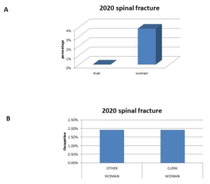
Figure 1. Displayed the percentage of spinal fractures among men and women in 2020 by employment.
Figure 2A displayed a variety of professions, including man's various professions. 2020 saw 3.84% of people, clerks 1.92%, retirees 1.92%, and women 1.92% suffer thoracic vertebral fractures. Technical and professional staff (1.92%), retirees (13.46%), workers (1.92%), clerks (1.92%), other (3.84%), and jobless In 2020, 1.92% of the population experienced a thoracic vertebral fracture. The percentage of thoracic vertebrae fractures in men and women in 2020 was shown in Figure 2B. In 2020, a quarter of women and a quarter of men suffered from thoracic vertebral fractures, respectively, according to Figure 1B.
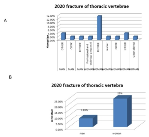
Figure 2. Demonstrated the fracture of thoracic vertebrae occupation and percentage between man and woman in the year of 2020.
The thoracic spine compression fracture that affected 3.84% of other men, 1.92% of retiree men, 11.76% of clerk women, 3.84% of retiree women, and 5.76% of other women in 2020 was noted in Figure 3A. According to Figure 3B, 21.15% of women and 5.76% of men experienced thoracic spine compression fractures in 2020.
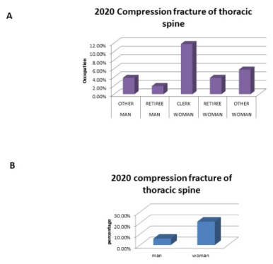
Figure 3. Demonstrated the compression fracture of thoracic spine occupations and percentage between man and woman in the year of 2020.
Figure 4. In 2020, the percentage of lumbar spinal compression fractures by occupation and the percentage of lumbar fractures by occupation were displayed.
In 2020, Figure 4A shows that 11.53% of women and 5.76% of men suffered from lumber vertebral compression fractures. A number of professions suffered lumbar vertebral compression fractures in 2020, as shown in Figure 4B (3.4% retire man, 1.92% clerk man, 5.76% retire woman, 1.92% clerk woman, and 1.92% other profession woman). In 2020, a lumbar fracture injured 5.76% of men and 13.46% of women, according to Figure 4C. Figure 4D shows the various occupations in 2020 with the number of people afflicted by lumbar fractures (3,84% retiree women, 3.84% retiree men, 3,84% other profession woman, 3.84% clerk woman, 1.92% other profession man, and 1.92% worker woman).
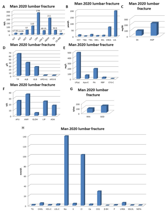
Figure 5. Man 2020's lumbar spinal compression fracture blood parameters (A-G).
In the year 2020, Figure 5A illustrated a lumbar vertebral compression fracture of a man. The blood profile revealed no significant changes in blood parameters such as ALT, AST, GGT, ALP, LDH-1, HBDH, CK, and CK-MB within the normal blood range, but there were significant changes in potential blood parameters such as LDH being higher than normal. Figure 5B shows that in the year 2020, a man suffered a lumbar vertebral compression fracture. The blood profile showed that there were no significant changes in blood parameters within the normal blood range, such as TBA, TBIL, UA, or IBIL, and that there were significant changes in potential blood parameters, such as HCY and CREA, which were higher than normal. The blood profile of a man with a lumbar spinal compression fracture in 2020, as shown in Figure 5C, indicates that there have been no notable changes in any relevant blood parameters, such as AGP or SA, within the normal blood range. The blood profile of a man with a lumbar spinal compression fracture in 2020, as shown in Figure 5D, indicated that there were no significant changes in any prospective blood parameters, such as TP, ALB, GLB, APO-A1, or APO-B, within the normal blood range. The blood profile of a man with a lumbar spinal compression fracture in 2020 is shown in Figure 5E. It shows that there are substantial changes in possible blood parameters, such as APO-E being greater than normal blood range, but no significant changes in LP(α), PA, or RBP. The blood range for CYS-C is normal. As seen in Figure 5F, which shows a man's blood profile following a lumbar vertebral compression fracture in 2020, there are no significant blood changes (AFU, AMY, GLDH, and ADA falling within the normal blood range) and significant blood changes in the potential blood parameters (LIP is higher than normal blood range). A man's blood profile following a lumbar spinal compression fracture in 2020 is depicted in Figure 5G. It indicates that there have been no notable changes to any relevant blood parameters, such as IMA or SOD, which are within normal range.
Figure 6. Man 2020 with a lumbar spinal compression fracture and blood parameters.
The blood profile of a man with a lumbar spinal compression fracture in 2020, as shown in Figure 6, revealed no significant changes in any potential blood parameters, including TG, CHOL, HDL-C, LDL-C, NEFA, SDLDL, β-BH, P, UREA, Na, K, Cl, Ca, and CO2, all of which were within the normal range.
Figure 7. Man's blood parameters for lumbar fracture in 2020 (A-H).
The blood profile of a man with a lumbar fracture in 2020, as shown in Figure 7A, showed no significant changes in any potential blood parameters, including ALT, AST, GGT, ALP, LDH, LDH-1, HBDH, CK, and CK-MB, within the normal blood range. In 2020, a lumbar fracture of the man's blood profile (Figure 7B) revealed no significant alterations in possible blood parameters, such as HCY, TBA, TBIL, DBIL, IBIL, CREA, and UA, which were all within normal range. In 2020, a man suffered a lumbar fracture. Figure 7C showed that there were notable alterations in the individual's blood profile, including elevated levels of SA and AGP levels above normal. As seen in Figure 7D, there were no appreciable alterations in blood parameters such as TP, ALB, GLB, APO-A1, and APO-B within the normal blood range following a lumbar fracture in a man in 2020. APO-E and LP are both greater than normal range in the blood profile of a man with a lumbar fracture in 2020, but PA, RBP, and CYS-C are all within normal range. Figure 7E illustrates these significant changes in the blood profile. Figure 7F demonstrated that, in 2020, a man's lumbar fracture blood profile indicated no appreciable changes in blood parameters, including AFU, AMY, GLDH, LIP, and ADA, which were all within normal range. Figure 7G highlights that, in 2020, a man's lumbar fracture blood profile showed no notable alterations, with parameters including IMA and SOD falling within normal range. Figure 7H illustrates a man's blood profile following a lumbar fracture in 2020; it shows that all parameters, including TG, HDL-C, Na, CL, CO2, P, and SDLDL, are within normal range. No notable changes were seen. Reference range: 9.0-50.0 IU/L ALT 15.0-40.0 IU/L AST 10.0-60.0 IU/L GGT 40.0-130.0 IU/L ALP 90.0-245.0 IU/L LDH 23.0-72.0 IU/L LDH-1 90.0-250.0 IU/L HBDH 24.0-194.0 IU/L CK 0-25 IU/L CK-MB.
The blood profile of a man who suffered a thoracic vertebrae fracture in 2020 is shown in Figure 8A. It shows that blood parameters such as GGT, ALP, LDH, and CK-MB were significantly altered and were higher than normal blood range, while blood parameters such as ALT, AST, LDH-1, HBDH, and CK were within normal blood range. APO-A1, APO-B, TP, ALB, and GLB are among the prospective blood parameters that are within the normal blood range, according to Figure 8B, which describes the blood profile of a thoracic vertebrae fracture in 2020As seen in Figure 8C, there were no notable changes in any prospective blood parameters, including AFU, GLDH, LIP, AMY, and ADA, within the normal blood range in the year 2020 for thoracic vertebrae fracture man. Blood parameters such as TBA, TBIL, DBIL, and HCY are significantly altered in the 2020 fracture of the thoracic vertebrae man, as Figure 8D illustrates. In contrast, blood parameters such as IBIL, CREA, and UA are within normal blood range and do not exhibit significant blood changes. The blood profile of the 2020 thoracic vertebrae fracture man, as described in Figure 8E, revealed no significant changes in any potential blood parameters, including LP(α), PA, RBP, Apo-E, and CYS-C, which are all within the normal blood range. Figure 8F highlights that, in 2020, a thoracic vertebrae fracture man's blood profile showed no significant changes in prospective blood parameters, such as IMA and SOD, within the normal blood range. The blood profile for the 2020 thoracic vertebrae fracture described in Figure 8G indicates that there are no noteworthy variations in possible blood parameters, such as SA and AGP, within the normal blood range. In 2020, a thoracic vertebrae fracture man's blood profile was shown in Figure 8H. It indicated that blood parameters such as CO2, β-BH, and CHOL, LDL-C, Na, and Cl were all significantly different from normal, and that no blood parameters, such as TG, HDL-C, K, Ca, UREA, P, SDLDL, and NEFA, were significantly different from normal.
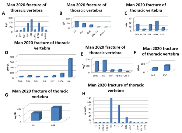
Figure 8. Blood parameter of man 2020 fracture of thoracic vertebrae (A-H).
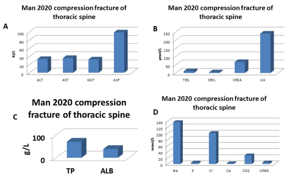
Figure 9. Blood parameter of man 2020 Compression fracture of thoracic spine (A-D).
The blood profile of a man with a thoracic spine compression fracture in 2020 is shown in Figure 9A, which indicates that there are no notable changes in any relevant blood parameters, such as ALT, AST, GGT, and ALP, within the normal blood range. Figure 9B, which depicts a compression fracture of the thoracic spine in 2020, indicates that blood parameters such as TBIL, DBIL, CREA, and UA are within normal range and there are no notable changes. The blood profile of a man with a thoracic spine compression fracture in 2020, as shown in Figure 9C, exhibited no significant alterations in prospective blood parameters, such as TP or ALB, within the normal blood range. Figure 9D indicates that in 2020, a compression fracture of the thoracic spine in a man, the individual's blood profile revealed no significant changes in possible blood parameters, including Na, K, Cl, CO2, and UREA, all of which were within the normal range.
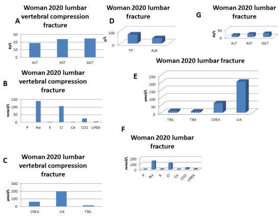
Figure 10. Blood parameter of woman 2020 lumbar vertebral compression fracture and Lumbar fracture (A-G).
The blood profile of the 2020 lumbar spinal compression fracture woman described in Figure 10A indicates that there are no notable variations in possible blood parameters, such as ALT, AST, or GGT, within the normal blood range. The blood profile of a lady with a lumbar spinal compression fracture in 2020, as illustrated in Figure 10B, revealed no significant changes in any relevant blood parameters, including P, Na, K, Cl, Ca, CO2, and UREA, all of which were within the normal blood range. The woman with a lumbar spinal compression fracture in 2020, as depicted in Figure 10C, had a blood profile that indicated no significant changes in possible blood parameters, such as CREA, UA, and TBIL, within the normal range. The blood profile of the 2020 lumbar fracture woman, as shown in Figure 10D, revealed no significant alterations in prospective blood parameters, such as TP or ALB, within the normal blood range. Figure 10E highlights that in 2020, a lady who suffered a lumbar fracture showed no significant changes in blood parameters such as TBIL, TBA, CREA, or UA within the normal blood range. Figure 10F illustrates a lumbar fracture woman's blood profile from the year 2020 and shows that all possible blood parameters, including P, Na, K, Cl, Ca, CO2, and UREA, are within the normal range and there are no noteworthy changes. Figure 10G highlights that the blood profile of a 2020 lumbar fracture woman revealed no notable changes in possible blood parameters, such as ALT, AST, or GGT, all falling within the normal blood range.
According to Figure 11A, a woman with a compression fracture to her thoracic spine in 2020 had blood changes in her blood profile that were not statistically significant. Her blood parameters, such as ALB, were found to be lower than normal. Her blood parameters, such as TP, were found to be within normal range. In the year 2020, a lady with a thoracic spine compression fracture, as depicted in Figure 11B, her blood parameters showed no notable alterations, with ALT, AST, GGT, and ALP all falling within the normal range. The woman's blood profile from a 2020 compression fracture of the thoracic spine, as shown in Figure 11C, indicates that there are no notable variations in any relevant blood parameters, including Na, K, Cl, Ca, CO2, and UREA, within the normal range. Figure 11D, which describes the woman's blood profile from a compression fracture of the thoracic spine in 2020, showed that prospective blood parameters such CREA, UA, P, and TBIL are all within normal range and have not changed significantly.
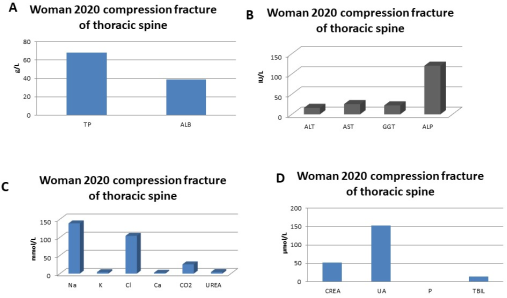
Figure 11. Blood parameter of woman 2020 Compression fracture of thoracic spine (A-D).
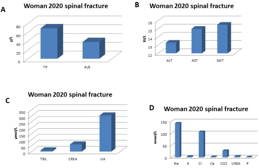
Figure 12. Blood parameter of women 2020 spinal fracture (A-D).
ALB is lower than normal blood range, and there are no significant changes in potential blood parameters like TP within normal blood range, as shown by Figure 12A, which depicts the blood profile of a woman who suffered a spinal fracture in 2020. A spinal fracture woman's blood profile in 2020, as shown in Figure 12B, revealed no appreciable variations in potentially dangerous blood parameters like ALT, AST, or GGT within normal blood range. According to Figure 12C, a spinal fracture woman's blood profile from 2020 indicated that no significant alterations had been observed in any prospective blood parameters, such as TBIL, CREA, or UA, which were all within the normal blood range. Figure 12D illustrates a spinal fracture woman's blood profile from 2020 and shows that there are no notable variations in any probable blood parameters, including Na, K, Cl, Ca, CO2, UREA, and P, within the normal blood range.
Figure 13. Blood parameter of woman 2020 fracture of thoracic vertebrae (A-C).
In 2020, a lady suffered a thoracic vertebrae fracture; Figure 13A indicated that her blood parameters, including ALT, AST, and GGT, were within normal range and that there were no notable alterations. The woman's blood profile for the 2020 thoracic vertebrae fracture is shown in Figure 13B. It indicates that there are no significant changes in potential blood parameters such as TP within normal blood range, and that there are significant changes in potential blood parameters such as ALB being lower than normal blood range. In 2020, a lady suffered a thoracic vertebrae fracture; Figure 13C illustrates that her blood profile, which includes prospective blood parameters including CREA, UA, and TBIL, was within normal range and did not exhibit any noteworthy alterations.
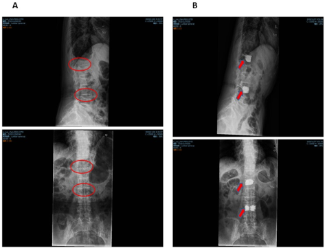
Figure 14. A. 2020 Lumbar vertebral compression, Fracture before surgery, (Female, age 88). B. 2020 Lumbar vertebral compression, Fracture after surgery, (Female, age 88).
Before Surgery, Figure 14A Conclusion drawn from the analysis: There was the lumbar spine's natural curvature. There were no visible anomalies in the vertebral appendages, and the lumbar 5/ sacral 1 vertebral gap was narrowed. The margins of the lumbar 1–5 vertebrae were sharp.
Comment on imaging: Degenerative alterations of the lumbar region. X-ray in Figure 14B Following surgery, the examination revealed the following: The lumbar and thoracolumbar vertebrae retained their natural curvatures. There was a thick filling shadow seen inside the flattened thoracic 10 and lumbar 1 vertebra. The lumber 3/4/5, lumbar 5/sacral 1 vertebrae were narrowed, the thoracic 12 vertebrae were somewhat altered into wedges, the thoracolumbar vertebrae's edges were pointed, and certain vertebral appendages were under-clear. There was calcium in the thoracic arch. Imaging comments: Thoracolumbar surgery, kindly perform a clinical self-review; take into account lumbar degenerative degeneration, mild wedging of the thoracic 12 vertebrae, chronic compression, and hardening of the aorta in conjunction with MRI diagnosis.
Figure 15. A. 2020 Lumbar fracture before surgery, Female, age 81. B. 2020 Lumbar fracture after surgery, Female, age 81.
Figure 15A: Preoperative X-ray report Conclusions drawn from the analysis: The lumbar spine had its normal curvature, the lumbar 1 vertebra was wedge-shaped, the rest of the vertebra was undamaged, there was varying degrees of bone hyperplasia at the margins of the lumbar 1–5 vertebrae, there was no discernible narrowing of the vertebral spaces, and there were no discernible anomalies in the vertebral appendages.
Commentary on imaging: 1. Compression fracture of lumbar and vertebral bodies; 2, lumbar spine bone hyperplasia.
After surgery, Figure 15B shows the X-ray report. Examined findings: There was no discernible narrowing of the vertebral spaces, no obvious abnormalities in the vertebral appendages, the lumbar spine's natural curvature was present, lumbar vertebrae 1 showed a high-density filling shadow, and the rest of the vertebrae were complete in their structure.
Imaging comments: 1. Postoperative lumbar and vertebral body alterations; 2. Hyperplasia of the lumbar spine bones.
X-ray in Figure 16A Prior to surgery, report: Results of the analysis: More intestines were present, the lumbar spine's transverse process was not clearly visible, the thoracic 12 vertebrae flattened, the lumbar spine had a physiological curvature, the lumbar vertebrae's edges were sharp, and the lumbar 3/4-lumbar 5/ sacrall intervertebral space narrowed.
Imagining remarks: 1. Compression fracture of the 12th thoracic vertebrae; more testing may be required. 2. Degenerative modifications to the lumbar region. X-ray in Figure 16B Report following surgery: Results of the analysis: More intestinal contents were present, the lumbar spine's transverse process was not clearly visible, the thoracic 12 vertebrae showed a high density of filling shadow, the lumbar spine had a physiological curvature, the lumbar vertebrae's bony edges sharpened, and the vertebral space between lumbar ¾ and lumbar 5/sacral 1 narrowed.
Imagining remarks: 1. Please evaluate the postoperative modifications to the thoracic 12 vertebrae by comparing them clinically to the prior video. 2. Degenerative modifications to the lower back.
Figure 16. A. 2020 fracture of thoracic vertebrae before surgery, Female, age 80. B. 2020 fracture of thoracic vertebrae after surgery, Female, age 80.
Figure 17. A. 2020 fracture of thoracic vertebrae before surgery, Male, age 57. B. 2020 fracture of thoracic vertebrae after surgery, Male, age 57.
Before surgery, Figure 17A shows the X-ray report: What was seen during the inspection: It was evident that the lumbar vertebrae 7, 9, 12, and 4 had flattened vertebral bodies. and a few of them had wedge-shaped shapes. There was no discernible narrowing of the intervertebral gaps, no visible abnormalities in the vertebral appendages, and a noticeable amount of bone hyperplasia along the borders of the first five lumbar vertebrae.
Commentary on imaging: If additional CT testing was required, compression fractures of the lumbar 4, 9, 12, and thoracic 7 vertebrae were taken into consideration; lumbar vertebral bone hyperplasia.
X-ray report following surgery (Figure 17B): Examining results revealed: The thoracic vertebrae 4-6 had somewhat flattened bodies, while the thoracic vertebrae 7 and 9 exhibited thick filling shadows on their bodies. The thoracic vertebrae 12 bodies were cuneiform. absence of a noticeable intervertebral narrowing No glaring anomalies were seen in the vertebral appendages, and spaces were seen. In the aortic arch, calcium arcs were seen.
Comments on the image: Clinical self-examination is requested after thoracic vertebrae 7 and 9. The degrees of flattening of thoracic vertebrae 4-6 and 12 were noted, a compression fracture was hypothesised, and more testing was required. the aorta hardening.
Figure 18. A. 2020 spinal fracture after surgery, Male, age 75. B. 2020 spinal fracture before surgery, Male, age 75.
X-ray report following surgery (Figure 18A). Conclusions drawn from the analysis: There was lumbar physiological curvature, thoracic 11–lumbar 4 vertebrae were cuneiform to varying degrees, the edges of the vertebral bodies became sharp, the lumbar 3 vertebrae appeared discontinuous, and a linear low density shadow was faintly visible. The space between the lumbar vertebrae narrowed, and certain vertebral appendages showed insufficient clarity. The thoracic 12 and lumbar 3 vertebrae showed high density filling shadows.
1. Postoperative thoracic and lumbar spine changes: Please conduct a clinical self-review. 2. Compression fractures were diagnosed as wedge-shaped alterations from thoracolumbar 11 to lumbar 4 vertebrae; suspicious degradation of the lumbar 3 vertebral arch; further testing is advised; 3. Degenerative modifications to the lumbar region.
Before surgery, the spine was examined using X-ray report (Figure 18B). spinal DR, lumbar anteroposterior Lateral lumbar spine Examined findings: There was lumbar vertebral physiological curvature. The wedge-shapedness of the thoracic-11-lumbo4 vertebrae varied. Some vertebral appendages are unclear, the borders of all ventebral bodies are pomtea, and the lumbar 3 vertebral arch appears discontinuous.
Remarks on the image: Consideration of compression fracture in thoracic 11-lumbar 4 vertebral wedge change; The lumbar arch in lumbar 3 appears to be suspiciously disintegrating, indicating that lumbar bone hyperplasia needs to be further investigated.
4. Discussion
Our research further demonstrates that an independent risk factor for nearby vertebral refracture following PVP is the uneven distribution of posterior spinal muscles, including the multifidus and erector spine. The erector spine and multifidus have a unique anatomical location, which may be the cause of this. Additionally, an uneven muscle distribution may make the spine more unstable. In conclusion, there is a significant chance of vertebral refracture following PVP, and there are a number of variables that can influence this risk. The posterior group of muscles is the most crucial group for maintaining the stability of the lumbar spine, hence their degeneration will result in a reduction in spinal stability. Our research demonstrated the impact of paraspinal muscle degeneration, particularly in the multifidus and erector spinae, on the recurrence of neighbouring vertebral fractures following PVP therapy for osteoporotic vertebral compression fractures . The purpose of this investigation was to assess the PVP/PKP Procedures' efficacy. We assessed how well the treatment's effects were examined on osteoporotic VCFs. We showed that PVP and PKP considerably reduce pain in patients with VCFs by evaluating the data gathered to evaluate patient outcomes . Our analysis included patient data from interventional treatment administered between January and December of 2020, with an average one-year gap between treatment and clinical evaluation. Since our study focused on long-term interventions in a geriatric population, only a subset of patients could be effectively reached and asked to return the appropriate questionnaire. A significant number of the patients passed away prior to the start of our investigation as a result of things like elderly age . The primary influencing elements of bone cement dispersion are thought to be the degree of osteoporosis, injection pressure, viscosity of bone cement, fracture degree, angle, and depth of puncture needle. While research on PVP from unilateral and bilateral pedicles has shown comparable long-term therapeutic results, this study came to the conclusion that the main goals of treating compressive vertebral fractures were pain relief, restoration of vertebral height, and healing of neighbouring fractures. Long-term pain alleviation is achievable with or without PVP surgery, although regaining vertebral height is challenging. Consider that unilateral PVP can benefit from less trauma, radiation, and operating time. However, the unequal distribution caused by unilateral bone cement entry prevents the bone cement from completely filling the fracture line in the vertebral body. This study therefore recommends that in PVP surgery, bilateral bone cement filling should be guaranteed to the greatest extent feasible. There is no appreciable difference in the clinical efficacy of unilateral and bilateral punctures when bone cement is equally distributed bilaterally across the vertebral body . The majority of patients with acute painful OVCF respond well to conservative medical therapy; however, in an elderly patient cohort with osteoporosis already present and severe pain, many patients do not receive enough pain relief, and their progress is stagnant, leading to serious complications and a protracted recovery . For this reason, early PVP procedures may be more clinically meaningful for an older patient group with established osteoporosis and severe pain. Since there have been reports of spinal cord compression following therapy, it is frequently believed that the existence of spinal canal impairment is a relative contraindication for PVP . We believe that the most significant operation failure is intraspinalmalpositioning, which has the potential to result in neurological complaints and consequently be a debilitating consequence. Nonetheless, there is a significant disparity between the two methods in the middle and upper thoracic spine. Once more, this implies that in this anatomical area, 3D imaging or 3D navigation may be acceptable. Undoubtedly, neurological symptoms wouldn't result from every intraspinalmalpositioning. The reported rate of clinical complications related to neurological impairments following vertebroplasty and kyphoplasty is extremely low; in fact, it is significantly lower than the rate of malpositioning observed in our study. Cement leaking was thought to be a potential cause of neurological complaints following cement augmentation procedures in the thoracic spine, rather than damage to the spinal cord during cannula insertion. The majority of cement extrusion is asymptomatic, and 8.5% of it leaksRadicular pain or compression of the thecal sac may occur in certain instances of intraforaminal leakage or radicular vein leaking. Though it seldom causes pulmonary emboli or cerebral infarct, the vascular leak is also typically asymptomatic . In 0.01% of kyphoplasty patients and 0.6 percent of vertebroplasty cases, there were serious issues. After surgery, postoperative imaging shows the majority of asymptomatic cement extrusions . According to Stoffel et al., there were approximately 33 asymptomatic instances of extra vertebral cement extrusion after kyphoplasty in 118 vertebral bodies. They also discovered a potential correlation between cement extrusions and cement volume and viscosity as a reason for the study's high rate of leaks . However, if the posterior wall of the vertebra is compromised by inserting the instruments through the spinal canal and establishing a connection between the cement depot and the spinal canal, there is a chance of both spinal cord injury and cement leakage into the spinal canal. PVP and PKP are currently utilised extensively in the treatment of OVCFs across the globe. Both surgical treatments have the potential to improve the stability and strength of the vertebral body, leading to rapid pain relief for patients. In the meanwhile, PKP can more effectively cure spinal kyphosis and restore compressed vertebral body height, allowing patients to begin walking as soon as possible. PKP can achieve a satisfactory clinical impact with an efficiency of up to 90%. . Relieving postoperative discomfort and restoring the patients' capacity to take care of themselves is the primary goal of these two surgeries. Patients' pain is primarily reduced by PKP and PVP surgery in the following ways. Patients can have their vertebrae fixed, their bones strengthened, and their pain reduced by a bone cement injection that lessens the stimulation of movement to the nerve . were considerably lower than before, showing that the procedure had a clear impact and that the discomfort had been successfully reduced. At one day, one week, and one month following surgery, the VASs of patients in the PKP group were lower than those in the PVP group. This suggests that PKP was more effective than PVP at relieving pain in the short term, which could allow the patients return to their regular lives as soon as feasible. Meanwhile, quality of life scores was better in the PKP group and the postoperative ODI index was significantly lower in the PVP group, suggesting that PKP was more effective than PVP at reducing pain and improving patients' ability to take care of themselves . According to this study, patients in the PKP group rested in bed for a shorter period of time after surgery than those in the PVP group, which may help avoid and lessen problems like pressure sores and hypostatic pneumonia, which are brought on by extended bed rest. Additionally, the PKP group experienced reduced intraoperative blood loss and fluoroscopy times than the PVP group, which may effectively lower the patient's risk of injury and operation while shielding the medical team from excessive radiation exposure. Studies on biomechanics have verified that 4-6 millilitres of bone cement are needed to restore the strength of the vertebrae. Therefore, enough bone cement should be injected into patients' spines during PKP or PVP surgery to ensure that there is no cement leakage . In posterior cervical puncture (PKP), a low-pressure injection environment is produced by using a balloon to make a hollow in the vertebral body before cement administration. Thus, in contrast to the high-pressure setting in PVP, it is feasible to inject a larger volume of bone cement and lower the rate at which bone cement leaks in PKP. Furthermore, the vertebral body's balloon expansion can compress the cancellous bone to increase its density, which closes venous access points and fractures in the bone and lessens the amount of bone cement that leaks out. In addition to reducing kyphosis and relieving discomfort, PKP and PVP can help raise spinal height and enhance function. . To summarise, PKP and PVP are effective treatments for older adults with osteoporotic spinal compression fractures. They enable patients to leave the bed sooner and minimise the risks associated with prolonged bed rest following a fracture. When it comes to restoring vertebral height and increasing Cobb's angle, PKP is clearly superior to PVP due to its reduced bone cement leakage rate, increased safety, and fewer problems. It can also more effectively relieve pain, which enhances the clinical therapeutic effects. To guarantee the efficacy of clinical treatment, the therapeutic approach chosen should still be related to the unique situation of the patient.
5. Conclusions
In the early stages, PVP is linked to more pain alleviation than conservative therapy. Moreover, PVP had no effect on the frequency of nearby vertebral fractures. The outcomes show that it is an efficient and safe treatment for OVCFs. These results should be regarded cautiously due to some restrictions. More research is required. Large, conclusive RCTs are required. Our research revealed a strong relationship between rising mobility and rising quality of life. Additionally, it was established that there was a strong correlation between a decline in VAS and an improvement in quality of life. To sum up, our research serves as an assessment of the outcomes of both PVP and PKP. Even after more than ten years, both approaches partially contribute to the intended therapy outcomes. Reduced VAS scores demonstrated the efficacy of PKP and PVP. Nevertheless, not every patient experienced the same degree of mild pain. Since these results are based on a limited clinical examination, more research is needed to fully understand long-term consequences and provide a prognostic assessment for PVP and PKP.
Abbreviations
6MWT: 6-Minute Walk Test
LDH: A Lactate Dehydrogenase Test
CK: A Creatine Kinase
CREA: A Creatinine Blood Test
GGT: A Gamma-Glutamyl Transferase
Na: A Sodium Test
TP: A Total Protein
ALP: Alkaline Phosphatase
AFU: Alpha-L-Fucosidase
ALT: Stands for Alanine Transaminase.
ALB: An Albumin Blood Test
HDL-C: An HDL Cholesterol Test
Apo-A1: Apolipoprotein A-I
apob: Apolipoprotein B
APOE: Apolipoprotein E
Ca: Calcium
CO2: Carbon Dioxide
Cl–: Chloride
CHOL: Cholesterol Test
CK-MB: CreatineKinase-Myocardial Band
ΔDBP: Iastolic Blood Pressure
DBIL: Direct Bilirubin
DXA: Dual Energy X Ray Absorptiometry
GLB: GlobulinTest
ΔHR: Heart Rate
Hcy: Homocysteine
HBDH: HydroxybutyrateDehydrogenase
IMA: Ischemia Modified Albumin
KP: Kyphoplasty,
LDH-1: Lactate dehydrogenase-1
LIP: Lipase Test
LP: Lipoprotein(a)
LDL-C: Low-densityLipoprotein Cholesterol Test
MRI: Magnetic Resonance Imaging
ovcfs: Osteoporosis Vertebral Compression Fracture
ODI: Oswestry Disability Index
PVP: Percutaneous Vertebroplasty
PKP: Percutaneous Kyphoplasty
VP: Percutaneous Vertebroplasty
NEFA: Plasma None Sterified Fatty Acid
p: Platelates
K: Potassium
PA: Prealbumin
RCT: Randomized Controlled Trial
RBP: Retinol Binding Protein
RMDQ: Roland-Morris Disability Questionnaire
ADA: Serum Adenosine Deaminase
AMY: Serum Amylase
SA: Serum Amylloid
UREA: Serum Blood Test
CYS-C: Serum CystatinC
SF-36: Short-Form 36 Health Survey
SDLDL: Small Dense Low Density Lipoprotein Cholesterol
Stryker: Spinejack Vertebral Implant (SJ) System
ΔSBP: Systolic Blood Pressure
SOD: The Activity of Superoxide Dismutase
AGP: The Ambulatory Glucose Profile
GLDH: The Glutamate Dehydrogenase
TBA: The Total Bilirubin
AST: The Aspartate Aminotransferase
"TBIL": Total Bilirubin
tbil: Total-Value Bilirubin
TG: Triglycerides Test
UA: Urinalysis, Urine Test
β-BH: Β-blood Hematology Test
Acknowledgments
We would like to thank the faculty members who collected the samples, assembled the data, Shandong University, and Jinan central hospital.
Author Contributions
Md. Zakaria Hossain: Conceptualization, participated in experiments, study design, manuscript preparation. carried out the study design, participated in experiments, manuscript preparation, and statistical analysis. checked the grammatical mistakes and corrected the final manuscript.
Bin Ning: Data curation, participated in experiments, study design, manuscript preparation. Supervising and directing the project.
Md. Reyad-ul-Ferdous: Formal Analysis, participated in experiments, study design, manuscript preparation. carried out the study design, participated in experiments, manuscript preparation, and statistical analysis. Supervising and directing the project. checked the grammatical mistakes and corrected the final manuscript. Md. Zakaria Hossain and Md. Reyad-ul-Ferdous are equally contributed.
All authors read and approved the final version of the manuscript.
Funding
This work is not supported by any external funding.
Data Availability Statement
The data is available from the corresponding author upon reasonable request.
Conflicts of Interest
The authors declare no conflicts of interest.
References
[1] Tsoumakidou, G., C. W. Too, G. Koch, et al., 2017. CIRSE Guidelines on Percutaneous Vertebral Augmentation. Cardiovascular and interventional radiology. 40, 331-342.
[2] Clark, W., P. Bird, P. Gonski, et al., 2016. Safety and efficacy of vertebroplasty for acute painful osteoporotic fractures (VAPOUR): a multicentre, randomised, double-blind, placebo-controlled trial. Lancet (London, England). 388, 1408-1416.
[3] Wang, H., F. Lin, G. Liang, et al., 2022. Percutaneous vertebroplasty in osteoporotic vertebral compression fracture with huge spinal epidural hematoma: A case report. Medicine. 101, e29340.
[4] Diamond, T., W. Clark, P. Bird, et al., 2020. Early vertebroplasty within 3 weeks of fracture for acute painful vertebral osteoporotic fractures: subgroup analysis of the VAPOUR trial and review of the literature. European spine journal: official publication of the European Spine Society, the European Spinal Deformity Society, and the European Section of the Cervical Spine Research Society. 29, 1606-1613.
[5] Schupfner, R., K. Koniarikova, C. Pfeifer, et al., 2021. An anatomical study of transpedicular vs. extrapedicular approach for kyphoplasty and vertebroplasty in the thoracic spine. Injury. 52 Suppl 5, S63-s69.
[6] Rebolledo, B. J., B. P. Gladnick, A. Unnanuntana, et al., 2013. Comparison of unipedicular and bipedicular balloon kyphoplasty for the treatment of osteoporotic vertebral compression fractures: a prospective randomised study. The bone & joint journal. 95-b, 401-406.
[7] Hinde, K., J. Maingard, J. A. Hirsch, et al., 2020. Mortality Outcomes of Vertebral Augmentation (Vertebroplasty and/or Balloon Kyphoplasty) for Osteoporotic Vertebral Compression Fractures: A Systematic Review and Meta-Analysis. Radiology. 295, 96-103.
[8] Zuo, X. H., Y. B. Chen, P. Xie, et al., 2021. Finite element analysis of wedge and biconcave deformity in four different height restoration after augmentation of osteoporotic vertebral compression fractures. Journal of orthopaedic surgery and research. 16, 138.
[9] Li, T., S. Pang, R. England, et al., 2023. Clinical Outcomes and Safety Comparison of Vertebroplasty, Balloon Kyphoplasty, and Vertebral Implant for Treatment of Vertebral Compression Fractures. AJNR. American journal of neuroradiology. 44, 1345-1351.
[10] Ding, X., Q. Zhang, Y. Zhao, et al., 2022. Location and Effect of Bone Cement in Percutaneous Vertebroplasty for Osteoporotic Vertebral Compression Fractures. BioMed research international. 2022, 6127620.
[11] Chen, M., C. Yang, Z. Cai, et al., 2022. Lumbar posterior group muscle degeneration: Influencing factors of adjacent vertebral body re-fracture after percutaneous vertebroplasty. Frontiers in medicine. 9, 1078403.
[12] Hackbarth, C. B., T. J. Vogl, N. Naguib, et al., 2021. Long-term evaluation of pain reduction after vertebroplasty and kyphoplasty. Acta radiologica open. 10, 20584601211028994.
[13] Sanli, I., S. M. J. van Kuijk, R. A. de Bie, et al., 2020. Percutaneous cement augmentation in the treatment of osteoporotic vertebral fractures (OVFs) in the elderly: a systematic review. European spine journal: official publication of the European Spine Society, the European Spinal Deformity Society, and the European Section of the Cervical Spine Research Society. 29, 1553-1572.
[14] Zhang, J., X. He, Y. Fan, et al., 2019. Risk factors for conservative treatment failure in acute osteoporotic vertebral compression fractures (OVCFs). Archives of osteoporosis. 14, 24.
[15] Ren, H., T. Feng, Y. Hu, et al., 2022. The Value of Dynamic Fracture Mobility in Determining the Optimum Operation Choice for Acute Osteoporotic Vertebral Compression Fracture. Journal of pain research. 15, 2327-2336.
[16] Dai, C., G. Liang, Y. Zhang, et al., 2022. Risk factors of vertebral re-fracture after PVP or PKP for osteoporotic vertebral compression fractures, especially in Eastern Asia: a systematic review and meta-analysis. Journal of orthopaedic surgery and research. 17, 161.
[17] He, C. J. and G. D. Liu, 2018. Comparison of the Efficacy and Safety of Bone-filling Mesh Container and Simple Percutaneous Balloon Kyphoplasty in the Treatment of Osteoporotic Vertebral Compression Fractures. Pain physician. 21, 259-268.
[18] Hu, K. Z., S. C. Chen and L. Xu, 2018. Comparison of percutaneous balloon dilation kyphoplasty and percutaneous vertebroplasty in treatment for thoracolumbar vertebral compression fractures. European review for medical and pharmacological sciences. 22, 96-102.
[19] Papanastassiou, I. D., F. M. Phillips, J. Van Meirhaeghe, et al., 2012. Comparing effects of kyphoplasty, vertebroplasty, and non-surgical management in a systematic review of randomized and non-randomized controlled studies. European spine journal: official publication of the European Spine Society, the European Spinal Deformity Society, and the European Section of the Cervical Spine Research Society. 21, 1826-1843.
[20] Liu, J., J. Tang, Y. Zhang, et al., 2019. Percutaneous Vertebral Augmentation for Osteoporotic Vertebral Compression Fracture in the Midthoracic Vertebrae (T5-8): A Retrospective Study of 101 Patients with 111 Fractured Segments. World neurosurgery. 122, e1381-e1387.
Cite This Article
  • APA Style

    Hossain, M. Z., Ning, B., Reyad-ul-Ferdous, M. (2024). Analysis of Clinical Outcomes of Percutaneous Kyphoplasty and Vertebroplasty in the Treatment of Osteoporosis-Induced Vertebral Compression Fracture . Journal of Surgery, 12(2), 45-65. https://doi.org/10.11648/j.js.20241202.15

    Copy | Download

    ACS Style

    Hossain, M. Z.; Ning, B.; Reyad-ul-Ferdous, M. Analysis of Clinical Outcomes of Percutaneous Kyphoplasty and Vertebroplasty in the Treatment of Osteoporosis-Induced Vertebral Compression Fracture . J. Surg. 2024, 12(2), 45-65. doi: 10.11648/j.js.20241202.15

    Copy | Download

    AMA Style

    Hossain MZ, Ning B, Reyad-ul-Ferdous M. Analysis of Clinical Outcomes of Percutaneous Kyphoplasty and Vertebroplasty in the Treatment of Osteoporosis-Induced Vertebral Compression Fracture . J Surg. 2024;12(2):45-65. doi: 10.11648/j.js.20241202.15

    Copy | Download

  • @article{10.11648/j.js.20241202.15,
      author = {Md. Zakaria Hossain and Bin Ning and Md. Reyad-ul-Ferdous},
      title = {Analysis of Clinical Outcomes of Percutaneous Kyphoplasty and Vertebroplasty in the Treatment of Osteoporosis-Induced Vertebral Compression Fracture
    },
      journal = {Journal of Surgery},
      volume = {12},
      number = {2},
      pages = {45-65},
      doi = {10.11648/j.js.20241202.15},
      url = {https://doi.org/10.11648/j.js.20241202.15},
      eprint = {https://article.sciencepublishinggroup.com/pdf/10.11648.j.js.20241202.15},
      abstract = {Percutaneous kyphoplasty (PKP) and Percutaneous vertebroplasty (PVP) have become very effective to treating the Osteoporosis vertebral compression fractures (OVCFs) and patients can get immediate relief from the pain and it can improve the motor functions of patient’s health status and widely used in clinical practice. Vertebroplasty and kyphoplasty are minimally invasive procedures for the treatment of painful vertebral compression fractures (VCF), which are fractures involving the vertebral bodies that make up the spinal column. Vertebroplasty are most often used to treat an injury called a compression fracture. This kind of injuries are most often caused by osteoporosis, which is the reason of bone weakness and osteoporosis are most common in older people. This study aimed to examine the osteoporosis vertebral compression fractures (OVCFs) of Jinan, Shandong province, China, between male and female participants who were over 50 years of age. There are very few pertinent data regarding the relative or absolute contraindication of percutaneous vertebroplasty (PVP) and percutaneous kyphoplasty (PKP) for severe osteoporotic vertebral compression fractures (OVCFs). The purpose of this research was to assess and contrast the effectiveness of traditional kyphoplasty and vertebroplasty using high-viscosity cement in the treatment of severe OVCFs. In aged people, osteoporotic vertebral compression fractures (OVCFs) are prevalent and can result in significant osteoporosis vertebral compression fractures. Treatment options for individuals with these types of fractures include conservative measures, percutaneous vertebroplasty (PVP), and percutaneous kyphoplasty (PKP). We compared the clinical effectiveness of PVP and PKP in treating osteoporotic vertebral compression fractures in this investigation. Therefore, we conducted a comparative study on Osteoporosis vertebral compression fractures (OVCFs) patients who received either PVP or PKP treatment in our hospitals from January 2020 to December 2020 to investigate the clinical efficacy, advantages and disadvantages of the surgical methods, and to provide a reference for clinical selection of treatment methods.
    },
     year = {2024}
    }
    

    Copy | Download

  • TY  - JOUR
    T1  - Analysis of Clinical Outcomes of Percutaneous Kyphoplasty and Vertebroplasty in the Treatment of Osteoporosis-Induced Vertebral Compression Fracture
    
    AU  - Md. Zakaria Hossain
    AU  - Bin Ning
    AU  - Md. Reyad-ul-Ferdous
    Y1  - 2024/04/17
    PY  - 2024
    N1  - https://doi.org/10.11648/j.js.20241202.15
    DO  - 10.11648/j.js.20241202.15
    T2  - Journal of Surgery
    JF  - Journal of Surgery
    JO  - Journal of Surgery
    SP  - 45
    EP  - 65
    PB  - Science Publishing Group
    SN  - 2330-0930
    UR  - https://doi.org/10.11648/j.js.20241202.15
    AB  - Percutaneous kyphoplasty (PKP) and Percutaneous vertebroplasty (PVP) have become very effective to treating the Osteoporosis vertebral compression fractures (OVCFs) and patients can get immediate relief from the pain and it can improve the motor functions of patient’s health status and widely used in clinical practice. Vertebroplasty and kyphoplasty are minimally invasive procedures for the treatment of painful vertebral compression fractures (VCF), which are fractures involving the vertebral bodies that make up the spinal column. Vertebroplasty are most often used to treat an injury called a compression fracture. This kind of injuries are most often caused by osteoporosis, which is the reason of bone weakness and osteoporosis are most common in older people. This study aimed to examine the osteoporosis vertebral compression fractures (OVCFs) of Jinan, Shandong province, China, between male and female participants who were over 50 years of age. There are very few pertinent data regarding the relative or absolute contraindication of percutaneous vertebroplasty (PVP) and percutaneous kyphoplasty (PKP) for severe osteoporotic vertebral compression fractures (OVCFs). The purpose of this research was to assess and contrast the effectiveness of traditional kyphoplasty and vertebroplasty using high-viscosity cement in the treatment of severe OVCFs. In aged people, osteoporotic vertebral compression fractures (OVCFs) are prevalent and can result in significant osteoporosis vertebral compression fractures. Treatment options for individuals with these types of fractures include conservative measures, percutaneous vertebroplasty (PVP), and percutaneous kyphoplasty (PKP). We compared the clinical effectiveness of PVP and PKP in treating osteoporotic vertebral compression fractures in this investigation. Therefore, we conducted a comparative study on Osteoporosis vertebral compression fractures (OVCFs) patients who received either PVP or PKP treatment in our hospitals from January 2020 to December 2020 to investigate the clinical efficacy, advantages and disadvantages of the surgical methods, and to provide a reference for clinical selection of treatment methods.
    
    VL  - 12
    IS  - 2
    ER  - 

    Copy | Download

Author Information
  • Department of Orthopedic, Cheeloo College of Medicine, Shandong University, Jinan, China; Department of Orthopedic, Jinan Central Hospital, Jinan, China

    Biography: Md. Zakaria Hossain has completed his Bachelor of medicine and Bachelor of surgery (MBBS) from Nanchang University, worked in Al-Haramain Hospital Sylhet, Bangladesh as an assistant doctor, 2021 awarded by Chinese government scholarship in Shandong University for master’s degree, at present in Shandong University Research works in orthopedic surgery related diseases. Bone and spinal injuries.

    Research Fields: Orthopedic surgery

  • Department of Orthopedic, Cheeloo College of Medicine, Shandong University, Jinan, China; Department of Orthopedic, Jinan Central Hospital, Jinan, China; Central Hospital Affiliated to Shandong First Medical University, Shandong First Medical University & Shandong Academy of Medical Sciences, Jinan, China

    Biography: Bin Ning Chief Physician MD, Phd. Associate Dean, Associate Director of Spine Surgery, Master and PhD Students Supervisor. Jinan central hospital vice president, Shandong University and Shandong First Medical University Professor. Deputy director of spinal surgery, Director of Jinan Key Lab of Biomechanics and Metabolism, member of the 14th Council of Shandong Medical Association, standing committee member of Jinan Youth Federation, and the chairman of Jinan Young Medical Workers Association, Shandong Provincial Committee of Spinal Surgery, Regeneration of nerve and the chairman of Jinan Medical Association, Natural Science Foundation of China and the disserta-tion reviewer of the Academic Degree and Graduate Education, BMC Musculobone Diseases, NRR and other SCI Journal Editor, published more than 30 papers in domestic nuclear journal and SCI magazine, Has been rated as Shandong Province Top Ten doctors, clinical research work of spine surgery for 18 years, govern-ment-sponsored study in Germany to learn spine surgery technology.

    Research Fields: Orthopedic surgery

  • Department of Orthopedic, Cheeloo College of Medicine, Shandong University, Jinan, China; Institute of Biopharmaceutical and Health Engineering, Tsinghua Shenzhen International Graduate School, Tsinghua University, Shenzhen, China

    Biography: Md. Reyad-ul-Ferdous B. Pharm. M. Pharm, PhD. He enrolled distance learning from The University of California San Francisco (USA); Course: Diabetes: Diagnosis, Treatment, and Opportunities, University of California San Francisco. University of Maryland, College Park (USA); Course: Genes and the Human Condition (From Behavior to Biotechnology). Vanderbilt University (USA); Case Studies in Personalized Medicine. He enrolled as a postdoctoral research scientist at department of Endocrinology and Metabolism, Shandong University. At present he also enrolls as a postdoctoral research scientist at Institute of Biopharmaceutical and Health Engineering, Tsinghua Shenzhen International Graduate School, Tsinghua University, Shenzhen, China. He has published more than 90 articles in international journals as a first and corresponding author. At present his research interests are Disease model, human genetic and metabolic diseases, Mitochondrial function and associated diseases, establishing mitochondrial uncoupling protein (UCP1) upregulated drug candidate to treat obesity related comorbidities, Pharmacology, Obesity, Metabolic diseases, Lipid metabolism in liver, Hormonal effect on NASH, Liver cancer (NASH, NAFALD, fibrosis, Liver cirrhosis), Ovarian Cancer, Neurodegenerative disease, Bone regeneration, formulation development, antioxidant, Herbal drugs and Epigenetic drugs, drug development, Phytochemistry and Phytomedicine.

    Research Fields: Pharmacology, Obesity, Metabolic diseases, Lipid metabolism in liver, Hormonal effect on NASH, Liver cancer (NASH, NAFALD, fibrosis, Liver cirrhosis), Ovarian Cancer, Neurodegenerative disease, Bone regeneration, formulation development, antioxidant, Herbal drugs and Epigenetic drugs, drug development, Phytochemistry and Phytomedicine.

  • Abstract
  • Keywords
  • Document Sections

    1. 1. Introduction
    2. 2. Materials and Methods
    3. 3. Results
    4. 4. Discussion
    5. 5. Conclusions
    Show Full Outline
  • Abbreviations
  • Acknowledgments
  • Author Contributions
  • Funding
  • Data Availability Statement
  • Conflicts of Interest
  • References
  • Cite This Article
  • Author Information