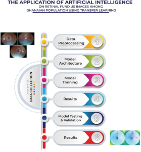
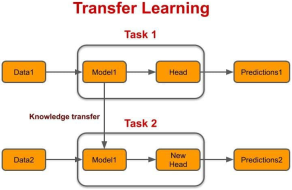
Artificial Intelligence is radically transforming various fields including the field of medical diagnosis and imaging especially for Computer-Aided Diagnosis (CAD). Automated disease detection from the retina has become increasingly important, especially in ophthalmology, where the eye offers a non-invasive way of visualizing and monitoring the progression of diseases. Early detection of these diseases is essential for preventing irreversible blindness. Although, various research have been carried out in Ghana in the area of artificial intelligence using convolutional neural network and machine learning, there is gap in literature on artificial intelligence focusing on local retinal fundus images using deep transfer learning techniques in Ghana. This study address the gap by using 184 retinal fundus images for patients between the ages of 10-70 years from Ghana using Artificial Intelligence Deep Transfer Learning (AIDL) techniques with the VGG-19 architecture augmentation to prepare them for training, testing, and validation, employing a deep transfer learning algorithm known as Convolutional Neural Network (CNN) due to the image size. After a two-stage classification approach enabled the distinction between healthy and unhealthy retinal images, and subsequently, classifying diverse retinal conditions from the unhealthy images including glaucoma, hypertensive and diabetic retinopathy, as well as chorio retinal and macular changes. The performance of the proposed solution was evaluated using various metrics such as accuracy, precision, recall, and AUC for the binary classification and the deep learning task. The results showed that, the proposed solution achieved high accuracy of 97.31%, precision of 96.85%, recall of 98.06%, and AUC of 0.993. This demonstrates the effectiveness in detecting various retina diseases. This solution enhance significant potential automated retinal disease screening, early diagnosis and tele optometry support services, contributing to the eradication of irreversible blindness especially for low resource communities in Ghana and Africa at large.
| Published in | American Journal of Artificial Intelligence (Volume 10, Issue 1) |
| DOI | 10.11648/j.ajai.20261001.22 |
| Page(s) | 136-147 |
| Creative Commons |
This is an Open Access article, distributed under the terms of the Creative Commons Attribution 4.0 International License (http://creativecommons.org/licenses/by/4.0/), which permits unrestricted use, distribution and reproduction in any medium or format, provided the original work is properly cited. |
| Copyright |
Copyright © The Author(s), 2026. Published by Science Publishing Group |
Artificial Intelligence, Deep Learning Techniques, Retinal Fundus, Ophthalmic Imaging, Diagnostic, Tele - Retinal Diseases Screening, Chronic Diseases
Items | Frequency (%) |
|---|---|
Gender | |
Male | 101 (74.8) |
Female | 34 (25.2) |
Type of Facility | |
Government | 37 (27.4) |
Private | 71 (52.6) |
CHA | 26 (19.3) |
Teaching hospital | 1 (0.71) |
Geographical Location | |
Urban | 112 (83.0) |
Rural | 23 (17.0) |
Health Cadre | |
Optometrist | 135 (100) |
Practicing Experience | |
1-3 years | 45 (33.3) |
4-6 years | 23 (17.0) |
7-9 years | 23 (17.0) |
10-15 years | 40 (29.7) |
16 years and above | 4 (3.0) |
Regional Location | |
Greater | 48 (35.5) |
Ashanti | 40 (29.6) |
Central | 10 (0.074) |
Western | 10 (0.074) |
Eastern | 7 (0.050) |
Bono | 7 (0.050) |
Volta | 3 (0.022) |
Ahafo | 2 (0.015) |
Bono East | 1 (0.007) |
Northern | 1 (0.007) |
Savanna | 1 (0.007) |
Upper West | 1 (0.007) |
Upper East | 1 (0.007) |
Western North | 1 (0.007) |
Oti | 1 (0.007) |
North East | 1 (0.007) |
Metric | Value |
|---|---|
Accuracy | 97.31% |
Precision | 96.85% |
Recall | 98.06% |
AUC | 0.993 |
AI | Artificial Intelligence |
CNN | Conventional Neural Network |
AIDL | Artificial Intelligence Deep Learning |
OCT | Optical Coherence Tomography |
PVD | Posterior Vitreous Detachment |
DR | Diabetic Retinopathy |
GPBBO | Gaussian Process-Based Bayesian Optimization |
ARMD | Age- Related Macular Degeneration |
SGD | Stochastic Gradient Descent |
| [1] | Atlan, F., & Pençe, İ. (2021). Overview of artificial intelligence and medical imaging technologies. Acta Infologica, 5(1), 207-230. |
| [2] | Mulè G, Vadalà M, Geraci G, Cottone S., (2018). Retinal vascular imaging in cardiovascular medicine: New tools for an old examination. Atherosclerosis. 268: 188-190. |
| [3] | Del Pinto, R., Mulè, G., Vadalà, M., Carollo, C., Cottone, S., Agabiti Rosei, C., & Muiesan, M. L. (2022). Arterial hypertension and the hidden disease of the eye: diagnostic tools and therapeutic strategies. Nutrients, 14(11), 2200. |
| [4] | Naidoo, K., Gichuhi, S., Basáñez, M.-G., Flaxman, S. R., Jonas, J. B., Keeffe, J., Leasher, J. L., Pesudovs, K., Price, H. & Smith, J. L. (2014). Prevalence and causes of vision loss in sub-Saharan Africa: 1990–2010. |
| [5] | Xulu-Kasaba, Z. N. & Kalinda, C. (2022). Prevalence of blindness and its major causes in sub-Saharan Africa in2020): A systematic review and meta-analysis. British Journal of Visual Impairment, 40, 563-577. |
| [6] | Ackland, P., Resnikoff, S. & Bourne, R. (2017). World blindness and visual impairment: despite many successes, the problem is growing. Community eye health, 30, 71. |
| [7] | Resnikoff, S., Pascolini, D., Etya Ale, D., Kocur, I., Pararajasegaram, R., Pokharel, G. P. & Mariotti, S. P. (2004). Global data on visual impairment in the year 2002. Bulletin of the world health organization, 82, 844-851. |
| [8] | Bourne, R., Price, H., Taylor, H., Leasher, J., Keeffe, J., Glanville, J., Sieving, P. C., Khairallah, M., Wong, T. Y., Zheng, Y., Mathew, A., Katiyar, S., Mascarenhas, M., Stevens, G. A., Resnikoff, S., Gichuhi, S., Naidoo, K., Wallace, D., Kymes, S., Peters, C., Pesudovs, K., Braithwaite, T., Limburg, H. & Global Burden Of Disease Vision Loss Expert, G. (2013). New systematic review methodology for visual impairment and blindness for the 2010 Global Burden of Disease study. Ophthalmic Epidemiol, 20, 33-9. |
| [9] | Eckert, K. A., Carter, M. J., Lansingh, V. C., Wilson, D. A., Furtado, J. M., Frick, K. D. & Resnikoff, S. (2015). A simple method for estimating the economic cost of productivity loss due to blindness and moderate to severe visual impairment. Ophthalmic epidemiology, 22, 349-355. |
| [10] | Fatima M, Pachauri P, Akram W, Parvez M, Ahmad S, Yahya Z (2024). Enhancing retinal disease diagnosis through AI: evaluating performance, ethical considerations, and clinical implementation. Inf Health. 1(2): 57–69. |
| [11] | Yap, A. Wilkinson, B. Chen E, Han L, Vaghefi E, Galloway C. (2022). Patients perceptions of artificial intelligence in diabetic eye screening. Asia Pac J Ophthalmol (Phila).11(3): 287–93. |
| [12] | Huang C, Sarabi M, Ragab A. E (2024). MobileNet-V2 /IFHO model for accurate detection ofearly-stage diabetic retinopathy. Heliyon. 10(17): e37293. |
| [13] | Shoukat A, Akbar S, Hassan SA, Iqbal S, Mehmood A, Ilyas Q. M (2023). Automatic diagnosis of glaucoma from retinal images using deep learning approach. Diagnostics (Basel). 13(10): 1738. |
| [14] | Opoku, M. P., Belbase, S., & Nsowah, F. A. (2023). Coronavirus Disease Vaccination among Persons with Disabilities. The Linacre Quarterly, 90(4), 452-471. |
| [15] | Duah G, Nyarko E, Lotsi A (2025) A comparative study of machine learning models for automated detection and classification of retinal diseases in Ghana. PLoS One 20(8): e0327743. |
| [16] | Ampong J, Agyekum S, Eisenbarth W, Andoh AKA, Osei Duah Junior I, Amankwah G, et al. (2025). Artificial intelligence applications in refractive error management: A systematic review and meta-analysis. PLOS Digit Health 4(9): e0000904. |
| [17] | Mensah-Debrah A, Amissah Arthur KN, Kumah DB, Akuffo KO, Osei Duah I, Bascaran C.(2021). Situational analysis of diabetic retinopathy treatment services in Ghana. BMC Health Serv Res. 21(1): 584. |
| [18] | Choi KJ, Choi JE, Roh HC (2021). Deep learning models for screening of high myopia using optical coherence tomography. Sci Rep. 11 (1): 21663. |
| [19] | Hall, J. J. & Taylor, R. (2003b). Health for all beyond 2000: the demise of the Alma-Ata Declaration and primary health care in developing countries. Med J Aust, 178, 17-20. |
| [20] | Aranda-Jan, C. B., Mohutsiwa-Dibe, N. & Loukanova, S. (2014). Systematic review on what works, what does not work and why of implementation of mobile health (mHealth) projects in Africa. BMC public health, 14, 1-15. |
| [21] | Li, J.-P. O., Liu, H., Ting, D. S., Jeon, S., Chan, R. P., Kim, J. E., Sim, D. A., Thomas, P. B., Lin, H. & Chen, Y. (2021). Digital technology, tele-medicine and artificial intelligence in ophthalmology: A global perspective. Progress in retinal and eye research, 82, 100900. |
| [22] | Bechange, S., Jolley, E., Virendrakumar, B., Pente, V., Milgate, J. & Schmidt, E. (2020). Strengths and weaknesses of eye care services in sub-Saharan Africa: a meta-synthesis of eye health system assessments. BMC Health Services Research, 20, 381. |
| [23] | Khan, S. M., Liu, X., Nath, S., Korot, E., Faes, L., Wagner, S. K., Keane, P. A., Sebire, N. J., Burton, M. J. & Denniston, A. K. (2021). A global review of publicly available datasets for ophthalmological imaging: barriers to access, usability, and generalisability. The Lancet Digital Health, 3, 51-66. |
| [24] | Cordeschi, R. (2007). AI turns fifty: revisiting its origins. Applied Artificial Intelligence, 21, 259-279. |
| [25] | Schmidt-Erfurth, U., Sadeghipour, A., Gerendas, B. S., Waldstein, S. M. & Bogunović, H. (2018). Artificial intelligence in retina. Progress in retinal and eye research, 67, 1-29. |
| [26] | Esteva, A., Kuprel, B., Novoa, R. A., Ko, J., Swetter, S. M., Blau, H. M. & Thrun, S. (2017). Dermatologist-level classification of skin cancer with deep neural networks. Nature, 542, 115-118. |
| [27] | Melendez, J., Philipsen, R., Chanda-Kapata, P., Sunkutu, V., Kapata, N. & Van Ginneken, B. (2017). Automatic versus human reading of chest X-rays in the Zambia National Tuberculosis Prevalence Survey. The International Journal of Tuberculosis and Lung Disease, 21, 880-886. |
| [28] | Wang, X., Peng, Y., Lu, L., Lu, Z., Bagheri, M. & Summers, R. M.,(2017) Chestx-ray8: Hospital-scale chest x-ray database and benchmarks on weakly-supervised classification and localization of common thorax diseases. Proceedings of the IEEE conference on computer vision and pattern recognition. 2097-2106. |
| [29] | Abràmoff MD, Lavin PT, Birch M, Shah N, Folk JC (2018). Pivotal trial of an autonomous AI-based diagnostic system for detection of diabetic retinopathy in primary care offices. NPJ Digit Med. 1: 39. |
| [30] | Gulshan V, Peng L, Coram M, Stumpe MC, Wu D, Nar ayanaswamy A. (2016). Development and validation of a deep learning algorithm for detection of diabetic retinopathy in retinal fundus photographs. JAMA. 316: 2402–10. |
| [31] | Walton OB, Garoon RB, Weng CY, Gross J, Young AK, Camero K. A. (2016). Evaluation of automated teleretinal screening program for diabetic retinopathy. JAMA Ophthalmol. 134: 204–9. 9. |
| [32] | Bhaskaranand M, Cuadros J, Ramachandra C, Bhat S, Nittala MG, Sadda S. R (2016). Automated diabetic retinopathy screening and monitoring using retinal fundus image analysis. J Diabetes Sci Technol. 10: 254–61. |
| [33] | Ramachandran Rajalakshmi, Radhakrishnan Subashini, Ranjit Mohan Anjana and Viswanathan Mohan (2018). Automated diabetic retinopahy detection in smartphone-based fundus photography using artificial intelligence. Eye 32: 1138–1144 |
| [34] | Rajkomar, A., Hardt, M., Howell, M. D., Corrado, G. & Chin, M. H. (2018). Ensuring fairness in machine learning to advance health equity. Annals of internal medicine, 169, 866-872. |
| [35] | Ting DS, Cheung CY, Lim G, Tan GS, Quang ND, Gan A. (2017). Development and validation of a deep learning system for diabetic retinopathy and related eye diseases using retinal images from multiethnic populations with diabetes. JAMA. 318: 2211-23. |
| [36] | Natarajan S, Jain A, Krishnan R, Rogye A, Sivaprasad S. (2019). Diagnostic accuracy of community-based diabetic retinopathy screening with an offline artificial intelligence system on a smartphone. JAMA Ophthalmol, 137: 1182-8. |
| [37] | Grzybowski A, Rao DP, Brona P, Negiloni K, Krzywicki T, Savoy FM (2023). Diagnostic accuracy of automated diabetic retinopathy image assessment softwares: IDx DR and medios artificial intelligence. Ophthalmic Res. 66: 1286-92. 11. |
| [38] | Aronson J. K. (2022). Artificial intelligence in pharmacovigilance: An introduction to terms, concepts, applications, and limitations. Drug Saf. 45: 407-18. |
| [39] | Gutierrez L, Lim JS, Foo LL, Ng WY, Yip M, Lim GY. (2022). Application of artificial intelligence in cataract management: Current and future directions. Eye Vis (Lond). 9: 3. |
| [40] | Yadav, S. S., Jadhav, S. M. Deep convolutional neural network based medical image classification for disease diagnosis. J Big Data 6, 113 (2019). |
| [41] | KHAN, S. M., LIU, X., NATH, S., KOROT, E., FAES, L., WAGNER, S. K., KEANE, P. A., SEBIRE, N. J., BURTON, M. J. & DENNISTON, A. K. 2021. A global review of publicly available datasets for ophthalmological imaging: barriers to access, usability, and eneralizability. The Lancet Digital Health, 3, e51-e66. |
| [42] | Anantha Krishnan, Ananya Dutta, Alok Srivastava, Nagaraju Konda & Ruby Kala Prakasam (2025). Artificial Intelligence in Optometry: Current and Future Perspectives. Clinical Optometry. 83-114, |
| [43] | OWOYEMI, A., OWOYEMI, J., OSIYEMI, A. & BOYD, A. 2020. Artificial Intelligence for Healthcare in Africa. Front Digit Health, 2, 6. |
| [44] | Mathenge, W., Whitestone, N., Nkurikiye, J., Partnaik, J. L., Piyasena, P., Uwaliraye, P., Lanouette, G., Kahook, M. Y., Chererwek,, D. H. & Condron, N. (2022). Impact of Artificial Intelligence Assessment of Diabetic Retinopathy on Referral Service Uptake in a Low-Resource Setting: The Raiders Randomized Trial. Ophthalmology Science, 2, 100-168. |
| [45] | Beare, N. A., Taylor, T. E., Harding, S. P., Lewallen, S. & Molyneux, M. E. (2006). Malarial retinopathy: a newly established diagnostic sign in severe malaria. The American journal of tropical medicine and hygiene, 75, 790-797. |
| [46] | Kivunja, C., & Kuyini, A. B. (2017). Understanding and applying research paradigms in educational contexts. International Journal of Higher Education, 6(5), 26-41. |
| [47] | Mackenzie, N., & Knipe, S. (2006). Research dilemmas: paradigms, methods and methodology. Issues in Educational Research, 16(2), 193-205. |
| [48] | Creswell, J. & Plano Clark, V. (2010). Designing and conducting mixed methods research. 2 edn CA. Thousand OaksSage Publications. |
| [49] | Johnson, R. B., & Onwuegbuzie, A. J. (2004). Mixed methods research: A research paradigm whose time has come. Educational Researcher, 33(7), 14-26. |
| [50] | Yoo TK, Ryu IH, Kim JK, Lee IS. Deep learning for predicting uncorrected refractive error using posterior segment optical coherence tomography images. Eye. 2022; 36(10): 1959–1965. |
| [51] | Pan, Y., Liu, J., Cai, Y., Yang, X., Zhang, Z., Long H., Zhao K., Yu X., Zeng, C., Duan, J., Xiao, P., Li, J., Cai. F., Yang, X. & Tan, Z. (2023), Fundus image classification using Inception V3 and ResNet-50 for the early diagnostics of fundus diseases. Frontier in Physiology. 14:1126780. |
APA Style
Adusei-Nsowah, M., Nsowah, F. A., Afari, S. A. (2026). VGG-19 Transfer Learning Technique for Automated Multi-Class Retinal Disease Detection: Model Development and Validation on a Ghanaian Fundus Image Dataset. American Journal of Artificial Intelligence, 10(1), 136-147. https://doi.org/10.11648/j.ajai.20261001.22
ACS Style
Adusei-Nsowah, M.; Nsowah, F. A.; Afari, S. A. VGG-19 Transfer Learning Technique for Automated Multi-Class Retinal Disease Detection: Model Development and Validation on a Ghanaian Fundus Image Dataset. Am. J. Artif. Intell. 2026, 10(1), 136-147. doi: 10.11648/j.ajai.20261001.22
@article{10.11648/j.ajai.20261001.22,
  author = {Michael Adusei-Nsowah and Fred Adusei Nsowah and Samuel Andy Afari},
  title = {VGG-19 Transfer Learning Technique for Automated Multi-Class Retinal Disease Detection: Model Development and Validation on a Ghanaian Fundus Image Dataset},
  journal = {American Journal of Artificial Intelligence},
  volume = {10},
  number = {1},
  pages = {136-147},
  doi = {10.11648/j.ajai.20261001.22},
  url = {https://doi.org/10.11648/j.ajai.20261001.22},
  eprint = {https://article.sciencepublishinggroup.com/pdf/10.11648.j.ajai.20261001.22},
  abstract = {Artificial Intelligence is radically transforming various fields including the field of medical diagnosis and imaging especially for Computer-Aided Diagnosis (CAD). Automated disease detection from the retina has become increasingly important, especially in ophthalmology, where the eye offers a non-invasive way of visualizing and monitoring the progression of diseases. Early detection of these diseases is essential for preventing irreversible blindness. Although, various research have been carried out in Ghana in the area of artificial intelligence using convolutional neural network and machine learning, there is gap in literature on artificial intelligence focusing on local retinal fundus images using deep transfer learning techniques in Ghana. This study address the gap by using 184 retinal fundus images for patients between the ages of 10-70 years from Ghana using Artificial Intelligence Deep Transfer Learning (AIDL) techniques with the VGG-19 architecture augmentation to prepare them for training, testing, and validation, employing a deep transfer learning algorithm known as Convolutional Neural Network (CNN) due to the image size. After a two-stage classification approach enabled the distinction between healthy and unhealthy retinal images, and subsequently, classifying diverse retinal conditions from the unhealthy images including glaucoma, hypertensive and diabetic retinopathy, as well as chorio retinal and macular changes. The performance of the proposed solution was evaluated using various metrics such as accuracy, precision, recall, and AUC for the binary classification and the deep learning task. The results showed that, the proposed solution achieved high accuracy of 97.31%, precision of 96.85%, recall of 98.06%, and AUC of 0.993. This demonstrates the effectiveness in detecting various retina diseases. This solution enhance significant potential automated retinal disease screening, early diagnosis and tele optometry support services, contributing to the eradication of irreversible blindness especially for low resource communities in Ghana and Africa at large.},
 year = {2026}
}
TY  - JOUR
T1  - VGG-19 Transfer Learning Technique for Automated Multi-Class Retinal Disease Detection: Model Development and Validation on a Ghanaian Fundus Image Dataset
AU  - Michael Adusei-Nsowah
AU  - Fred Adusei Nsowah
AU  - Samuel Andy Afari
Y1  - 2026/03/17
PY  - 2026
N1  - https://doi.org/10.11648/j.ajai.20261001.22
DO  - 10.11648/j.ajai.20261001.22
T2  - American Journal of Artificial Intelligence
JF  - American Journal of Artificial Intelligence
JO  - American Journal of Artificial Intelligence
SP  - 136
EP  - 147
PB  - Science Publishing Group
SN  - 2639-9733
UR  - https://doi.org/10.11648/j.ajai.20261001.22
AB  - Artificial Intelligence is radically transforming various fields including the field of medical diagnosis and imaging especially for Computer-Aided Diagnosis (CAD). Automated disease detection from the retina has become increasingly important, especially in ophthalmology, where the eye offers a non-invasive way of visualizing and monitoring the progression of diseases. Early detection of these diseases is essential for preventing irreversible blindness. Although, various research have been carried out in Ghana in the area of artificial intelligence using convolutional neural network and machine learning, there is gap in literature on artificial intelligence focusing on local retinal fundus images using deep transfer learning techniques in Ghana. This study address the gap by using 184 retinal fundus images for patients between the ages of 10-70 years from Ghana using Artificial Intelligence Deep Transfer Learning (AIDL) techniques with the VGG-19 architecture augmentation to prepare them for training, testing, and validation, employing a deep transfer learning algorithm known as Convolutional Neural Network (CNN) due to the image size. After a two-stage classification approach enabled the distinction between healthy and unhealthy retinal images, and subsequently, classifying diverse retinal conditions from the unhealthy images including glaucoma, hypertensive and diabetic retinopathy, as well as chorio retinal and macular changes. The performance of the proposed solution was evaluated using various metrics such as accuracy, precision, recall, and AUC for the binary classification and the deep learning task. The results showed that, the proposed solution achieved high accuracy of 97.31%, precision of 96.85%, recall of 98.06%, and AUC of 0.993. This demonstrates the effectiveness in detecting various retina diseases. This solution enhance significant potential automated retinal disease screening, early diagnosis and tele optometry support services, contributing to the eradication of irreversible blindness especially for low resource communities in Ghana and Africa at large.
VL  - 10
IS  - 1
ER  -
Department of Optometry and Visual Science, Kwame Nkrumah University of Science and Technology, Kumasi, Ghana
Biography: Michael Adusei-Nsowah is a Doctor of Optometry and Vision Scientist the Kwame Nkrumah University of Science and Technology (KNUST), Ghana. He has certificate in Global Innovation & Entrepreneurship and trained in Human–Centred Innovation Designs (HCID) at Ashesi University, in YISD Challenge & Amref Health Africa. His work also focuses on developing solutions and leading a team of Artificial Intelligence researchers and innovators, for African communities. He has a certificate in "Introduction to Artificial Intelligence by IBM, Coursera". Currently, Michael Adusei-Nsowah is the Managing Director, Innovation Consultant Policies and Strategies for Innovation & Enterprise Development (Youth, Women, PwDs, Rural & Grassroots Communities), at PHG Health Foundation Africa a Community Innovation & Research Development Project Centre, in Ghana, Africa, with active work across 4 regions in Ghana. Michael has worked on projects with UNDP and GIZ, all in Ghana.
Research Fields: Innovation Consultant Policy, Innovation and Enterprise Development, Grassroots Innovation Scouting, Community Engagement
Faculty of Education, Pentecost University, Accra, Ghana
Research Fields: Assessment for Learning, Secondary Education, Grassroots Innovation Scouting, Methods of Teaching Mathematics.
Department of Optometry and Visual Science, Kwame Nkrumah University of Science and Technology, Kumasi, Ghana
Research Fields: Data Scientist, Responsible Artificial Intelligence Engineer, Grassroots Innovation Scouting, Data Analyst
Information