Abstract
Radiation is valuable for diagnosis and treatment, but excessive exposure can harm human health. To ensure safety, measuring radiation absorbed by human tissue during imaging is critical. Scientists use phantoms models simulating human bodies or organs, for this purpose. Designing X-ray phantoms requires careful material selection. Most commercial phantoms mimic neonates or adults, leaving a gap for pediatric models (ages 1-15 years). This study aimed to fabricate a low-cost pediatric phantom. The methodology involved creating a lung phantom using dark cork tiles (30.4 × 30.4 × 1.0 cm), cut into coronal plates and stacked to mimic lung anatomy (density: 0.33 g/cm³). The body frame, based on a 3-year-old’s dimensions, was constructed using a wood-and-iron skeletal structure shaped with sea mud. After drying, aluminum barriers and gypsum molds were made for the front and back body parts. The cork lungs and skeletal were placed inside the mold, and a soft tissue-equivalent mixture (STEM) (2.8% calcium carbonate, urethane PMC 121/30 Dry) was poured into the cavity. Once dried, the molds were removed, yielding a complete phantom for testing. Density and Hounsfield Units (HU) were evaluated to simulate human tissue properties. Soft tissue-equivalent material’s (STEM) average density was 1.032±0.007 g/cm³, aligning with the reference range (1.04 g/cm³). Lung density measured 0.441 g/cm³, close to the expected 0.33 g/cm³. HU values for soft tissue ranged from -17 to 44, encompassing muscle (10-40) and adipose tissue (-50 to -100). Lung HU values (-600 to -1014) fell within the standard range (-500 to -1000), validating the material’s efficacy. A full X-ray image of the trunk confirmed the phantom’s structural accuracy and absorption efficiency. Traditional phantoms, often based on Computer tomography (CT) slices, struggle to depict skeletal complexities like vertebrae. In contrast, this model clearly visualizes spinal vertebrae and skeletal details, aiding precise dosage calculations. The study demonstrates the successful fabrication of a pediatric phantom using cost-effective materials, with density and HU values closely matching human tissue. This advancement addresses the critical need for pediatric phantoms in radiation research and clinical applications.
|
Published in
|
European Journal of Biophysics (Volume 13, Issue 1)
|
|
DOI
|
10.11648/j.ejb.20251301.12
|
|
Page(s)
|
10-18 |
|
Creative Commons
|

This is an Open Access article, distributed under the terms of the Creative Commons Attribution 4.0 International License (http://creativecommons.org/licenses/by/4.0/), which permits unrestricted use, distribution and reproduction in any medium or format, provided the original work is properly cited.
|
|
Copyright
|
Copyright © The Author(s), 2025. Published by Science Publishing Group
|
Keywords
Soft Tissue Equivalent Material, Computed Tomography, Hounsfield Unit, Two-part Urethane Rubber Compound “PMC 121/30 Dry”, Phantom, Pediatric, Mold
1. Introduction
The necessity of pediatric computed tomography, which is helpful imaging equipment, has been increasing because CT surviving as tool for diagnosing diseases and injury in children. However, because of the potential for increased radiation exposure to children undergoing these scans, pediatric CT is a public health concern. For every one child, the risks of CT are small and the individual risk-benefit compromised favors the benefit when used appropriately
| [1] | Radiation Risks and Pediatric Computed Tomography (CT): A Guide for Health Care Providers, US department of health and human surface, National institute of health, National cancer institute, USA, September 4, 2018. |
[1]
. Despite the many benefits of CT, a disadvantage is the inevitable radiation exposure. Radiation exposure is a concern in both adults and children
| [2] | Howard, Anthony, West, Ropert M. et al., Should Radiation Exposure be an Issue of Concern in Children with Multiple Trauma? Annual of Surgery, 275(3), p. 596-601, march 2022. |
[2]
. However, there are three unique considerations in children
| [1] | Radiation Risks and Pediatric Computed Tomography (CT): A Guide for Health Care Providers, US department of health and human surface, National institute of health, National cancer institute, USA, September 4, 2018. |
[1]
. The first one is that children are considerably more sensitive to radiation than adults, as demonstrated in epidemiologic studies of exposed populations. Also have a longer life expectancy than adults, resulting in a larger window of opportunity for expressing radiation damage; the last thing is that children may receive a higher radiation dose than necessary if CT settings are not adjusted for their smaller body size
| [1] | Radiation Risks and Pediatric Computed Tomography (CT): A Guide for Health Care Providers, US department of health and human surface, National institute of health, National cancer institute, USA, September 4, 2018. |
[1]
. Also because the risks of stochastic effects are higher than for adults, because of the wide range of their weight, which makes standardization of procedure more complicated; and because of the number of pediatric diagnostic procedures conducted annually, currently above 250 million throughout the world
| [3] | H. G. Ringertz, S. Bremmer from Radiological Protection of Patients in Diagnostic and Interventional Radiology book, International Atomic Energy Agency VIENNA, 2001. |
[3]
. As a result, the risk for developing a radiation-related cancer can be several times higher for a young child compared with an adult exposed to an identical CT scan
| [1] | Radiation Risks and Pediatric Computed Tomography (CT): A Guide for Health Care Providers, US department of health and human surface, National institute of health, National cancer institute, USA, September 4, 2018. |
[1]
.
In the last decade improvements in CT equipment have allowed for better images at lower doses. The use of appropriate settings has also become much more widespread, resulting in reductions in doses for children. There is no need for higher doses in children, and appropriate settings should always be used
| [1] | Radiation Risks and Pediatric Computed Tomography (CT): A Guide for Health Care Providers, US department of health and human surface, National institute of health, National cancer institute, USA, September 4, 2018. |
[1]
. The use of more than one scan during a single examination will further increase the radiation dose., single scan should be sufficient during pediatric CT
| [1] | Radiation Risks and Pediatric Computed Tomography (CT): A Guide for Health Care Providers, US department of health and human surface, National institute of health, National cancer institute, USA, September 4, 2018. |
[1]
. Major national and international organizations responsible for evaluating radiation risks agree that no amount of radiation should be considered absolutely safe.
The first study to assess directly the risk of cancer after CT scans in childhood found a clear dose-response relationship for both leukemia and brain tumors: risk increased with increasing cumulative radiation dose
| [1] | Radiation Risks and Pediatric Computed Tomography (CT): A Guide for Health Care Providers, US department of health and human surface, National institute of health, National cancer institute, USA, September 4, 2018. |
[1]
.
There are issues relating to optimization of the protection associated with the procedure and CT scanner. Particular problems that were noted include poor beam collimation, inadequate devices for immobilization, lack of adequate quality control (QC) and the need for age-specific exposure factors based on appropriate anatomical parameters, especially with CT examinations. It was estimated that, with appropriate care, doses to pediatric patients could be reduced by 35-75% without affecting image quality. To achieve better patient care quality control test must be done for the CT scanner
| [3] | H. G. Ringertz, S. Bremmer from Radiological Protection of Patients in Diagnostic and Interventional Radiology book, International Atomic Energy Agency VIENNA, 2001. |
[3]
. A phantom in the medical field means a replica of a body part or an organ for teaching purposes or experiments
| [4] | Dudenredaktion, EditorDuden: Die deutsche Rechtschreibung, 26th edn. Dudenverlag; 2014. Dudenredaktion, Editor Duden: Die deutsche Rechtschreibung, 26th edn. Dudenverlag; 2014. |
[4]
, it was designed to mimic human anatomy or tissue properties
.
It was constructed using materials that mimic the physical properties of human tissues, such as water-based gels, plastics, or other specialized materials. They are often shaped to resemble specific body parts or anatomical structures, such as heads, chests, or limbs, allowing for accurate simulation and evaluation of imaging or treatment techniques
.
In medical imaging, phantoms are used to assess the performance and accuracy of imaging modalities, such as X-ray, CT. By placing the phantom in the imaging field, researchers or technicians can evaluate image quality, spatial resolution, contrast, and other important parameters. Phantoms also play a role in calibrating imaging equipment to ensure accurate and consistent measurements.
In therapy, phantoms are used to measure and verify radiation dose delivery to specific target areas or organs
.
Tissue equivalent materials (TEM) in phantom mimics the photon properties of human tissue, which means the material, will respond in a similar manner to how human tissues and organs would act under the specific imaging modality.
Tissue analogues materials. Usually contained within phantoms, have been used for many years in imaging and therapeutic applications.
Unfortunately, there are not enough materials to mimic all the different tissue properties
| [6] | McGarry, C. K., Grattan, L. J., Ivory, A. M., Leek, F., Liney, G. P., Liu, Y., Miloro, P., Rai, R., Robinson, A., Shih, A. J., Zeqiri, B., & Clark, C. H. (2020, Dec 16). Tissue mimicking materials for imaging and therapy phantoms: a review Physics in Medicine and Biology, 65(23), Article 23TR01. https://doi.org/10.1088/1361-6560/abbd17 |
[6]
. TEM used within physical phantoms are critical to the success of their application. Development of physical phantoms materials is a flourishing area of research. Very few tissue equivalent materials have been developed for research use at the low photon energies encountered in diagnostic radiology
| [7] | A K Jones, D E Hintenlang, W E Bolch, Tissue-equivalent materials for construction of tomographic dosimetry phantoms in pediatric radiology, National Liberty of Medicine, National Center for Biotechnology Information, medical Physics 2003 Aug; 30(8): 2072-81. https://doi.org/10.1118/1.1592641 |
[7]
.
The main purpose of this study was to design body frame and lung for pediatric phantom using law cost tissue equivalent materials. Because there was a huge lack of dosimeter phantoms tissue equivalent materials in Sudan and the cost of purchase is too high from outside country.
The QC. tests and dose assessment was mostly done with commercially available anthropomorphic phantoms such as RANDO (The Phantom Laboratory, Salem, NY) or ATOM phantoms (Computerized Imaging Reference Systems, Inc, Norfolk, VA) the proposed material can have used in construction of these phantoms. In order to give a representation of the human body shape, these phantoms typically use three tissue equivalent materials imitating bone, lung, and soft tissue.
Materials with particular soft tissue properties have been widely investigated for CT, Solids such as Polymethyl methacrylate (PMMA) and resins can be used to mimic soft tissue
| [8] | Hatamikia, S., Jaksa, L., Kronreif, G., Birkfellner, W., … Lorenz, A. (2022). Silicone phantoms fabricated with multi‑material extrusion 3D printing technology mimicking imaging properties of soft tissues in CT. |
[8]
. Liquids mixed with varying concentrations of contrast agents have allowed the development of liquid phantoms with similar imaging contrast properties to a variety of organs and tissues. Gelatin materials have been shown to have similar properties to soft tissue albeit with radiological changes observed over time; although methods to preserve these properties have been developed. Synthetic polymers, such as polyvinyl chloride (PVC) and silicone, do not have water within the structure and therefore generally have more stable properties, as well as a longer shelf life
| [9] | De Oliveira, R. C. E., et al. (2022). Gelatin-based liver phantoms for training purposes: A cookbook. J. Clin. Med., 13(12), 3440. |
[9]
. PVC with different softener ratios can result in different HU, allowing the creation of a calibration curve to replicate many organ densities. Silicone and urethane materials have been used to investigate soft tissue with different silicone types and mixes giving possibilities to tune the concentrations to mimic the relevant HU
| [7] | A K Jones, D E Hintenlang, W E Bolch, Tissue-equivalent materials for construction of tomographic dosimetry phantoms in pediatric radiology, National Liberty of Medicine, National Center for Biotechnology Information, medical Physics 2003 Aug; 30(8): 2072-81. https://doi.org/10.1118/1.1592641 |
[7]
.
Numerous studies have been carried out in the area of soft tissue elastic materials, the first study where a series of tissue-equivalent materials designed to radiographically mimic human tissue at diagnostic photon energies. this include (newborn soft tissue substitute), (newborn bone tissue substitute), (newborn as well as a child/adult lung tissue substitute), (child/adult soft tissue substitute), and (child/adult bone tissue substitute). For each material, reference values of mass density, mass attenuation coefficients (10-150 keV), and mass energy-absorption coefficients (10-150 keV) were matched as closely as permitted by material selection and manufacturing constraints. Simple calculations of x-ray fluency attenuation under narrow-beam geometry using a 66 kVp spectrum typical of newborn CR radiographs indicate that the tissue-equivalent materials presented here yield estimates of absorbed dose at depth to within 3.6% for newborn soft tissue substitute, 3.2% for newborn bone tissue substitute, and 1.2% for lung tissue substitute of the doses assigned to reference newborn soft, bone, and lung tissue, respectively
| [7] | A K Jones, D E Hintenlang, W E Bolch, Tissue-equivalent materials for construction of tomographic dosimetry phantoms in pediatric radiology, National Liberty of Medicine, National Center for Biotechnology Information, medical Physics 2003 Aug; 30(8): 2072-81. https://doi.org/10.1118/1.1592641 |
[7]
.
Second study which provides a review on different types of phantom materials, it says that Water was the first tissue equivalent material which was used for radiation measurement as water had unit density similar to human body; in reality anatomy of human body is completely different. It is made up of heterogeneous medium of different densities like bone, tissue, muscle, air cavities and teeth. These heterogeneities in human body produce perturbation in dose-distribution during radiation therapy. Therefore, the exact evaluation of energy imparted to the human body may not be feasible by water based measurements which is highly desirable for understanding the effect of energy absorbed. Thus, there is need to develop a phantom composed of such materials which could absorb radiation like human body structures and the selected phantom material also remains stable. Its overall efficacy is to improve the accuracy of dosimetry and hence radiation treatment outcomes
| [10] | Steidl, J. H., Liao, H., Ehrbar, S., & colleagues (2020-2021). Review of dynamic and deformable phantoms including heterogeneous tissue‑equivalent materials (e.g. PVC‑softener, silicon) for dose accuracy evaluation. Published in Tissue‑mimicking materials for imaging and therapy phantoms: a review. |
[10]
.
An additional research where pediatric chest phantom was designed constructed and evaluated to optimize chest CT examinations for infants. The phantom was designed to enable dosimetry and image measurements within the anthropomorphic structure. It was constructed using tissue equivalent materials to mimic thoracic structures of infants, aged 0-6 months. The phantom materials were validated across a range of diagnostic tube voltages with resulting CT numbers found equivalent to pediatric tissues observed via a survey of clinical pediatric chest studies. The phantom has been successfully used to measure radiation dose and evaluate various image quality parameters for pediatric specific protocols
| [11] | Hutchinson, R., Smith, J., & Jones, A. (2023). Construction and validation of an infant chest phantom for pediatric computed tomography. Physical and Engineering Sciences in Medicine. |
[11]
.
This study has recently developed methodology to construct pediatric body frame with low cost tissue equivalent materials that are simply prepared in the laboratory to help in designing pediatric phantoms with diagnostic energy 120 kVp. To construct the body frame, lung and bone tissue equivalent materials was used to give the complete shape of phantom to see the efficiency of soft tissue equivalent materials when imaging with x-ray machine. Three objectives were taken into consideration when developing the suggested material: to approximate the physical characteristics of human tissue, including density, attenuation coefficients, and physical dimensions. Soft tissue-equivalent substitute is based on two‐part urethane rubber compound “PMC 121/30 Dry”, (Smooth‐On, Easton, PA) calcium carbonate mixture that forms a pliable compound, the phantom body frame was designed to mimic the body frame of 3 to 4 years old patient.
2. Materials and Method
2.1. Materials
The proposed tissue-equivalent substitutes were selected depend on three goals, the first one is to similar physical properties to human tissue, such as density and attenuation coefficients, the second one was to create an easy and simple method in the phantom body frame manufacturing process, the final one was to simulate the same physical dimensions of pediatric frame.
The fabricated tissue-equivalent materials were assessed by measuring the material density and attenuation properties (HU). The attenuation coefficient of the soft tissue equivalent materials was evaluated by measuring the Hounsfield value from multiple thicknesses of material using a narrow beam geometry generated by clinical radiographic unit.
For each soft tissue and lung tissue samples, the density was calculated using Archimedes’ principle.
2.1.1. Soft Tissue-equivalent Substitute (STEM)
Soft tissue equivalent materials were designed to be a homogeneous soft-tissue analog that represents skeletal muscle as well as organs, connective tissue, and adipose tissue.
The materials used in the fabrication was urethane-based soft tissue equivalent materials and was manufactured to match the X-ray attenuation and density of human soft tissue (1.04 g/cm
3) within the diagnostic energy range 130 kVp and to achieve a target X-ray attenuation coefficient based on the ICRU-44 reference soft tissue composition
| [12] | Fisher RF. Tissue equivalent phantoms for the evaluation of tube‑current modulated CT dose and image quality [master’s thesis]. Gainesville (FL): University of Florida; 2006. |
| [13] | International Commission on Radiation Units and Measurement. Tissue substitutes in radiation dosimetry and measurement. Report No. 44. Bethesda, MD: ICRU, 1989. |
[12, 13]
.
The commercially available, two-part urethane rubber compound “PMC 121/30 Dry”, (Smooth-On, Easton, PA), was mixed with 2.8% by weight of powdered Calcium Carbonate (CaCO3) (Fisher Scientific, Hanover Park, IL) to acquire the comparable substance. Using an electric mixer, the calcium carbonate was added to the two parts of urethane and blended until a homogenous mixture was achieved with no undissolved CaCO3. The commercially available urethane-based material is straightforward to prepare and handle, and its elastic properties make it easy to remove from molds once it has cured. The soft tissue equivalent material was designed to be a homogeneous soft tissue analog that includes skeletal muscle with organs, connective tissue, and adipose tissue.
2.1.2. Lung-tissue Equivalent Substitute (LTEM)
Commercially available dark cork tiles measuring 30.4 × 30.4 × 1.0 cm (ArtMinds, Michaels, Irving, TX) were integrated into the proposed materials using the same methodology as Nima Kasraie et al.
| [14] | NimaKasraie, Amie Robinson, and Sherwin Chan, Construction of an Anthropomorphic Phantom for Use in Evaluating Pediatric Airway Digital Tomosynthesis Protocols, Department of Radiology, Children’s Mercy Hospital, 2401 Gillham Rd., Kansas City, MO 64108, USA Radiology Research and Practice. Volume 2018, Article ID 3835810, February 2018. |
[14]
. They were then cut into coronal plates in four different quadrants (anterior left, anterior right, posterior left, and posterior right) and adhered to one another with transparent adhesive in stacks of 2.0 cm superior lobe width, 4.0 cm inferior lobe width, 8.0 cm lateral length, 2.0, 4.0 in medial area length and 3 cm midsection thicknesses. The superior lobes were sliced to a transverse length of 2.0 cm, and they grew wider as we approached the inferior lobes. When the stacks were aligned, the overall shape of the lung changed stepwise from anterior to posterior, in accordance with the sloped shape of the cost diaphragmatic recess area of the lungs. This was because each of the six plates had a slightly different contour in the bottom (inferior) section when they were cut. To reflect typical anatomical structure, the right lung was also purposefully slightly raised in relation to the left. The amorphous texture, light density, attenuation qualities, and ease of handling of the cork material led to its selection. Since lung tissue has a density of 0.33 g/cm
3, which is equivalent to the density of a fully inspired lung, it was selected as a lung equivalent alternative
| [15] | CRU, Tissue substitutes in radiation dosimetry and measurement. Report 44. In. ICRU report. Bethesda, Md., U.S.A.: International Commission on Radiation Units and Measurements, 1989. |
[15]
.
2.2. Samples Preparation
2.2.1. Soft and Lung Tissue Equivalent Materials
The methodology was based on designing of complete trunk for pediatric patient as one body unit. This construction methodology can be used for pediatric and adult when phantom design was needed. This method involved preparation of samples from 2.8% weight of calcium carbonate and then added to the two parts of urethane PMC 121/30 Dry”, (Smooth-On, Easton, PA) and mixed with an electric mixer, with care being taken to ensure a homogeneous mixture with no undissolved CaCO3 and this mixture for soft tissue equivalent materials. The resulting material was divided into 3 cubic 3 cm3 samples to measure the density and was left to completely cure for 1 day.
The cubic sample that the prepared materials fill with was made and designed from red wax that was usually used in the field of dentistry for taking the dimension of the patient teeth with wax mold.
After the samples were dry, the density of each sample was measured by the mathematical formula of density. Each sample was weighed on a digital balance that has scale with 0.001/gram precision and the mass of each piece was found. The density of each sample was calculated using equation (
1).
where 𝜌 is density of material, v is volume of the object, and m is mass of the object. Tissue equivalent material was selected to give a density similar to that of human tissue and to obtain x-ray attenuation coefficient based on the ICRU-44 reference for soft tissue composition
| [14] | NimaKasraie, Amie Robinson, and Sherwin Chan, Construction of an Anthropomorphic Phantom for Use in Evaluating Pediatric Airway Digital Tomosynthesis Protocols, Department of Radiology, Children’s Mercy Hospital, 2401 Gillham Rd., Kansas City, MO 64108, USA Radiology Research and Practice. Volume 2018, Article ID 3835810, February 2018. |
[14]
.
The same test for the density measurements was done for the lung tissue equivalent materials, where dark cork tiles measuring 30.4 × 30.4 × 1.0 cm was weighted and the density of the dark cork whole piece was found by dividing the mass over the volume of the piece.
2.2.2. The Attenuation Coefficient
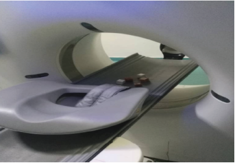
To determine the attenuation coefficient, (HU) of suggested tissue equivalent materials, CT images of developed material were investigated using a calibrated 128 slice Philip CT scanner at the Ibn ALhaytham diagnostic center, ALhawadith Street, Khartoum, Sudan operated at a tube voltage of 130 kVp. The mean HU was determined from three selected regions of interest (ROI) at different axial positions in the samples, using areas of approximately 200 mm2.
2.3. Phantom Body Frame and Lung Construction Methodology
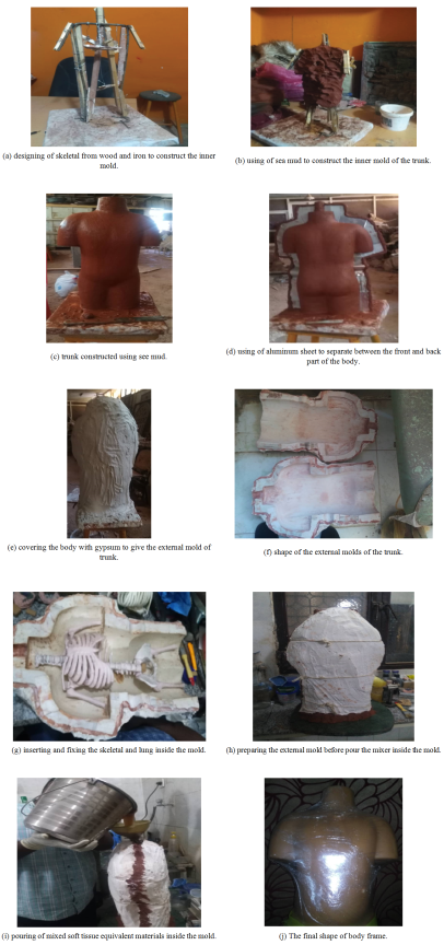
Two molds were made for the body frame. One to the front part and the other one for the back part of the body. The body frame dimension depends on the measurements of external dimension of pediatric of 3 years old age. The external dimension measurements include length and diameter of the neck, the distance between two shoulders from front and back, the length and diameter of the arms and legs, the diameter of the pelvic area and the waist, the diameter around chest and stomach.
The first step in the fabrication methodology was to prepare a skeletal from wood and iron just like the shape of body; the skeletal include neck, part of arms and legs, and pelvic area. the second step is to use a certain type of sea mud where this mud was put in to the skeletal from inside to outside, then start to sculpt the body shape using mud until the final shape of the body (trunk) with its specific dimension appear with all its details, then it leaves for three days until it become correlated and consistent. The trunk must be smooth and very sleek.
After the trunk frame is ready, aluminum was used to create barrier between the front body part and the back part and above the neck area a hole was made using mud also. White gypsum was used to make an external mold for the front and back part of the body, it should separate between the two molds parts with mud, then it tied up with thread until it held together and leaved to completely dry.
The front and back mold was taken after the gypsum was dry and the inside body was taking out, it was used with lung constructed from dark cork and skeletal that prepared before from Silicon Rubber RTV (683) and Calcium Carbonate to figure out how the body frame can work with the other tissue equivalent material and the shape of the body frame in the construction of phantom and for measurement purposes.
The skeletal and lung was arranged and fixed carefully inside the hollow back part of the mold like the arrangement in human body, then covered with the front part of the mold and a mixer of the tissue equivalent materials was prepared (which contain 2.8% weight of calcium carbonate and two parts of urethane PMC 121/30 Dry”, (Smooth-On, Easton, PA) was added together and mixed with an electric mixer, with care being taken to ensure a homogeneous mixture with no undissolved CaCO3) and poured through the hole in the neck until the hollow was full, and leave to complete dry and integrated. When the molds were removed, a complete frame of body or (phantom) can obtained.
3. Results
To know the degree of the useful and benefit of the chosen material which are urethane rubber mixed with CaCO3 as tissue equivalent material, the density of the material and the Hounsfield value was tested using computed tomography unit to figure out the agreement degree where it was compared with a reading of real patient soft tissue and with the slandered (reference level of density and HU) and the results of the density was as follow:
3.1. Density Measurements
3.1.1. Density of Soft Tissue
Table 1. The density values of the bone equivalent material based on the volume and the mass measurements.
Samples | Mass (g) | Volume () | Density (G/) |
1 | 28 | 27 | 1.037 |
2 | 27 | 26 | 1.038 |
3 | 23 | 22.5 | 1.022 |
Average density = (1.032±0.007) g/.
3.1.2. Density of Lung Tissue
The density was Taken by Weight of the whole piece dark cork tiles measuring 30.4 × 30.4 × 1.0 cm divided by its volume as follow:
ρ=
3.2. Attenuation Coefficient Measurements
Table 2. The value of the attenuation coefficients (Hounsfield value HU) for each sample of soft equivalent materials.
Region of Interest in CT Image | Samples 1 HU values | Samples 2 Hu Values | Samples 3 Hu Values |
1 | 10 | 15 | 36 |
2 | 26 | 17 | 16 |
3 | 17 | 19 | 21 |
4 | 13 | 44 | 29 |
5 | 43 | 15 | 39 |
6 | -17 | 23 | 44 |
Table 3. The value of the attenuation coefficients (Hounsfield value HU) for the lung equivalent materials sample.
Region of Interest in CT Image | Randum Measurment on the Samples | Randum Measurment on the Samples |
1 | -800 | -1000 |
2 | -764 | -1014 |
3 | -600 | -795 |
4 | -807 | 888 |
5 | 960 | -832 |
6 | -995 | -758 |
3.3. The Figures for the Samples and Body Frame Constructions
Figure 1. Image of the samples during measurements of HU using CT scanner.
Figure 2. Images illustrate the preparation steps for the designing of the mold.
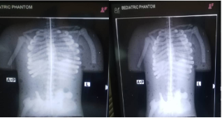
Figure 3. Image quality pictures using x-ray machine for body frame (trunk) assist with lung and skeletal.
4. Discussion
The paper focused on developing a methodology to construct body frames and lungs for phantoms using inexpensive, easily prepared tissue-equivalent materials. For the purpose of simulating the physical characteristics of human bone using CT scans, the density and HU were tested; density was determined by dividing mass by volume. The resulting average density of the tissue equivalent material was (1.032±0.007) g/cm3. It was found that it falls within the density range of tissue (1.04 g/cm3) and the density of the lung was 0.441 g/cm3, so the measurements of density were agreeing with reference level (0.33 g/cm3).
To account for tissue homogeneities inside the human body, CT scanners employ HU. To ensure that the results are comparable to those at the reference level, the linear attenuation coefficient value in CT images of the soft equivalent material was assessed from various positions of the testing sample. The resulting HU for soft tissue was swinging in a range from (-17 up to 44) HU. The goal of the soft tissue equivalent material was to create a homogeneous soft tissue analog that contained adipose tissue, connective tissue, and skeletal muscle with organs, so the range of HU slandered value for which muscle tissue was (10-40) and for the adipose tissue (-50 to -100) therefore the range of the results were fluctuated between (-50 to 40). The resulting HU for lung tissue in a range from (-600 up to -1014) HU, which fall in the range of HU slandered value (-500 up to -1000), Therefore the value of CT UH is passed the test and accepted.
This study offers a new composition manufacturing methodology and approval of the efficacy of the two-part urethane rubber compound “PMC 121/30 Dry” with CaCO3 as a soft tissue-equivalent material to construct body frame as one unit with the help of other part of body (bone and lung) to help in the construction of pediatric phantom. The methodology of molds construction is easily and simple to handle. And the final shape of the body was clear with all its details illustrate in the x-ray image as in human body.
5. Conclusion
A new methodology and simple way for designing body frame to be used in the construction of pediatric CT phantoms for quality control and dosimetry studies purposes was successfully constructed. Two-part urethane rubber compound “PMC 121/30 Dry”, was used with CaCO3 to fabricate tissue equivalent material, the urethane rubber compound was mixed with 2.8% by weight of powdered CaCO3 to be as low-cost soft tissue materials and dark cork was used as lung tissue equivalent materials.
The methodology and the material used was passed the tests of the density and the attenuation coefficient measurements and achieved the required results, and when they compared with the slandered it was found that the density of the fabricated material ((1.032±0.007) g/cm3) fall within the range of human soft tissue concentration which are (1.04) g/cm3, and the density of the lung tissue equivalent materials are 0.441 g/cm3 also fall within the range of human lung tissue concentration which are 0.3 g/cm3, the attenuation coefficients (HU) range from (-50 to 40) HU and (-600-1014 HU) on different CT images of soft tissue and equivalent materials also similar to that in reference level.
The majority of the conventional phantoms used in quality control testing were constructed using CT slices, which may have made it difficult to see all of the body details and the skeletal vertebrae. Nonetheless, the suggested model allows for a clear view of the spine, organs, vertebrae, and every other component of the body with all of its features, which can be useful in determining the radiation exposure to the human body.
Abbreviations
STEM | Soft Tissue Equivalent Materials |
HU | Hounsfield Units |
CT | Computed Tomography |
QC | Quality Control |
TEM | Tissue Equivalent Materials |
PMMA | Polymathic Methacrylate |
PVC | Polyvinyl Chloride |
KVP | Kilo Voltage Peak |
CaCO3 | Calcium Carbonate |
LTEM | Lung Tissue Equivalent Materials |
ROI | Regions of Interest |
Author Contributions
Esra Jafar Elameen: Conceptualization, Formal Analysis, Methodology, Writing - original draft, Writing - review & editing
Mona Ahmed Mohamed: Conceptualization, Formal Analysis, Investigation, Methodology, Resources, Supervision, Writing - review & editing
Suhaib Alameen: Investigation, Resources, Supervision, Writing - review & editing
Yousif Hassan Alsheikh: Writing - review & editing
Conflicts of Interest
The authors declare no conflicts of interest.
References
| [1] |
Radiation Risks and Pediatric Computed Tomography (CT): A Guide for Health Care Providers, US department of health and human surface, National institute of health, National cancer institute, USA, September 4, 2018.
|
| [2] |
Howard, Anthony, West, Ropert M. et al., Should Radiation Exposure be an Issue of Concern in Children with Multiple Trauma? Annual of Surgery, 275(3), p. 596-601, march 2022.
|
| [3] |
H. G. Ringertz, S. Bremmer from Radiological Protection of Patients in Diagnostic and Interventional Radiology book, International Atomic Energy Agency VIENNA, 2001.
|
| [4] |
Dudenredaktion, EditorDuden: Die deutsche Rechtschreibung, 26th edn. Dudenverlag; 2014. Dudenredaktion, Editor Duden: Die deutsche Rechtschreibung, 26th edn. Dudenverlag; 2014.
|
| [5] |
Phantoms, Mercuriushealth, advanced Oncology Solutions 2021,
https://mercuriushealth.com/glossary/phantom/
|
| [6] |
McGarry, C. K., Grattan, L. J., Ivory, A. M., Leek, F., Liney, G. P., Liu, Y., Miloro, P., Rai, R., Robinson, A., Shih, A. J., Zeqiri, B., & Clark, C. H. (2020, Dec 16). Tissue mimicking materials for imaging and therapy phantoms: a review Physics in Medicine and Biology, 65(23), Article 23TR01.
https://doi.org/10.1088/1361-6560/abbd17
|
| [7] |
A K Jones, D E Hintenlang, W E Bolch, Tissue-equivalent materials for construction of tomographic dosimetry phantoms in pediatric radiology, National Liberty of Medicine, National Center for Biotechnology Information, medical Physics 2003 Aug; 30(8): 2072-81.
https://doi.org/10.1118/1.1592641
|
| [8] |
Hatamikia, S., Jaksa, L., Kronreif, G., Birkfellner, W., … Lorenz, A. (2022). Silicone phantoms fabricated with multi‑material extrusion 3D printing technology mimicking imaging properties of soft tissues in CT.
|
| [9] |
De Oliveira, R. C. E., et al. (2022). Gelatin-based liver phantoms for training purposes: A cookbook. J. Clin. Med., 13(12), 3440.
|
| [10] |
Steidl, J. H., Liao, H., Ehrbar, S., & colleagues (2020-2021). Review of dynamic and deformable phantoms including heterogeneous tissue‑equivalent materials (e.g. PVC‑softener, silicon) for dose accuracy evaluation. Published in Tissue‑mimicking materials for imaging and therapy phantoms: a review.
|
| [11] |
Hutchinson, R., Smith, J., & Jones, A. (2023). Construction and validation of an infant chest phantom for pediatric computed tomography. Physical and Engineering Sciences in Medicine.
|
| [12] |
Fisher RF. Tissue equivalent phantoms for the evaluation of tube‑current modulated CT dose and image quality [master’s thesis]. Gainesville (FL): University of Florida; 2006.
|
| [13] |
International Commission on Radiation Units and Measurement. Tissue substitutes in radiation dosimetry and measurement. Report No. 44. Bethesda, MD: ICRU, 1989.
|
| [14] |
NimaKasraie, Amie Robinson, and Sherwin Chan, Construction of an Anthropomorphic Phantom for Use in Evaluating Pediatric Airway Digital Tomosynthesis Protocols, Department of Radiology, Children’s Mercy Hospital, 2401 Gillham Rd., Kansas City, MO 64108, USA Radiology Research and Practice. Volume 2018, Article ID 3835810, February 2018.
|
| [15] |
CRU, Tissue substitutes in radiation dosimetry and measurement. Report 44. In. ICRU report. Bethesda, Md., U.S.A.: International Commission on Radiation Units and Measurements, 1989.
|
Cite This Article
-
APA Style
Elameen, E. J., Mohamed, M. A., Alameen, S., Alsheikh, Y. H. (2025). Designing of External Body Frame (Trunk Area) Part for Pediatric Phantom from Law Cost Soft Tissue Equivalent Materials. European Journal of Biophysics, 13(1), 10-18. https://doi.org/10.11648/j.ejb.20251301.12
 Copy
|
Copy
|
 Download
ACS Style
Elameen, E. J.; Mohamed, M. A.; Alameen, S.; Alsheikh, Y. H. Designing of External Body Frame (Trunk Area) Part for Pediatric Phantom from Law Cost Soft Tissue Equivalent Materials. Eur. J. Biophys. 2025, 13(1), 10-18. doi: 10.11648/j.ejb.20251301.12
Copy
|
Download
AMA Style
Elameen EJ, Mohamed MA, Alameen S, Alsheikh YH. Designing of External Body Frame (Trunk Area) Part for Pediatric Phantom from Law Cost Soft Tissue Equivalent Materials. Eur J Biophys. 2025;13(1):10-18. doi: 10.11648/j.ejb.20251301.12
Copy
|
Download
-
@article{10.11648/j.ejb.20251301.12,
  author = {Esra Jafar Elameen and Mona Ahmed Mohamed and Suhaib Alameen and Yousif Hassan Alsheikh},
  title = {Designing of External Body Frame (Trunk Area) Part for Pediatric Phantom from Law Cost Soft Tissue Equivalent Materials
},
  journal = {European Journal of Biophysics},
  volume = {13},
  number = {1},
  pages = {10-18},
  doi = {10.11648/j.ejb.20251301.12},
  url = {https://doi.org/10.11648/j.ejb.20251301.12},
  eprint = {https://article.sciencepublishinggroup.com/pdf/10.11648.j.ejb.20251301.12},
  abstract = {Radiation is valuable for diagnosis and treatment, but excessive exposure can harm human health. To ensure safety, measuring radiation absorbed by human tissue during imaging is critical. Scientists use phantoms models simulating human bodies or organs, for this purpose. Designing X-ray phantoms requires careful material selection. Most commercial phantoms mimic neonates or adults, leaving a gap for pediatric models (ages 1-15 years). This study aimed to fabricate a low-cost pediatric phantom. The methodology involved creating a lung phantom using dark cork tiles (30.4 × 30.4 × 1.0 cm), cut into coronal plates and stacked to mimic lung anatomy (density: 0.33 g/cm³). The body frame, based on a 3-year-old’s dimensions, was constructed using a wood-and-iron skeletal structure shaped with sea mud. After drying, aluminum barriers and gypsum molds were made for the front and back body parts. The cork lungs and skeletal were placed inside the mold, and a soft tissue-equivalent mixture (STEM) (2.8% calcium carbonate, urethane PMC 121/30 Dry) was poured into the cavity. Once dried, the molds were removed, yielding a complete phantom for testing. Density and Hounsfield Units (HU) were evaluated to simulate human tissue properties. Soft tissue-equivalent material’s (STEM) average density was 1.032±0.007 g/cm³, aligning with the reference range (1.04 g/cm³). Lung density measured 0.441 g/cm³, close to the expected 0.33 g/cm³. HU values for soft tissue ranged from -17 to 44, encompassing muscle (10-40) and adipose tissue (-50 to -100). Lung HU values (-600 to -1014) fell within the standard range (-500 to -1000), validating the material’s efficacy. A full X-ray image of the trunk confirmed the phantom’s structural accuracy and absorption efficiency. Traditional phantoms, often based on Computer tomography (CT) slices, struggle to depict skeletal complexities like vertebrae. In contrast, this model clearly visualizes spinal vertebrae and skeletal details, aiding precise dosage calculations. The study demonstrates the successful fabrication of a pediatric phantom using cost-effective materials, with density and HU values closely matching human tissue. This advancement addresses the critical need for pediatric phantoms in radiation research and clinical applications.},
 year = {2025}
}
Copy
|
Download
-
TY  - JOUR
T1  - Designing of External Body Frame (Trunk Area) Part for Pediatric Phantom from Law Cost Soft Tissue Equivalent Materials

AU  - Esra Jafar Elameen
AU  - Mona Ahmed Mohamed
AU  - Suhaib Alameen
AU  - Yousif Hassan Alsheikh
Y1  - 2025/08/12
PY  - 2025
N1  - https://doi.org/10.11648/j.ejb.20251301.12
DO  - 10.11648/j.ejb.20251301.12
T2  - European Journal of Biophysics
JF  - European Journal of Biophysics
JO  - European Journal of Biophysics
SP  - 10
EP  - 18
PB  - Science Publishing Group
SN  - 2329-1737
UR  - https://doi.org/10.11648/j.ejb.20251301.12
AB  - Radiation is valuable for diagnosis and treatment, but excessive exposure can harm human health. To ensure safety, measuring radiation absorbed by human tissue during imaging is critical. Scientists use phantoms models simulating human bodies or organs, for this purpose. Designing X-ray phantoms requires careful material selection. Most commercial phantoms mimic neonates or adults, leaving a gap for pediatric models (ages 1-15 years). This study aimed to fabricate a low-cost pediatric phantom. The methodology involved creating a lung phantom using dark cork tiles (30.4 × 30.4 × 1.0 cm), cut into coronal plates and stacked to mimic lung anatomy (density: 0.33 g/cm³). The body frame, based on a 3-year-old’s dimensions, was constructed using a wood-and-iron skeletal structure shaped with sea mud. After drying, aluminum barriers and gypsum molds were made for the front and back body parts. The cork lungs and skeletal were placed inside the mold, and a soft tissue-equivalent mixture (STEM) (2.8% calcium carbonate, urethane PMC 121/30 Dry) was poured into the cavity. Once dried, the molds were removed, yielding a complete phantom for testing. Density and Hounsfield Units (HU) were evaluated to simulate human tissue properties. Soft tissue-equivalent material’s (STEM) average density was 1.032±0.007 g/cm³, aligning with the reference range (1.04 g/cm³). Lung density measured 0.441 g/cm³, close to the expected 0.33 g/cm³. HU values for soft tissue ranged from -17 to 44, encompassing muscle (10-40) and adipose tissue (-50 to -100). Lung HU values (-600 to -1014) fell within the standard range (-500 to -1000), validating the material’s efficacy. A full X-ray image of the trunk confirmed the phantom’s structural accuracy and absorption efficiency. Traditional phantoms, often based on Computer tomography (CT) slices, struggle to depict skeletal complexities like vertebrae. In contrast, this model clearly visualizes spinal vertebrae and skeletal details, aiding precise dosage calculations. The study demonstrates the successful fabrication of a pediatric phantom using cost-effective materials, with density and HU values closely matching human tissue. This advancement addresses the critical need for pediatric phantoms in radiation research and clinical applications.
VL  - 13
IS  - 1
ER  -
Copy
|
Download