Case Report | | Peer-Reviewed

Delayed Third Molar Development: Report of a Case

Received: 26 August 2025     Accepted: 5 September 2025     Published: 9 October 2025
Views:       Downloads:
Abstract

Introduction: The delayed development of third molar teeth occurs due to a variety of genetic, evolutionary, environmental, nutritional, and systemic medical factors, as well as maxillary and mandibular developmental skeletofacial disorders. These factors, either alone or in combination, have the potential to influence the complex growth and development of the dentition, including the third molar teeth. Objective: This case report presents an unusual occurrence of differential third molar development in a seventeen-year-old Caucasian female. Tooth #1, the maxillary right third molar tooth, developed in delayed fashion compared to the other three third molars. The purpose of this report is to draw attention to the importance of obtaining contemporaneous panoramic radiographic imaging as a critical component of the examination and treatment planning process when third molar teeth are being consider for removal. Case Presentation: This report presents a case of a seventeen-year-old Caucasian female who had originally presented at age fifteen for examination and consultation regarding proposed removal of her impacted and developing third molar teeth, due to a history of recently completed orthodontic treatment. Her panoramic radiograph at that time demonstrated the absence of #1, and was considered to be a result of agenesis. The parents elected to defer removal of her third molars at that time. She subsequently returned for another consultation appointment at seventeen years of age, at which time panoramic radiographic evaluation demonstrated a minimally developed tooth #1, as compared to her other three third molar teeth. Conclusions: Delayed development of a given tooth, in particular a third molar tooth, can be easily misdiagnosed as agenesis, depending upon the age of the patient when the radiograph was taken. In this case, early removal of the patient’s third molars would have resulted in inadvertent surgical omission, thereby necessitating additional surgical intervention.

Published in International Journal of Clinical and Developmental Anatomy (Volume 9, Issue 1)
DOI 10.11648/j.ijcda.20250901.12
Page(s) 7-11
Creative Commons

This is an Open Access article, distributed under the terms of the Creative Commons Attribution 4.0 International License (http://creativecommons.org/licenses/by/4.0/), which permits unrestricted use, distribution and reproduction in any medium or format, provided the original work is properly cited.

Copyright

Copyright © The Author(s), 2025. Published by Science Publishing Group

Keywords

Third Molar, Delayed Development, Agenesis, Panoramic Radiograph, Cone-Beam Computed Tomography, Surgical Removal

1. Introduction
The delayed development of third molar teeth occurs due to a variety of factors, including genetic, evolutionary, nutritional, and environmental influences, as well as systemic medical disorders, including hormonal and developmental skeletofacial disorders. These factors, either alone or in combination, have the potential to influence the complex growth and development of the dentition, including the third molar teeth. It is accepted that an impacted tooth is one that fails to fully erupt in the dental arch within the usual range of expected time, and which can be a result of abnormal tooth orientation, adjacent teeth, dense overlying bone, or genetic anomaly . Additionally, all third molars, maxillary and mandibular, demonstrate a greater degree of variation in development than any of the other teeth in the oral cavity . Interestingly, females tend to demonstrate a higher frequency of hypodontia and microdontia than males, while males demonstrate a greater frequency of supernumerary teeth and megadontia . On the cellular level, the influence of various which affect tooth organ formation is a complex phenomenon. While it has been demonstrated that mesenchymal condensation is critical for organogenesis, little is known about the exact nature of how the process is controlled, and it is believed that induction by antagonistic epithelial morphogens manifest their pattern-generating actions mechanically via changes in mesenchymal cell shape and altered mechanotransduction .
In this case report, the critical importance of appropriate radiographic examination, performed contemporaneously, cannot be overstated. It is additionally important to appreciate that while radiographic evaluation of third molar development in relation to chronological age is important for examination and treatment planning purposes, the presence of agenesis or delayed development of a tooth must be confirmed by sequential and contemporaneous radiographic examination. Studies have shown that the developmental stages of third molar teeth are more advanced in the maxillary arch than the mandibular arch, and that third molars develop earlier in the maxillary arch than the mandibular arch and are developed earlier in males than in females .
It should also be appreciated that various systems for third molar development have been developed and utilized for accurate classification of the developmental stages for third molar development, and are useful for diagnosis, treatment planning, age assessment, and forensic applications. As determination of tooth development stages or dental age holds profound significance, in particular with the use of Demirjian classification system, it is well known that some individuals may have teeth that develop and mature faster or slower than their peers, resulting in variations between dental age and chronological age .
This report presents a case of a seventeen-year -old Caucasian female with delayed development of her maxillary right third molar tooth #1. The purpose of this report is to draw attention to the importance of obtaining contemporaneous panoramic radiographs, along with adjunctive cone-beam computed tomography (CBCT) imaging, in order to facilitate comprehensive and accurate treatment planning.
2. Case Report
A seventeen-year-old Caucasian female presented for examination and consultation regarding removal of her presumed three impacted third molar teeth #’s 16, 17, 32. She had undergone orthodontic treatment at an early age, which was completed at fifteen years of age. A panoramic radiograph was taken at that time, which demonstrated the presence of three impacted teeth, #’s 16, 17, 32, and the absence of tooth #1 (Figure 1).
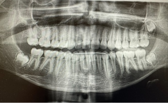
Figure 1. Panoramic radiograph taken at fifteen years of age, demonstrating absence of tooth #1.
Upon inspection of this radiograph, it was presumed at that time to be indicative of agenesis of tooth #1. Although the examination and consultation was performed at fifteen years of age, the parents deferred surgical treatment at that time. Subsequent to this, the patient returned at seventeen years of age for re-examination and consultation regarding removal of her third molar teeth, due to her history of orthodontic treatment, the presence of impacted third molar teeth, and intermittent pain. Her past medical history was essentially benign, with no significant medical problems or surgeries noted upon intake. Physical examination demonstrated a healthy seventeen-year-old Caucasian female with her dentition in a good state of repair. Her occlusion was Class I, her oral hygiene was good, and no soft tissue pathology was identified. Her peritonsillar tissues were atrophic. She demonstrated mandibular asymmetry to the right, with her chin point approximately 5 millimeters to the right of her maxillary midline. A contemporaneous panoramic radiograph was obtained that demonstrated four impacted third molars being present in various stages of development and impaction (Figure 2).
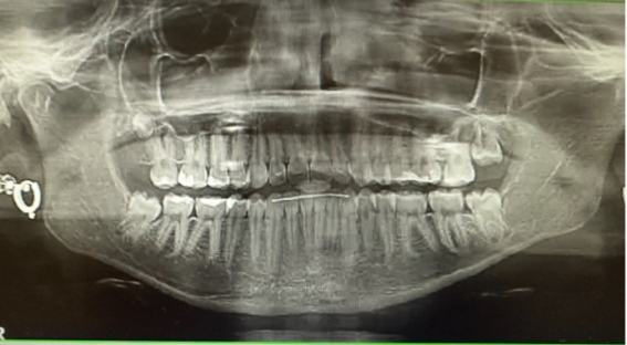
Figure 2. Panoramic radiograph taken at seventeen years of age, demonstrating the presence of tooth #1.
Upon comparison of the prior and contemporaneous panoramic radiographs, it was evident that tooth #1 was in a state of delayed development, as compared to teeth #’s 16, 17, and 32. Comparative radiographs of her right posterior mandibular and maxillary regions further demonstrate the comparison of between the initial apparent agenesis of tooth #1 and the subsequent development of this tooth (Figures 3, 4). Additionally, the contemporary panoramic radiograph was suggestive of possible pathology inferiorly to the developing tooth #1, and as such CBCT was obtained in order to rule out or confirm the presence associated of pathology, and which indicated that no specific pathology was present, and that a possible hyperplastic dental follicle was associated with this developing tooth (Figure 5).
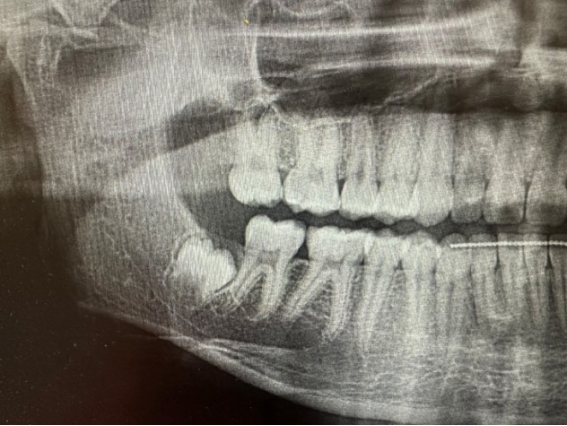
Figure 3. Magnified image of panoramic radiograph demonstrating absence of tooth #1.
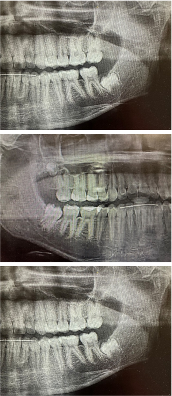
Figure 4. Magnified image of radiograph demonstrating presence of tooth #1.
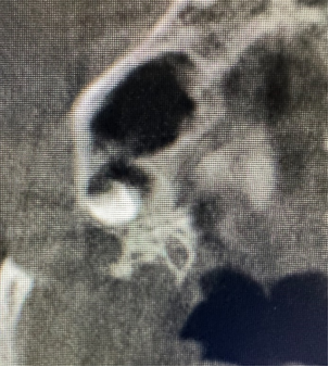
Figure 5. Magnified CBCT scan demonstrating developing and impacted tooth #1, with pericoronal radiolucency suggestive of a possible hyperplastic follicle.
The patient was scheduled for removal of her four third molar teeth, and subsequently underwent removal of these teeth under general anesthesia. Tooth #1 was removed without complication, and the follicular tissue was curettage and removed along with the tooth. No sinus exposure was observed, and no specimen was submitted for histopathologic evaluation. In comparison to the other three third molars which were removed (#’s 16, 17, and 32), tooth #1 was noted to have smaller dimensions and root development (Figure 6). The patient experienced an uncomplicated postoperative course, and was noted to have regained masticatory function as expected for her age and health.
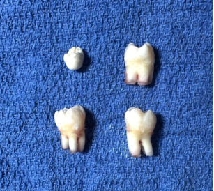
Figure 6. Postoperative comparison of the four third molar teeth #’s 1, 16, 17, 32, demonstrating significantly decreased dimensions and root development of tooth #1, as compared to teeth #’s 16, 17, and 32.
3. Discussion
The decision to observe or remove third molar teeth presents challenges to the astute clinician, in particular with respect to whether or not the proposed removal of these teeth are prophylactic or therapeutic. While the ability to accurately predict third molar development and eruption would allow clinicians to continue to improve and appropriately define third molar treatment strategies, it is important to appreciate that the natural course of an asymptomatic third molar is uncertain, with change in position or eruption reported even with advancing age . Understanding that accurate estimation of chronological age based upon the stages of third molar development, following the eight stages method of Demirjian, the astute clinician must consider that age estimation must take into consideration a variety of biologic factors, and variation can be expected to occur . Given this, the occurrence of agenesis versus delayed development must be considered by the clinician during the treatment planning process, in particular when orthodontic treatment is ongoing or has been completed.
In this case report, the patient completed orthodontic therapy at chronological age fifteen, and at that time was referred for examination and consultation regarding removal of her third molar teeth. The post-orthodontic radiograph at that time demonstrated the presence of teeth #’s 16, 17, and 32, and was suggestive of agenesis of the maxillary right third molar tooth #1. While it is accepted that genetic factors which may result in agenesis of one or more third molar teeth may affect the dentition as a whole, the third molar teeth are more often vulnerable to the factors involved in agenesis of the dentition and can be associated with the evolutionary trend in humans toward reduced molar number . In this regard, it is worthwhile to show that as there is a significant component of genetic variation present in the regulatory mechanisms of third molar development, this variability is of great clinical significance for making decisions on the timing of third molar removal, in particular with respect to orthodontic treatment planning and chronological age estimation for medico-legal purposes . Important with respect to this case report is that females are significantly more likely than males to have agenesis one or more third molars, and that maxillary agenesis was also significantly more likely than mandibular agenesis in both genders .
In this report, the patient in question and her parents deferred removal of her third molars at fifteen years of age. She subsequently returned at 17 years of age for re-examination and consultation of her third molars teeth. A contemporaneous panoramic radiograph was obtained at this time, which demonstrated the presence of developing third molar tooth #1, as well as her remaining third molar teeth #’s 16, 17, and 32, in various degrees of impaction. This was confirmation that the prior diagnosis of agenesis of tooth #1 was erroneous, and that this represented a case of delayed third molar development. It has been observed that third molar genesis may occur up to the age of sixteen years, and that individual variability in the stage of development of third molars increases when genesis is delayed beyond ten years of age . As the multifactorial etiology of third molar development remains a challenging field of study, it has been hypothesized that developmental skeletofacial deformities, such a maxillary and mandibular hypoplasia, are positively correlated with delayed third molar development . As the patient in this report demonstrated mandibular asymmetry to the right side, it may be a possible etiologic factor with respect to the delayed development of her right maxillary third molar tooth.
Ultimately, the decision to remove third molar teeth is based upon clinical and radiographic evaluation, clinical judgment, and appropriate consideration of a variety of factors including angulation, degree of impaction, and the presence of symptoms . As the prophylactic removal of third molar teeth following completion of orthodontic treatment is an accepted and clinically indicated therapeutic modality, the timing of removal of these teeth following orthodontic treatment should be considered based upon the knowledge that delayed development of third molars can occur. In the case presented in this report, delayed treatment and contemporaneous radiographic imaging were critical with respect to appropriate surgical management. While decisions regarding removal or observation of third molars should optimally be made before the middle of the third decade, it is important to appreciate the potential for variability in third molar development, and the need for contemporaneous radiographic imaging for appropriate treatment planning and surgical management .
4. Conclusion
This case report discusses the delayed development of a maxillary right third molar tooth, which was discovered upon radiographic evaluation, approximately two years following the completion of orthodontic therapy, and which earlier panoramic radiographic examination was suggestive of agenesis. This case demonstrates the critical importance of contemporaneous radiographic examination, as well as consideration of delaying this molar extraction from the earlier teenage years when agenesis is suspected. As contemporary orthodontic treatment is being completed in many cases at younger ages, the possibility of delayed development of one or more third molar teeth should be considered for treatment planning purposes. It is recommended that this is an appropriate opportunity for research for the dental profession.
Abbreviations

CBCT

Cone-Beam Computed Tomography

Author Contributions
Daniel Joseph Traub is the sole author. The author read and approved the final manuscript.
Conflicts of Interest
The author declares no conflict of interest.
References
[1] Hupp, J. R, Ellis, E., Tucker, M. R. Contemporary Oral and Maxillofacial Surgery, Seventh Edition (Elsevier, 2019).
[2] Nelson, S. J., Ash, M. M. Wheeler’s Dental Anatomy, Physiology, and Occlusion, Ninth Edition (Saunders Elsevier, 2010).
[3] Brook, A. H. A unifying aetiological explanation for anomalies of human tooth number and size. Archives of Oral Biology 29, 373-379 (1984).
[4] Mammato, T., et al. Mechanochemical control of mesenchymal condensation and embryonic tooth organ formation. Developmental Cell 21, 758-769 (2011).
[5] Jung, Y., et al. Radiographic evaluation of third molar development in 6-to-24-year olds. Imaging Science in Dentistry 17, 185-191 (2014).
[6] Milani, O. H., et al. A fully automated classification of third molar development stages using deep learning. Scientific Reports 14, Article number: 13082 (2024).
[7] American Association of Oral and Maxillofacial Surgeons. White Paper on Third Molar Data (2014).
[8] Lewis, A. J. Demirjian’s method in the estimation of age: A study on human third molars. Journal of Forensic Dental Sciences 7, 153-157 (2015).
[9] Scheiwiller, M., et al. Third molar agenesis in modern humans with and without agenesis of other teeth. Peer J (2020).
[10] Trakiniene, G., et al. Impact of genetics on third molar agenesis. Scientific Reports 8: 8307 (2018).
[11] Carter, K. W., et al. Morphologic and Demographic Predictors of Third Molar Agenesis: A Systematic Review and Meta-analysis. Journal of Dental Research 94, 886-894 (2015).
[12] Richardson, M. Late third molar genesis: its significance in orthodontic treatment. Angle Orthodontist 50, 121-128 (1980).
[13] Marchiori, D. F. et al. Initial third molar development is delayed in jaws with short distal space: An early impaction sign? Archives of Oral Biology 106, (2019).
[14] D’Amico, C., et al. Clinical and Surgical Indications and Current Guidelines on Surgical Removal of Third Molars. Engineering Proceedings 87 (2015).
[15] American Association of Oral and Maxillofacial Surgeons. White Paper on Management of Third Molar Teeth (2016).
Cite This Article
  • APA Style

    Traub, D. J. (2025). Delayed Third Molar Development: Report of a Case. International Journal of Clinical and Developmental Anatomy, 9(1), 7-11. https://doi.org/10.11648/j.ijcda.20250901.12

    Copy | Download

    ACS Style

    Traub, D. J. Delayed Third Molar Development: Report of a Case. Int. J. Clin. Dev. Anat. 2025, 9(1), 7-11. doi: 10.11648/j.ijcda.20250901.12

    Copy | Download

    AMA Style

    Traub DJ. Delayed Third Molar Development: Report of a Case. Int J Clin Dev Anat. 2025;9(1):7-11. doi: 10.11648/j.ijcda.20250901.12

    Copy | Download

  • @article{10.11648/j.ijcda.20250901.12,
      author = {Daniel Joseph Traub},
      title = {Delayed Third Molar Development: Report of a Case},
      journal = {International Journal of Clinical and Developmental Anatomy},
      volume = {9},
      number = {1},
      pages = {7-11},
      doi = {10.11648/j.ijcda.20250901.12},
      url = {https://doi.org/10.11648/j.ijcda.20250901.12},
      eprint = {https://article.sciencepublishinggroup.com/pdf/10.11648.j.ijcda.20250901.12},
      abstract = {Introduction: The delayed development of third molar teeth occurs due to a variety of genetic, evolutionary, environmental, nutritional, and systemic medical factors, as well as maxillary and mandibular developmental skeletofacial disorders. These factors, either alone or in combination, have the potential to influence the complex growth and development of the dentition, including the third molar teeth. Objective: This case report presents an unusual occurrence of differential third molar development in a seventeen-year-old Caucasian female. Tooth #1, the maxillary right third molar tooth, developed in delayed fashion compared to the other three third molars. The purpose of this report is to draw attention to the importance of obtaining contemporaneous panoramic radiographic imaging as a critical component of the examination and treatment planning process when third molar teeth are being consider for removal. Case Presentation: This report presents a case of a seventeen-year-old Caucasian female who had originally presented at age fifteen for examination and consultation regarding proposed removal of her impacted and developing third molar teeth, due to a history of recently completed orthodontic treatment. Her panoramic radiograph at that time demonstrated the absence of #1, and was considered to be a result of agenesis. The parents elected to defer removal of her third molars at that time. She subsequently returned for another consultation appointment at seventeen years of age, at which time panoramic radiographic evaluation demonstrated a minimally developed tooth #1, as compared to her other three third molar teeth. Conclusions: Delayed development of a given tooth, in particular a third molar tooth, can be easily misdiagnosed as agenesis, depending upon the age of the patient when the radiograph was taken. In this case, early removal of the patient’s third molars would have resulted in inadvertent surgical omission, thereby necessitating additional surgical intervention.},
     year = {2025}
    }
    

    Copy | Download

  • TY  - JOUR
    T1  - Delayed Third Molar Development: Report of a Case
    AU  - Daniel Joseph Traub
    Y1  - 2025/10/09
    PY  - 2025
    N1  - https://doi.org/10.11648/j.ijcda.20250901.12
    DO  - 10.11648/j.ijcda.20250901.12
    T2  - International Journal of Clinical and Developmental Anatomy
    JF  - International Journal of Clinical and Developmental Anatomy
    JO  - International Journal of Clinical and Developmental Anatomy
    SP  - 7
    EP  - 11
    PB  - Science Publishing Group
    SN  - 2469-8008
    UR  - https://doi.org/10.11648/j.ijcda.20250901.12
    AB  - Introduction: The delayed development of third molar teeth occurs due to a variety of genetic, evolutionary, environmental, nutritional, and systemic medical factors, as well as maxillary and mandibular developmental skeletofacial disorders. These factors, either alone or in combination, have the potential to influence the complex growth and development of the dentition, including the third molar teeth. Objective: This case report presents an unusual occurrence of differential third molar development in a seventeen-year-old Caucasian female. Tooth #1, the maxillary right third molar tooth, developed in delayed fashion compared to the other three third molars. The purpose of this report is to draw attention to the importance of obtaining contemporaneous panoramic radiographic imaging as a critical component of the examination and treatment planning process when third molar teeth are being consider for removal. Case Presentation: This report presents a case of a seventeen-year-old Caucasian female who had originally presented at age fifteen for examination and consultation regarding proposed removal of her impacted and developing third molar teeth, due to a history of recently completed orthodontic treatment. Her panoramic radiograph at that time demonstrated the absence of #1, and was considered to be a result of agenesis. The parents elected to defer removal of her third molars at that time. She subsequently returned for another consultation appointment at seventeen years of age, at which time panoramic radiographic evaluation demonstrated a minimally developed tooth #1, as compared to her other three third molar teeth. Conclusions: Delayed development of a given tooth, in particular a third molar tooth, can be easily misdiagnosed as agenesis, depending upon the age of the patient when the radiograph was taken. In this case, early removal of the patient’s third molars would have resulted in inadvertent surgical omission, thereby necessitating additional surgical intervention.
    VL  - 9
    IS  - 1
    ER  - 

    Copy | Download

Author Information