Introduction: Lumbosacral transitional anomaly is a very common congenital malformation that is diagnosed incidentally. Our objective was to study lumbosacral transitional anomalies using computed tomography. Method: Our study was conducted in the medical imaging department of the Pr Bocar Sidy SALL University Hospital in Kati and the Amitie Medical Clinic in Kati. It was a cross-sectional study with retrospective and prospective recruitment from January 2023 to December 2024. All patients who underwent lumbosacral computed tomography and in whom a lumbosacral transitional anomaly was ob-served were included. The sampling was consecutive. We used the Castellvi classification to identify the types of anomaly. Anonymity and confidentiality were respected. Results: The overall prevalence of lumbosacral transitional anomalies was 3.99%, including 3.6% in the imaging department of the Pr Bocar Sidy SALL University Hospital in Kati and 5.03% at the Amitie medical clinic in Kati. The average age was 39±9 years, with extremes ranging from 15 to 48 years. Males were predominant (53.3%). Lumbar sciatica was the main clinical finding in 52% of cases. Computed tomography revealed sacralization in 74.7% of cases, 25% of which were Castellvi type IIIa. Lumbalization was noted in 25.3%, of which 36.8% were Castellvi type IIa. Conclusion: Lumbosacral transition anomalies remain common, and knowledge of them could help reduce the incidence of surgical interventions at the wrong site.
This is an Open Access article, distributed under the terms of the Creative Commons Attribution 4.0 International License (http://creativecommons.org/licenses/by/4.0/), which permits unrestricted use, distribution and reproduction in any medium or format, provided the original work is properly cited.
Lumbosacral transitional anomaly is a congenital malformation characterized by the presence of mega-transverse processes on the last mobile lumbar vertebra, these mega-processes coming into contact with the sacrum and/or iliac wing, sometimes forming an equivalent of an articular space
[1]
Bertolotti M. Contributo alla conoscenze dei vizi di differenzazione regionale del rachide con speciale riguardo alla assimilazione sacrale della V. lombare. Radiologique Medica [A Contribution to the Understanding of Regional Variations in Spinal Differentiation, with Special Reference to the Sacral Assimilation of the Lumbar Vertebrae. Radiologique Medica]. Turin, 1917; 4: 113-44.
[1]
. It corresponds to a morphological variation ranging from partial or complete sacralization to partial or complete Lombalization of the affected vertebra
[2]
Mahato NK. Redefining lumbosacral transitional vertebrae (LSTV) classification: integrating the full spectrum of morphological alterations in a biomechanical continuum. Medical hypotheses, Kanchipuram, 2013; 81: 76-81.
[2]
. Lumbalization of the S1 vertebrae presents as an abnormal joint, with well-formed lumbar-type facet joints and a well-defined disc of normal size, while sacralization of the L5 vertebra is characterized by enlarged and elongated transverse processes that are fused to the sacrum
[3]
French HD, Somasundaram AJ, Schaefer NR, Laherty RW. Lumbosacral Transitional Vertebrae and Its Prevalence in the Australian Population. Glob Spine J. déc 2014; 4(4): 229‑32.
[3]
.
It is a very common congenital malformation, with a global prevalence ranging from 4% to 36% and a marked preference in males
[4]
Nardo L, Alizai H, Virayavanich W, Liu F, Hernandez A, Lynch JA, et al. Lumbosacral Transitional Vertebrae: Associ-ation with Low Back Pain. Radiology. nov 2012; 265(2): 497‑503.
[5]
Uçar D, Uçar BY, Coşar Y, Emrem K, Gümüşsuyu G, Mutlu S, et al. Retrospective Cohort Study of the Prevalence of Lumbosacral Transitional Vertebra in a Wide and Well-Represented Population. Arthritis. 2013; 2013: 461425.
[4, 5]
. In Europe, it is 28.6% according to a study of a Caucasian population
[6]
Hanhivaara J, Määttä JH, Niinimäki J, & Nevalainen MT. Lumbosacral transitional vertebrae are associated with lum-bar degeneration: retrospective evaluation of 3855 consecutive abdominal CT scans. European Radiology, Oulo, 2020 ; 30 : 3409-3416.
[6]
. In China, its prevalence varies between 12% and 30%, and it is 4 to 19% in India
[7]
Gopalan B, Yerramshetty JS. Lumbosacral Transitional Vertebra-Related Low Back Pain: Resolving the Controversy. Asian Spine J. juin 2018; 12(3): 407‑15.
[8]
Sekharappa V, Amritanand R, Krishnan V, David KS. Lumbosacral Transition Vertebra: Prevalence and Its Signifi-cance. Asian Spine J. 2014; 8(1): 51‑8.
[7, 8]
. However, the frequency of lumbosacral transitional anomaly in Africa is patchy. According to a study conducted in Cameroon, its prevalence was 13.6%
[6]
Hanhivaara J, Määttä JH, Niinimäki J, & Nevalainen MT. Lumbosacral transitional vertebrae are associated with lum-bar degeneration: retrospective evaluation of 3855 consecutive abdominal CT scans. European Radiology, Oulo, 2020 ; 30 : 3409-3416.
[6]
. In Mali, a prevalence of 12.84% was reported in a recent study conducted in the medical imaging department of the Pr Bocar Sidy SALL University Hospital in Kati in 2023
[9]
Tangara MS. Aspects scanographiques des anomalies transitionnelles du rachis lombo-sacré au service d‘imagerie médicale du CHU Pr Bocar Sidy Sall de Kati [Imaging Findings of Transitional Abnormalities of the Lumbosacral Spine at the Medical Imaging Department of the Pr. Bocar Sidy Sall University Hospital in Kati]. USTTB, Bamako, 2023: 85p.
[9]
.
Most of these malformations go unrecognized, either because they are asymptomatic or because they are not recognized or misdiagnosed. Some attract attention because they cause back pain or because they are associated with other abnormalities or even pathologies. Others attract attention because spinal mobility is limited or because there is spinal instability
[8]
Sekharappa V, Amritanand R, Krishnan V, David KS. Lumbosacral Transition Vertebra: Prevalence and Its Signifi-cance. Asian Spine J. 2014; 8(1): 51‑8.
[9]
Tangara MS. Aspects scanographiques des anomalies transitionnelles du rachis lombo-sacré au service d‘imagerie médicale du CHU Pr Bocar Sidy Sall de Kati [Imaging Findings of Transitional Abnormalities of the Lumbosacral Spine at the Medical Imaging Department of the Pr. Bocar Sidy Sall University Hospital in Kati]. USTTB, Bamako, 2023: 85p.
[8, 9]
.
Over the last decade, the problem of diagnosing and classifying lumbosacral transitional anomalies has shifted from the clinical domain to that of imaging. Computed tomography (CT) allows for a comprehensive morphological analysis of the lumbosacral spine and provides an etiological orientation for low back pain
[9]
Tangara MS. Aspects scanographiques des anomalies transitionnelles du rachis lombo-sacré au service d‘imagerie médicale du CHU Pr Bocar Sidy Sall de Kati [Imaging Findings of Transitional Abnormalities of the Lumbosacral Spine at the Medical Imaging Department of the Pr. Bocar Sidy Sall University Hospital in Kati]. USTTB, Bamako, 2023: 85p.
[9]
.
Knowledge and detection of anomalies are very important in order to prevent serious secondary complications and assist in preoperative assessment and postoperative follow-up. It is important to note that unusual patient characteristics and anatomy are major risk factors and must be taken into account when performing lumbar-sacral spine surgery in order to avoid surgery on the wrong site. To our knowledge, there are no studies on lumbosacral transitional anomalies in patients under 50 years of age. It is in this context that we initiated this study to investigate the prevalence of this anomaly at the Pr Bocar Sidi Sall University Hospital and the Amitie Medical Clinic in Kati.
The objective of this study was to investigate lumbosacral transitional anomalies in subjects under 50 years of age at the medical imaging department of the Pr Bocar Sidy Sall University Hospital in Kati and at the Amitie Medical Clinic in Kati.
2. Materials and Methods
This was a retrospective and prospective cross-sectional study of lumbosacral transitional anomalies during the period from January 1, 2023, to December 2024, i.e., 24 months.
The examinations were performed using 16-slice SIEMENS CT scanners at the Pr BSS University Hospital and 16-slice GENERAL ELECTRIC scanners at the clinic.
This study included all patients who underwent a thoracolumbar CT scan.
All patients under the age of 50 admitted to both departments for lumbosacral computed tomography and diagnosed with a lumbosacral transitional anomaly were included.
The sampling was consecutive
Data collection was performed using the Google FORMS application based on CT scan reports. Data analysis was performed using SPSS version 25 software. Descriptive statistics included frequencies, percentages, and means.
The variables studied were: gender, age, indications for the examination; transition anomalies (sacralization and lumbalization), and different types according to Castellvi.
Anonymity and confidentiality were respected. The results of this study will be used for scientific purposes only. This work has not been reviewed by an ethics committee.
3. Results
During the study period, we recorded 75 cases of lumbosacral transitional abnormalities in patients under 50 years of age out of 1,880 CT scans of the lumbosacral spine performed, including 378 at the L’Amitie Clinic and 1,564 at the Pr Bocar Sidy SALL University Hospital in Kati. This represents a frequency of:
3.99% (75/1,880) of all CT scans of the lumbosacral spine performed in the two imaging departments.
3.6% (56/1,564) of all CT scans of the lumbosacral spine performed in the imaging department of the Pr Bocar Sidy SALL University Hospital in Kati;
5.03% (19/378) of all CT scans of the lumbosacral spine performed in the L’Amitie Clinical Imaging Department in Kati;
The age group of 36 years and older accounted for 65.3% of cases. The average age was 39±9 years, with extremes ranging from 15 to 48 years. Males accounted for 53.3% of cases. They were manual workers in 33.3% of cases (Table 1).
Table 1. Sociodemographic characteristics of patients.
Sociodemographic characteristics
(n = 75)
%
Tranche d’âge (years old)
≤ 19
6
8.0
20 to 35
20
26.7
≥ 36
49
65.3
Sex
Male
40
53.3
Female
35
46.7
Profession
Manual laborer
25
33.3
Housewife
20
26.7
Military personnel
9
12.0
Student
8
10.7
Civil servant
7
9.3
Shopkeeper
3
4.0
Farmer
2
2.7
Doctor
1
1.3
Clinical information was dominated by lumbosciatica (52%), followed by low back pain (37.3%) and post-traumatic assessment in 13.3% (Table 2).
Table 2. Distribution of patients according to clinical information.
Clinical information
n
%
Lumbosciatica
39
52.0
Low back pain
28
37.3
Post-traumatic assessment
10
13.3
Functional impotence
4
5.3
Coxalgia
3
4.0
Hemiparesis
1
1.3
In this table, multiple selections were allowed.
Sacralization was found in 74.7% (56 patients), with type IIIa being the most common (n=14, or 25%). Lumbalization accounted for 25.3%, of which 36.8% was type IIa (Table 3).
Table 3. Distribution of patients according to Castellvi's classification.
Castellvi classification
n
%
Sacralization according to Castellvi classification
Type Ib
9
16.1
Type IIa
5
8.9
Type IIb
13
23.2
Type IIIa
14
25.0
Type IIIb
12
21.4
Type IV
3
5.4
Lombalization according to Castellvi's classification
Type Ib
2
10.5
Type IIa
7
36.8
Type IIb
6
31.6
Type IIIb
4
21.1
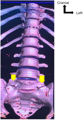
Figure 1 shows a CT scan of the lumbosacral spine in volumetric reconstruction, revealing type IIb sacralization according to the Castellvi classification.
Figure 1. CT scan of a type IIb sacralization according to Castellvi (arrow).
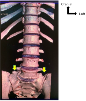
Figure 2 shows a CT scan of the lumbosacral spine in volumetric reconstruction, showing a type IIIb sacralization according to the Castellvi classification.
Figure 2. CT scan of a type IIIb sacralization according to Castellvi (Arrow).
Figure 3 shows a CT scan of the lumbosacral spine in volumetric reconstruction, showing type Ib lumbalization according to the Castellvi classification.
Figure 3. CT scan of Castellvi type Ib lumbalization (Arrow).
4. Discussion
4.1. Frequency of lumbosacral Transitional Anomalies
In this study, the overall prevalence of lumbosacral transitional anomalies was 3.99%. Verhaegen et al.
[10]
Verhaegen JCF, Alves Batista N, Horton I, Rakhra K, Beaulé PE, Michielsen J, et al. Prevalence of Lumbosacral Tran-sitional Vertebral Anomalies Among Healthy Volunteers and Patients with Hip Pathology. JBJS Open Access. 22 2023; 8(1): e22.00095.
[10]
noted a frequency of 8.5% of lumbosacral transitional anomalies in 2023. Similarly, in Saudi Arabia, Khashoggi et al.
[11]
Khashoggi KG, Hafiz RM, Bock YM, Kaki AM. Determination of lumbosacral transitional vertebrae in kidney urinary bladder x-ray films in the Saudi population. Saudi Med J. août 2017; 38(8): 794‑7.
[11]
found a prevalence of 7.6% of lumbosacral transitional anomalies in 2017. In Australia, French et al.
[3]
French HD, Somasundaram AJ, Schaefer NR, Laherty RW. Lumbosacral Transitional Vertebrae and Its Prevalence in the Australian Population. Glob Spine J. déc 2014; 4(4): 229‑32.
[3]
noted a prevalence of 9.9% in 2014.
This difference in proportions between studies could be explained by the sampling technique, the study period considered, and also the choice of medical imaging techniques.
4.2. Sociodemographic Data of Patients
In the present study, the age group of 36 years and older was the most common (65.3%). The mean age was 39±9 years, with extremes ranging from 15 to 48 years. Tangara
[9]
Tangara MS. Aspects scanographiques des anomalies transitionnelles du rachis lombo-sacré au service d‘imagerie médicale du CHU Pr Bocar Sidy Sall de Kati [Imaging Findings of Transitional Abnormalities of the Lumbosacral Spine at the Medical Imaging Department of the Pr. Bocar Sidy Sall University Hospital in Kati]. USTTB, Bamako, 2023: 85p.
[9]
found a mean age of 46.25 years. Byvaltsev et al.
[12]
Byvaltsev VA, Kalinin AA, Shepelev VV, Pestryakov YYa, Aliyev MA, Hozeev DV, et al. Prevalence of lumbosacral transitional vertebra among 4816 consecutive patients with low back pain: A computed tomography, magnetic reso-nance imaging, and plain radiographic study with novel classification schema. J Craniovertebral Junction Spine. 2023; 14(1): 35‑43.
[12]
found an average age of 42.3 years in 2023. This increase in the prevalence of lumbosacral abnormalities in this age group could be explained by the fact that they are more likely to have low back pain, prompting them to seek medical advice and undergo lumbosacral computed tomography.
Male patients accounted for 53.3% of the total, with a sex ratio of 1.1. This result is comparable to that of Byvaltsev et al.
[12]
Byvaltsev VA, Kalinin AA, Shepelev VV, Pestryakov YYa, Aliyev MA, Hozeev DV, et al. Prevalence of lumbosacral transitional vertebra among 4816 consecutive patients with low back pain: A computed tomography, magnetic reso-nance imaging, and plain radiographic study with novel classification schema. J Craniovertebral Junction Spine. 2023; 14(1): 35‑43.
[12]
, who reported 59.9% of men in 2023. Similarly, a previous study found 71.6% of males in 2017
[11]
Khashoggi KG, Hafiz RM, Bock YM, Kaki AM. Determination of lumbosacral transitional vertebrae in kidney urinary bladder x-ray films in the Saudi population. Saudi Med J. août 2017; 38(8): 794‑7.
[11]
. We found a higher frequency of lumbosacral transitional anomalies in men, with a predominance of sacralization. This is consistent with data in the literature, which has shown that sacralization is more prevalent in men than in women, while lumbalization is more prevalent in women than in men
[4]
Nardo L, Alizai H, Virayavanich W, Liu F, Hernandez A, Lynch JA, et al. Lumbosacral Transitional Vertebrae: Associ-ation with Low Back Pain. Radiology. nov 2012; 265(2): 497‑503.
[8]
Sekharappa V, Amritanand R, Krishnan V, David KS. Lumbosacral Transition Vertebra: Prevalence and Its Signifi-cance. Asian Spine J. 2014; 8(1): 51‑8.
[13]
Mahato NK. Relationship of sacral articular surfaces and gender with occurrence of lumbosacral transitional vertebrae. Spine J Off J North Am Spine Soc. oct 2011; 11(10): 961‑5.
[4, 8, 13]
.
4.3. Clinical Information
The clinical information mainly consisted of lumbosciatica in 52.0% of cases, followed by low back pain in 37.3% and post-traumatic assessment in 13.3% of cases. This result is comparable to that of Tangara.
[9]
Tangara MS. Aspects scanographiques des anomalies transitionnelles du rachis lombo-sacré au service d‘imagerie médicale du CHU Pr Bocar Sidy Sall de Kati [Imaging Findings of Transitional Abnormalities of the Lumbosacral Spine at the Medical Imaging Department of the Pr. Bocar Sidy Sall University Hospital in Kati]. USTTB, Bamako, 2023: 85p.
[9]
, who noted lumbosciatica (L5 and S1) in 50% of cases, followed by low back pain in 26.25%. It has been reported that people with lumbosacral transitional anomalies are more prone to developing low back pain
[14]
Dai L. Lumbosacral transitional vertebrae and low back pain. Bull Hosp Jt Dis N Y N. 1999; 58(4): 191‑3.
[15]
Quinlan JF, Duke D, Eustace S. Bertolotti’s syndrome. A cause of back pain in young people. J Bone Joint Surg Br. sept 2006; 88(9): 1183‑6.
[14, 15]
. This could be explained by the fact that there are several etiologies of low back pain related to lumbosacral transitional anomalies, such as disc pathology, extraforaminal stenosis secondary to the presence of an enlarged transverse process of the transitional vertebra, decreased paraspinal muscle volume, and increased lumbar lordosis
[16]
Bahadir Ulger FE, Illeez OG. The Effect of Lumbosacral Transitional Vertebrae (LSTV) on Paraspinal Muscle Volume in Patients with Low Back Pain. Acad Radiol. juill 2020; 27(7): 944‑50.
[16]
.
4.4. Computed Tomography Data
4.4.1. Sacralization
In this study, sacralization was found in 74.7% of cases, with Castellvi type IIIa being the most common in 25% of cases. This result is consistent with that of Tangara.
[9]
Tangara MS. Aspects scanographiques des anomalies transitionnelles du rachis lombo-sacré au service d‘imagerie médicale du CHU Pr Bocar Sidy Sall de Kati [Imaging Findings of Transitional Abnormalities of the Lumbosacral Spine at the Medical Imaging Department of the Pr. Bocar Sidy Sall University Hospital in Kati]. USTTB, Bamako, 2023: 85p.
[9]
who noted sacralization in 70% of cases, with Castellvi type IIb predominating in 32.14%. The same finding was made by Khashoggi et al.
[11]
Khashoggi KG, Hafiz RM, Bock YM, Kaki AM. Determination of lumbosacral transitional vertebrae in kidney urinary bladder x-ray films in the Saudi population. Saudi Med J. août 2017; 38(8): 794‑7.
[11]
, with sacralization present in 96.6% of patients, 28.5% of whom were classified as Castellvi type 2b. A proportion of 4.1% of sacralization, including 2.7% of Castellvi type II, was reported in the study by French et al.
[3]
French HD, Somasundaram AJ, Schaefer NR, Laherty RW. Lumbosacral Transitional Vertebrae and Its Prevalence in the Australian Population. Glob Spine J. déc 2014; 4(4): 229‑32.
[3]
in Australia in 2014. A sacralization rate of 5.1% was reported by Doo et al.
[17]
Doo AR, Lee J, Yeo GE, Lee KH, Kim YS, Mun JH, et al. The prevalence and clinical significance of transitional ver-tebrae: a radiologic investigation using whole spine spiral three-dimensional computed tomographic images. Anesth Pain Med. 31 janv 2020; 15(1): 103‑10.
[17]
in a radiological investigation study using three-dimensional spiral computed tomography images of the entire spine in 2020. Tokgoz et al.
[18]
Tokgoz N, Ucar M, Erdogan AB, Kilic K, Ozcan C. Are Spinal or Paraspinal Anatomic Markers Helpful for Vertebral Numbering and Diagnosing Lumbosacral Transitional Vertebrae? Korean J Radiol. 2014; 15(2): 258‑66.
[18]
reported a sacralization rate of 10.0% in their 2014 study.
4.4.2. Lumbalization
Lumbalization abnormalities were found in 25.3% of patients, with type IIa being the most common (n=7, or 36.8%). This result is comparable to that of Basel et al.
[19]
Basel SK, Barakoti RK, Chaudhary RK, Shrestha BK, Kaucha D, Rijal S. Lumbosacral Transitional Vertebra among Patients Visiting the Department of Orthopaedics in a Tertiary Care Centre: A Descriptive Cross-sectional Study. JNMA J Nepal Med Assoc. 2023; 61(258): 102‑5.
[19]
, who noted a l Lombalization rate of 29.47% in 2023. Similarly, Khashoggi et al.
[11]
Khashoggi KG, Hafiz RM, Bock YM, Kaki AM. Determination of lumbosacral transitional vertebrae in kidney urinary bladder x-ray films in the Saudi population. Saudi Med J. août 2017; 38(8): 794‑7.
[11]
found lombalization in 3.2% of patients, 60% of whom were type IIb according to Castellvi's classification. In Tangara's study
[9]
Tangara MS. Aspects scanographiques des anomalies transitionnelles du rachis lombo-sacré au service d‘imagerie médicale du CHU Pr Bocar Sidy Sall de Kati [Imaging Findings of Transitional Abnormalities of the Lumbosacral Spine at the Medical Imaging Department of the Pr. Bocar Sidy Sall University Hospital in Kati]. USTTB, Bamako, 2023: 85p.
[9]
, lombalization accounted for 30%, 50% of which were type IIb. French et al.
[3]
French HD, Somasundaram AJ, Schaefer NR, Laherty RW. Lumbosacral Transitional Vertebrae and Its Prevalence in the Australian Population. Glob Spine J. déc 2014; 4(4): 229‑32.
[3]
reported 5.8% lombalization, 50% of whom were type IIb.
[9]
Tangara MS. Aspects scanographiques des anomalies transitionnelles du rachis lombo-sacré au service d‘imagerie médicale du CHU Pr Bocar Sidy Sall de Kati [Imaging Findings of Transitional Abnormalities of the Lumbosacral Spine at the Medical Imaging Department of the Pr. Bocar Sidy Sall University Hospital in Kati]. USTTB, Bamako, 2023: 85p.
[9]
, lombalization accounted for 30%, of which 50% were type IIb. In contrast, French et al.
[3]
French HD, Somasundaram AJ, Schaefer NR, Laherty RW. Lumbosacral Transitional Vertebrae and Its Prevalence in the Australian Population. Glob Spine J. déc 2014; 4(4): 229‑32.
[3]
reported 5.8% lombalization, of which 3.4% were type III in their study. Jagannathan et al.
[20]
Jagannathan D, Indiran V, Hithaya F, Alamelu M, Padmanaban S. Role of Anatomical Landmarks in Identifying Nor-mal and Transitional Vertebra in Lumbar Spine Magnetic Resonance Imaging. Asian Spine J. 2017; 11(3): 365‑79.
[20]
noted 3.8% S1 Lombalization in their 2017 study. In the study by Karki et al.
[21]
Karki S, Paudel R, Phuyal A, Bhandari A. Lumbosacral Transitional Vertebrae amongst the Individuals Undergoing Magnetic Resonance Imaging of the Whole Spine in a Tertiary Care Hospital: A Descriptive Cross-sectional Study. JNMA J Nepal Med Assoc. 2021; 59(242): 975‑8.
[21]
, S1 Lombalization accounted for 33.78% of cases.
5. Conclusion
Lumbosacral transitional anomalies remain a common spinal anomaly identified incidentally. Three-dimensional computed tomography images are suitable for detecting transitional vertebrae, and our findings could help reduce the incidence of medical and surgical procedures performed on the wrong site.
Abbreviations
LSTV
Lumbosacral Transitional Vertebrae
SPSS
Statistical Package for the Social Sciences
CT
Computed Tomography
Author Contributions
Aldjouma Yanogue: Conceptualization, Resources, Writing – original draft
Abdoulaye Kante: Supervision, Validation
Ilias Guindo: Supervision, Validation
Tata Toure: Supervision, Validation
Mody Abdoulaye Camara: Supervision, Validation
Abou Sogodogo: Supervision, Methodology
Mamadou Simpara: Investigation
Mamadou H Konate: Investigation
Oumar Diallo: Supervision, Validation
Conflicts of Interest
The authors declare no conflicts of interest.
References
[1]
Bertolotti M. Contributo alla conoscenze dei vizi di differenzazione regionale del rachide con speciale riguardo alla assimilazione sacrale della V. lombare. Radiologique Medica [A Contribution to the Understanding of Regional Variations in Spinal Differentiation, with Special Reference to the Sacral Assimilation of the Lumbar Vertebrae. Radiologique Medica]. Turin, 1917; 4: 113-44.
[2]
Mahato NK. Redefining lumbosacral transitional vertebrae (LSTV) classification: integrating the full spectrum of morphological alterations in a biomechanical continuum. Medical hypotheses, Kanchipuram, 2013; 81: 76-81.
[3]
French HD, Somasundaram AJ, Schaefer NR, Laherty RW. Lumbosacral Transitional Vertebrae and Its Prevalence in the Australian Population. Glob Spine J. déc 2014; 4(4): 229‑32.
[4]
Nardo L, Alizai H, Virayavanich W, Liu F, Hernandez A, Lynch JA, et al. Lumbosacral Transitional Vertebrae: Associ-ation with Low Back Pain. Radiology. nov 2012; 265(2): 497‑503.
[5]
Uçar D, Uçar BY, Coşar Y, Emrem K, Gümüşsuyu G, Mutlu S, et al. Retrospective Cohort Study of the Prevalence of Lumbosacral Transitional Vertebra in a Wide and Well-Represented Population. Arthritis. 2013; 2013: 461425.
[6]
Hanhivaara J, Määttä JH, Niinimäki J, & Nevalainen MT. Lumbosacral transitional vertebrae are associated with lum-bar degeneration: retrospective evaluation of 3855 consecutive abdominal CT scans. European Radiology, Oulo, 2020 ; 30 : 3409-3416.
[7]
Gopalan B, Yerramshetty JS. Lumbosacral Transitional Vertebra-Related Low Back Pain: Resolving the Controversy. Asian Spine J. juin 2018; 12(3): 407‑15.
[8]
Sekharappa V, Amritanand R, Krishnan V, David KS. Lumbosacral Transition Vertebra: Prevalence and Its Signifi-cance. Asian Spine J. 2014; 8(1): 51‑8.
[9]
Tangara MS. Aspects scanographiques des anomalies transitionnelles du rachis lombo-sacré au service d‘imagerie médicale du CHU Pr Bocar Sidy Sall de Kati [Imaging Findings of Transitional Abnormalities of the Lumbosacral Spine at the Medical Imaging Department of the Pr. Bocar Sidy Sall University Hospital in Kati]. USTTB, Bamako, 2023: 85p.
[10]
Verhaegen JCF, Alves Batista N, Horton I, Rakhra K, Beaulé PE, Michielsen J, et al. Prevalence of Lumbosacral Tran-sitional Vertebral Anomalies Among Healthy Volunteers and Patients with Hip Pathology. JBJS Open Access. 22 2023; 8(1): e22.00095.
[11]
Khashoggi KG, Hafiz RM, Bock YM, Kaki AM. Determination of lumbosacral transitional vertebrae in kidney urinary bladder x-ray films in the Saudi population. Saudi Med J. août 2017; 38(8): 794‑7.
[12]
Byvaltsev VA, Kalinin AA, Shepelev VV, Pestryakov YYa, Aliyev MA, Hozeev DV, et al. Prevalence of lumbosacral transitional vertebra among 4816 consecutive patients with low back pain: A computed tomography, magnetic reso-nance imaging, and plain radiographic study with novel classification schema. J Craniovertebral Junction Spine. 2023; 14(1): 35‑43.
[13]
Mahato NK. Relationship of sacral articular surfaces and gender with occurrence of lumbosacral transitional vertebrae. Spine J Off J North Am Spine Soc. oct 2011; 11(10): 961‑5.
[14]
Dai L. Lumbosacral transitional vertebrae and low back pain. Bull Hosp Jt Dis N Y N. 1999; 58(4): 191‑3.
[15]
Quinlan JF, Duke D, Eustace S. Bertolotti’s syndrome. A cause of back pain in young people. J Bone Joint Surg Br. sept 2006; 88(9): 1183‑6.
[16]
Bahadir Ulger FE, Illeez OG. The Effect of Lumbosacral Transitional Vertebrae (LSTV) on Paraspinal Muscle Volume in Patients with Low Back Pain. Acad Radiol. juill 2020; 27(7): 944‑50.
[17]
Doo AR, Lee J, Yeo GE, Lee KH, Kim YS, Mun JH, et al. The prevalence and clinical significance of transitional ver-tebrae: a radiologic investigation using whole spine spiral three-dimensional computed tomographic images. Anesth Pain Med. 31 janv 2020; 15(1): 103‑10.
[18]
Tokgoz N, Ucar M, Erdogan AB, Kilic K, Ozcan C. Are Spinal or Paraspinal Anatomic Markers Helpful for Vertebral Numbering and Diagnosing Lumbosacral Transitional Vertebrae? Korean J Radiol. 2014; 15(2): 258‑66.
[19]
Basel SK, Barakoti RK, Chaudhary RK, Shrestha BK, Kaucha D, Rijal S. Lumbosacral Transitional Vertebra among Patients Visiting the Department of Orthopaedics in a Tertiary Care Centre: A Descriptive Cross-sectional Study. JNMA J Nepal Med Assoc. 2023; 61(258): 102‑5.
[20]
Jagannathan D, Indiran V, Hithaya F, Alamelu M, Padmanaban S. Role of Anatomical Landmarks in Identifying Nor-mal and Transitional Vertebra in Lumbar Spine Magnetic Resonance Imaging. Asian Spine J. 2017; 11(3): 365‑79.
[21]
Karki S, Paudel R, Phuyal A, Bhandari A. Lumbosacral Transitional Vertebrae amongst the Individuals Undergoing Magnetic Resonance Imaging of the Whole Spine in a Tertiary Care Hospital: A Descriptive Cross-sectional Study. JNMA J Nepal Med Assoc. 2021; 59(242): 975‑8.
Yanogue, A., Kante, A., Guindo, I., Toure, T., Camara, M. A., et al. (2026). Transitional Anomalies of the Lumbosacral Spine in Subjects Under 50 Years of Age: 75 Cases in Kati. International Journal of Medical Imaging, 14(1), 1-6. https://doi.org/10.11648/j.ijmi.20261401.11
Yanogue, A.; Kante, A.; Guindo, I.; Toure, T.; Camara, M. A., et al. Transitional Anomalies of the Lumbosacral Spine in Subjects Under 50 Years of Age: 75 Cases in Kati. Int. J. Med. Imaging2026, 14(1), 1-6. doi: 10.11648/j.ijmi.20261401.11
Yanogue A, Kante A, Guindo I, Toure T, Camara MA, et al. Transitional Anomalies of the Lumbosacral Spine in Subjects Under 50 Years of Age: 75 Cases in Kati. Int J Med Imaging. 2026;14(1):1-6. doi: 10.11648/j.ijmi.20261401.11
@article{10.11648/j.ijmi.20261401.11,
  author = {Aldjouma Yanogue and Abdoulaye Kante and Ilias Guindo and Tata Toure and Mody Abdoulaye Camara and Abou Sogodogo and Mamadou Simpara and Mamadou H Konate and Oumar Diallo},
  title = {Transitional Anomalies of the Lumbosacral Spine in Subjects Under 50 Years of Age: 75 Cases in Kati},
  journal = {International Journal of Medical Imaging},
  volume = {14},
  number = {1},
  pages = {1-6},
  doi = {10.11648/j.ijmi.20261401.11},
  url = {https://doi.org/10.11648/j.ijmi.20261401.11},
  eprint = {https://article.sciencepublishinggroup.com/pdf/10.11648.j.ijmi.20261401.11},
  abstract = {Introduction: Lumbosacral transitional anomaly is a very common congenital malformation that is diagnosed incidentally. Our objective was to study lumbosacral transitional anomalies using computed tomography. Method: Our study was conducted in the medical imaging department of the Pr Bocar Sidy SALL University Hospital in Kati and the Amitie Medical Clinic in Kati. It was a cross-sectional study with retrospective and prospective recruitment from January 2023 to December 2024. All patients who underwent lumbosacral computed tomography and in whom a lumbosacral transitional anomaly was ob-served were included. The sampling was consecutive. We used the Castellvi classification to identify the types of anomaly. Anonymity and confidentiality were respected. Results: The overall prevalence of lumbosacral transitional anomalies was 3.99%, including 3.6% in the imaging department of the Pr Bocar Sidy SALL University Hospital in Kati and 5.03% at the Amitie medical clinic in Kati. The average age was 39±9 years, with extremes ranging from 15 to 48 years. Males were predominant (53.3%). Lumbar sciatica was the main clinical finding in 52% of cases. Computed tomography revealed sacralization in 74.7% of cases, 25% of which were Castellvi type IIIa. Lumbalization was noted in 25.3%, of which 36.8% were Castellvi type IIa. Conclusion: Lumbosacral transition anomalies remain common, and knowledge of them could help reduce the incidence of surgical interventions at the wrong site.},
 year = {2026}
}
TY  - JOUR
T1  - Transitional Anomalies of the Lumbosacral Spine in Subjects Under 50 Years of Age: 75 Cases in Kati
AU  - Aldjouma Yanogue
AU  - Abdoulaye Kante
AU  - Ilias Guindo
AU  - Tata Toure
AU  - Mody Abdoulaye Camara
AU  - Abou Sogodogo
AU  - Mamadou Simpara
AU  - Mamadou H Konate
AU  - Oumar Diallo
Y1  - 2026/03/31
PY  - 2026
N1  - https://doi.org/10.11648/j.ijmi.20261401.11
DO  - 10.11648/j.ijmi.20261401.11
T2  - International Journal of Medical Imaging
JF  - International Journal of Medical Imaging
JO  - International Journal of Medical Imaging
SP  - 1
EP  - 6
PB  - Science Publishing Group
SN  - 2330-832X
UR  - https://doi.org/10.11648/j.ijmi.20261401.11
AB  - Introduction: Lumbosacral transitional anomaly is a very common congenital malformation that is diagnosed incidentally. Our objective was to study lumbosacral transitional anomalies using computed tomography. Method: Our study was conducted in the medical imaging department of the Pr Bocar Sidy SALL University Hospital in Kati and the Amitie Medical Clinic in Kati. It was a cross-sectional study with retrospective and prospective recruitment from January 2023 to December 2024. All patients who underwent lumbosacral computed tomography and in whom a lumbosacral transitional anomaly was ob-served were included. The sampling was consecutive. We used the Castellvi classification to identify the types of anomaly. Anonymity and confidentiality were respected. Results: The overall prevalence of lumbosacral transitional anomalies was 3.99%, including 3.6% in the imaging department of the Pr Bocar Sidy SALL University Hospital in Kati and 5.03% at the Amitie medical clinic in Kati. The average age was 39±9 years, with extremes ranging from 15 to 48 years. Males were predominant (53.3%). Lumbar sciatica was the main clinical finding in 52% of cases. Computed tomography revealed sacralization in 74.7% of cases, 25% of which were Castellvi type IIIa. Lumbalization was noted in 25.3%, of which 36.8% were Castellvi type IIa. Conclusion: Lumbosacral transition anomalies remain common, and knowledge of them could help reduce the incidence of surgical interventions at the wrong site.
VL  - 14
IS  - 1
ER  -