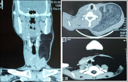
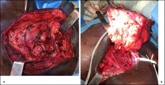
Background: Giant lipomas of the neck are rare, slow-growing benign tumours of adipose tissue that may pose significant diagnostic and therapeutic challenges. Their large size may result in cosmetic deformities or compression of adjacent vital cervical structures, making early diagnosis and appropriate surgical management essential. Objective: This study aims to describe the clinical, radiological, and surgical features of giant cervical lipomas based on a case series, and to provide a brief review of the relevant literature. Methods: We retrospectively analysed six cases of giant cervical lipomas managed in the ENT Department of the Regional University Hospital of Ouahigouya. Data collected included demographic characteristics, clinical presentation, tumour dimensions, radiological findings, surgical techniques, and outcomes. Results: The patients, aged between 11 and 50 years, presented with slow-growing, painless cervical masses, with the largest tumour measuring 194 mm in its greatest diameter. The lipomas were predominantly located in the posterior cervical triangle. CT suggested a benign appearance of the tumours and provided detailed information about their extent and relationship to surrounding structures. All patients underwent complete surgical excision under general anaesthesia. No perioperative complications or recurrences were observed during the follow-up period. Conclusion: Giant cervical lipomas, though rare, should be considered in the differential diagnosis of large cervical swellings. Detailed clinical and radiological evaluation followed by meticulous surgical excision can ensure favourable outcomes. This case series emphasises the importance of early intervention and the role of multidisciplinary collaboration in management.
| Published in | International Journal of Otorhinolaryngology (Volume 11, Issue 2) |
| DOI | 10.11648/j.ijo.20251102.14 |
| Page(s) | 24-27 |
| Creative Commons |
This is an Open Access article, distributed under the terms of the Creative Commons Attribution 4.0 International License (http://creativecommons.org/licenses/by/4.0/), which permits unrestricted use, distribution and reproduction in any medium or format, provided the original work is properly cited. |
| Copyright |
Copyright © The Author(s), 2025. Published by Science Publishing Group |
Lipoma, Giant, Neck, Cervical Tumour
Case | Sex | Age (years) | Disease duration (months) | Size (largest diameter) mm) |
|---|---|---|---|---|
1 | F | 32 | 52 | 121 |
2 | M | 40 | 108 | 125 |
3 | M | 36 | 54 | 114 |
4 | M | 50 | 180 | 194 |
5 | F | 42 | 62 | 142 |
6 | F | 11 | 84 | 185 |
CT | Computed Tomography |
| [1] | Bancroft LW, Kransdorf M, Peterson J, O’Connor M. Benign fatty tumors: classification, clinical course, imaging appearance, and treatment. Skeletal Radiol. 35: 719-33. |
| [2] | Cukic O. Giant Lipoma of the Anterior Neck Causing Dyspnea. The Journal of Craniofacial Surgery. 2020; 31(6): 553-5. |
| [3] | Derin A, Güney K, Turhan M, Erdoğan A, Ağırdır B. Giant cervical lipoma invading carotid artery: a case report. Kulak Burun Bogaz Ihtis Derg. 2009; 19(1): 28-39. |
| [4] | Eryılmaz M, Yücel A, Yücel H, Arıcıgil M. Cervico-Thoracic Giant Lipoma in a Child. Turk Arch Otorhinolaryngol. 2016; 54: 82-5. |
| [5] | Medina C, Schneider S, Mitra A, Spears J, Mitra A. Giant submental lipoma: Case report and review of the literature. Can J Plast Surg. 2007; 15(4): 219-22. |
| [6] | Li Y, Tsai W, Chen Y. Huge lipoma in the left submandibular region. J Dent Sci. 2019; 14(3): 330-1. |
| [7] | Petruzzi G, Trozzi L, Moretto S, De Virgilio A, Melis E, Gallina F, et al. An unusual approach for a cervical mass - sternotomy for the treatment of a giant cervico-thoracic lipoma. Acta Biomed. 2021; 92(6): 1-6. |
| [8] | Rana K, Meher R, Singh I, Narula V, Sura J. Lipoma in unusual head and neck region: Case series of 4 patients. International Journal of Pediatric Otorhinolaryngology Extra. 2013; 8(3): 75-8. |
| [9] | Najaf Y, Cartier C, Favier V, Garrel R. Symptomatic head and neck lipomas. Eur Ann Otosssrhinolaryngol Head Neck Dis. 2019; 136(2): 127-9. |
| [10] | Ndukwe K, Ugboko V, Somotun G, Adebiyi K. A clinicopathological study of lipomas of the head and neck. Nigerian Journal of Surgical Research. 2003; 5: 12-7. |
| [11] | Schranz A, Riordan F, Dolan R, Lawlor C, Morrison C, O’Toole G, et al. Retrospective analysis of radiological investigation of surgically excised head and neck lipomas. Eur Arch Otorhinolaryngol. 2024; 281(8): 4333-9. |
| [12] | Ralli M, De Vincentiis M, Greco A. First, Rule Out Cancer: Giant Lipoma. The American Journal of Medicine. 2018; 131(2): 146-7. |
| [13] | Woo S, Hong K, Chang H. Giant Ventral Lipoma of the Neck. J Craniofac Surg. 2020; 31(5): e501-3. |
| [14] | Du A, Wang H, Dai J, Dong Q, Yuan G, Pan Y. Case report: a case report of excision of giant lipoma in the posterior neck. Front Oncol. 2024; 14: 1395130. |
| [15] | Elakhiri M, Darouassi Y, Oukabli M, Jahidi A, Benariba F. Prise en charge d’un lipome cervical géant: à propos d’un cas et revue de la littérature. Pan Afr Med J. 2021; 39: 100. |
APA Style
Lengane, N. I., Savadogo, I., Somé, J. M. M., Coulibaly, A., Zaghre, N., et al. (2025). Giant Cervical Lipomas: Case Series and Literature Review. International Journal of Otorhinolaryngology, 11(2), 24-27. https://doi.org/10.11648/j.ijo.20251102.14
ACS Style
Lengane, N. I.; Savadogo, I.; Somé, J. M. M.; Coulibaly, A.; Zaghre, N., et al. Giant Cervical Lipomas: Case Series and Literature Review. Int. J. Otorhinolaryngol. 2025, 11(2), 24-27. doi: 10.11648/j.ijo.20251102.14
AMA Style
Lengane NI, Savadogo I, Somé JMM, Coulibaly A, Zaghre N, et al. Giant Cervical Lipomas: Case Series and Literature Review. Int J Otorhinolaryngol. 2025;11(2):24-27. doi: 10.11648/j.ijo.20251102.14
@article{10.11648/j.ijo.20251102.14,
  author = {Nogognan Ignace Lengane and Ibrahim Savadogo and Judicaël Marouruana Milkisedek Somé and Arsène Coulibaly and Noe Zaghre and Bertin Priva Ouedraogo},
  title = {Giant Cervical Lipomas: Case Series and Literature Review
},
  journal = {International Journal of Otorhinolaryngology},
  volume = {11},
  number = {2},
  pages = {24-27},
  doi = {10.11648/j.ijo.20251102.14},
  url = {https://doi.org/10.11648/j.ijo.20251102.14},
  eprint = {https://article.sciencepublishinggroup.com/pdf/10.11648.j.ijo.20251102.14},
  abstract = {Background: Giant lipomas of the neck are rare, slow-growing benign tumours of adipose tissue that may pose significant diagnostic and therapeutic challenges. Their large size may result in cosmetic deformities or compression of adjacent vital cervical structures, making early diagnosis and appropriate surgical management essential. Objective: This study aims to describe the clinical, radiological, and surgical features of giant cervical lipomas based on a case series, and to provide a brief review of the relevant literature. Methods: We retrospectively analysed six cases of giant cervical lipomas managed in the ENT Department of the Regional University Hospital of Ouahigouya. Data collected included demographic characteristics, clinical presentation, tumour dimensions, radiological findings, surgical techniques, and outcomes. Results: The patients, aged between 11 and 50 years, presented with slow-growing, painless cervical masses, with the largest tumour measuring 194 mm in its greatest diameter. The lipomas were predominantly located in the posterior cervical triangle. CT suggested a benign appearance of the tumours and provided detailed information about their extent and relationship to surrounding structures. All patients underwent complete surgical excision under general anaesthesia. No perioperative complications or recurrences were observed during the follow-up period. Conclusion: Giant cervical lipomas, though rare, should be considered in the differential diagnosis of large cervical swellings. Detailed clinical and radiological evaluation followed by meticulous surgical excision can ensure favourable outcomes. This case series emphasises the importance of early intervention and the role of multidisciplinary collaboration in management.},
 year = {2025}
}
TY  - JOUR
T1  - Giant Cervical Lipomas: Case Series and Literature Review

AU  - Nogognan Ignace Lengane
AU  - Ibrahim Savadogo
AU  - Judicaël Marouruana Milkisedek Somé
AU  - Arsène Coulibaly
AU  - Noe Zaghre
AU  - Bertin Priva Ouedraogo
Y1  - 2025/08/13
PY  - 2025
N1  - https://doi.org/10.11648/j.ijo.20251102.14
DO  - 10.11648/j.ijo.20251102.14
T2  - International Journal of Otorhinolaryngology
JF  - International Journal of Otorhinolaryngology
JO  - International Journal of Otorhinolaryngology
SP  - 24
EP  - 27
PB  - Science Publishing Group
SN  - 2472-2413
UR  - https://doi.org/10.11648/j.ijo.20251102.14
AB  - Background: Giant lipomas of the neck are rare, slow-growing benign tumours of adipose tissue that may pose significant diagnostic and therapeutic challenges. Their large size may result in cosmetic deformities or compression of adjacent vital cervical structures, making early diagnosis and appropriate surgical management essential. Objective: This study aims to describe the clinical, radiological, and surgical features of giant cervical lipomas based on a case series, and to provide a brief review of the relevant literature. Methods: We retrospectively analysed six cases of giant cervical lipomas managed in the ENT Department of the Regional University Hospital of Ouahigouya. Data collected included demographic characteristics, clinical presentation, tumour dimensions, radiological findings, surgical techniques, and outcomes. Results: The patients, aged between 11 and 50 years, presented with slow-growing, painless cervical masses, with the largest tumour measuring 194 mm in its greatest diameter. The lipomas were predominantly located in the posterior cervical triangle. CT suggested a benign appearance of the tumours and provided detailed information about their extent and relationship to surrounding structures. All patients underwent complete surgical excision under general anaesthesia. No perioperative complications or recurrences were observed during the follow-up period. Conclusion: Giant cervical lipomas, though rare, should be considered in the differential diagnosis of large cervical swellings. Detailed clinical and radiological evaluation followed by meticulous surgical excision can ensure favourable outcomes. This case series emphasises the importance of early intervention and the role of multidisciplinary collaboration in management.
VL  - 11
IS  - 2
ER  -
Department of Otorhinolaryngology, Ouahigouya University Hospital, Ouahigouya, Burkina Faso. School of Medecine, Lédea Bernard Ouédraogo University, Ouahigouya, Burkina Faso
Research Fields: rhinology, otology, head and neck surgery, laryngology, pediatric Otolaryngology
Department of Anatomic Pathology, Ouahigouya University Hospital, Ouahigouya, Burkina Faso. School of Medecine, Lédea Bernard Ouédraogo University, Ouahigouya, Burkina Faso
Research Fields: head and neck pathology, Breast Pathology, Liver Cancer, Prostate Cancer, Melanoma
Department of Radiology, Ouahigouya University Hospital, Ouahigouya, Burkina Faso. School of Medecine, Lédea Bernard Ouédraogo University, Ouahigouya, Burkina Faso
Research Fields: Oncologic Imaging, Interventional Radiology, Neuroimaging, Emergency Radiology, Tele-radiology
Department of Maxillofacial Surgery, Ouahigouya University Hospital, Ouahigouya, Burkina Faso. School of Medecine, Lédea Bernard Ouédraogo University, Ouahigouya, Burkina Faso
Research Fields: facial plastic surgery, salivary glands, Temporomandibular Joint Disorders, Facial Injuries and Deformities, Oral Head & Neck Cancer
Department of Otorhinolaryngology, Tingandogo University Hospital, Ouagadougou, Burkina Faso. School of Medecine, Joseph Ki-Zerbo University, Ouagdougou, Burkina Faso
Research Fields: rhinology, Reconstructive and Plastic Surgery, head and neck surgery, laryngology, pediatric Otolaryngology
Department of Otorhinolaryngology, Tingandogo University Hospital, Ouagadougou, Burkina Faso. School of Medecine, Joseph Ki-Zerbo University, Ouagdougou, Burkina Faso
Research Fields: rhinology, Reconstructive and Plastic Surgery, head and neck surgery, laryngology, pediatric Otolaryngology
Information