2. Material and Methods
Statement
The study is reported in accordance with ARRIVE guidelines. All methods are performed in accordance with the relevant guidelines and regulations.
2.1. Differential Analysis of SLC7A5 Gene Expression
Downloaded the standardized pan-cancer datasets from TCGA and GTEx databases (PANCAN, N=19131, G=60499). Selected samples from primary solid cancers, normal tissues and primary blood derived cancers. Extracted the expression data of ENSG0000103257 (SLC7A5) gene in various samples. Each data underwent a log2 (x+0.001) transformation. Then, cancer types with less than three samples are excluded. Finally the expression of SLC7A5 in different cancers are visualized and analyzed.
2.2. Prognostic Analysis of SLC7A5 Gene Expression
Downloaded the standardized pan-cancer datasets from TCGA and GTEx databases. Selected samples from primary solid cancers, normal tissues, metastatic cancers and primary blood derived cancers. Extracted the expression data of SLC7A5 gene in various samples. A log2 (x+0.001) transformation is performed on each expression data. Then, cancer types with less than ten samples are excluded. Finally, the prognosis of different cancer patients with abnormal SLC7A5 expression are visually analyzed.
2.3. Mutation and Expression Analysis of SLC7A5 Gene
Downloaded the standardized pan-cancer datasets from TCGA and GTEx databases (PANCAN, N=10535, G=60499). Selected samples from primary cancers and primary blood derived cancers. Extracted the expression data of SLC7A5 gene in various samples. Integrated the mutation data and gene expression data of the samples, and filtered out samples with synonymous mutations. A log2 (x+0.001) transformation is performed on each expression data. Then, cancer types with less than three samples are excluded. Finally, the mutation of different cancer patients with abnormal SLC7A5 expression are visually analyzed.
2.4. Immune Infiltration Analysis of SLC7A5 Gene
Downloaded the standardized pan-cancer datasets from TCGA and GTEx databases. Selected samples from primary solid cancers, normal tissues, metastatic cancers and primary blood derived cancers. Extracted the expression data of SLC7A5 gene in various samples and gene expression profiles for each cancer separately. A log2 (x+0.001) transformation is performed on each expression data. R software (version 3.6.4) is used to estimate the immune scores of each cancer. Finally, the immune infiltration of different cancer patients with abnormal SLC7A5 expression are visually analyzed.
2.5. Immune Cells Analysis of SLC7A5 Gene
Downloaded the standardized pan-cancer datasets from TCGA and GTEx databases. Selected samples from primary solid cancers, metastatic cancers and primary blood derived cancers. Extracted the expression data of SLC7A5 gene in various samples and gene expression profiles for each cancer separately. A log2 (x+0.001) transformation is performed on each expression data. Through R software the immune infiltration score of each patient is reevaluated based on cell expression by lots of methods. Including CIBERSOR method, EPIC method, IPS method, MCPCounter method, QUANTISEQ method, TIMER method and xCell method. Finally, the immune cells of different cancer patients with abnormal SLC7A5 expression are visually analyzed.
2.6. Genomic Heterogeneity and Clinical Staging Analysis of SLC7A5 Gene
Downloaded the standardized pan-cancer datasets from TCGA and GTEx databases. Selected samples from primary cancers and primary blood derived cancers. Extracted the expression data of SLC7A5 gene in various samples. R software is used to calculate the TMB (cancer mutation border) for each cancer. Integrated the TMB and gene expression data of the samples. A log2 (x+0.001) transformation is performed on each integrated data. Then, cancer types with less than three samples are excluded. Finally, the genomic heterogeneity and clinical staging of different cancer patients with abnormal SLC7A5 expression are visually analyzed.
2.7. Animal Experiment
2.7.1. Rats
Sixty Wistar rats (male, 180±5 g) are provided by the Animal Experimental Center of Southwest Medical University. Based on statistical power analysis (including significance level, statistical efficiency, effect size and attrition rate adjustment), they are randomly divided into three groups (treatment group, model group and control group, n=20 each).
2.7.2. Experimental Methods
The materials used include the following: Microsyringes (Tianjin Chenhang Keyuan Technology Development Co., Ltd), Pentobarbital sodium (Beijing Younikang Biotechnology Co., Ltd.), Diethylnitrosamine (DEN)(Sichuan Vicki Biotechnology Co., Ltd), JPH203 (Amgicam Biomedicine), anti-SLC7A5 antibody (WUHAN SANYING Biotechnology Co., Ltd.), Real-time PCR kits (SR1100) and ABI 7500 real-time PCR detection system (Singapore). Tissue sections are prepared using ethanol, xylene, paraffin and a sectioning mechanism. The sense and antisense primers used to detect SLC7A5 mRNA levels are as follows: 5'-GGAACATTG TGCTGGCATTATACAG-3' and 5'-CCAGGTGATAGTTCCCGA AGTCC-3'. The sense and antisense primers used to detect GAPDH mRNA levels are as follows: 5'-AGGCCGGCTTCGCGGGCGAC-3' and 5'-CTCGGGAGC CACACGCAGCTC-3'. The 2-∆∆Ct method is used to normalize the data.
Establishment of the animal model All Wistar rats are fed at 25°C and 60% relative humidity. Provide a 12 hour change period of light and shade. After a week of stable feeding, the rats in treatment group and model group are given 0.2% DEN by gavage with a dose of 10mg/kg, 3 times a week, and it stopped at 10 weeks. The control group rats are given physiological saline by gavage. At 10 weeks, one rat in the treatment group and one rat in the model group are euthanized by CO2 and the changes of livers are recorded. The procedure is repeated at 12 weeks. After 12 weeks, confirming the formation of the cancer through pathological examination, the rats in treatment group are intravenously injected with JPH203 at a concentration of 25mg/kg for 20 consecutive days. One week after injection, the rats in three groups are euthanized. Liver tissues and cancer tissues are taken from the three groups. HE staining showed the tissues of HCC in treatment group and model group, normal liver tissues in control group. RT-PCR is used to detect the expression of SLC7A5 mRNA (RNA is extracted from cancer tissues and normal liver tissues), with GAPDH as the internal control. Immunohistochemical SP method is used to detect the expression of SLC7A5 proteins. Under an optical microscope, SLC7A5 protein reacted with its antibody and exhibited granular expression. Every pathological section is equally divided into 6 regions and 150 cells are observed in each region. The dividing line between positive and negative is whether the number of positive cells exceeds 10%. If it is greater than this, it is positive, otherwise it is negative.
4. Results
Comparison chart of cancer abbreviations and full names (Abbreviations).
4.1. Differential Analysis Results of SLC7A5 Gene Expression
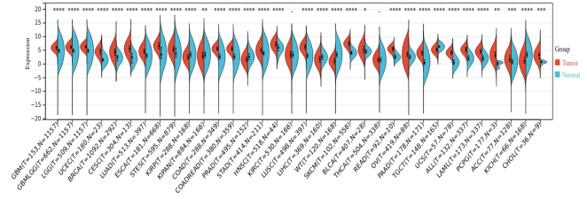
The expression differences of SLC7A5 are obtained between normal and cancer tissues in 34 cancer species. Significant upregulation is observed in 25 types of cancers. They are GBM (Tumor: 5.88±0.88, Normal: 4.73±1.36, p=8.8e-41), GBMLGG (Tumor: 6.19±0.74, Normal: 4.73±1.36, p=6.1e-184), LGG (Tumor: 6.28±0.66, Normal: 4.73±1.36, p=4.5e-172), UCEC (Tumor: 4.43±1.39, Normal: 1.73±1.52, p=2.4e-10), BRCA (Tumor: 4.37±1.60, Normal: 2.16±1.42, p=1.8e-75), CESC (Tumor: 5.65±1.66, Normal: 2.18±2.06, p=6.0e-7), LUAD (Tumor: 4.65±1.41, Normal: 2.99±1.20, p=2.0e-66), ESCA (Tumor: 6.68±1.48, Normal: 3.91±2.36, p=2.0e-41), STES (Tumor: 5.47±1.62, Normal: 3.93±2.24, p=1.8e-40), COAD (Tumor: 5.53±1.02, Normal: 2.64±1.81, p=6.4e-85), COADREAD (Tumor: 5.51±1.02, Normal: 2.64±1.80, p=6.4e-99), STAD (Tumor: 4.94±1.38, Normal: 3.99±1.82, p=1.9e-15), HNSC (Tumor: 7.15±1.28, Normal: 5.55±1.63, p=8.8e-11), LUSC (Tumor: 6.10±1.31, Normal: 2.99±1.20, p=2.7e-125), SKCM (Tumor: 7.41±1.15, Normal: 4.08±0.88, p=4.3e-56), BLCA (Tumor: 5.22±1.65, Normal: 4.59±1.13, p=0.03), READ (Tumor: 5.43±1.00, Normal: 2.64±1.14, p=1.6e-6), OV (Tumor: 3.42±1.44, Normal: 2.68±1.15, p=5.3e-8), PAAD (Tumor: 4.01±1.18, Normal: 0.75±1.90, p=1.9e-44), UCS (Tumor: 4.12±1.00, Normal: 0.72±1.41, p=6.0e-20), ALL (Tumor: 5.19±1.20, Normal: 2.16±1.16, p=6.1e-54), LAML (Tumor: 4.51±1.19, Normal: 2.16±1.16, p=1.4e-53), PCPG (Tumor: 3.23±1.88, Normal: -0.11±1.05, p=9.4e-3), ACC (Tumor: 1.71±2.08, Normal: 0.94±1.68, p=7.3e-4) and CHOL (Tumor: 3.08±1.76, Normal: 0.57±0.55, p=1.2e-4. Significant downregulation is observed in 7 types of cancers. They are KIRP (Tumor: 2.65±1.48, Normal: 3.27±1.64, p=1.3e-9), KIPAN (Tumor: 2.98±1.80, Normal: 3.27±1.64, p=1.5e-3), PRAD (Tumor: 1.64±1.27, Normal: 2.71±1.58, p=2.5e-13), LIHC (Tumor: 1.69±1.65, Normal: 2.52±1.27, p=3.9e-8), WT (Tumor: 0.92±1.51, Normal: 3.27±1.64, p=4.3e-29), TGCT (Tumor: 5.26±1.03, Normal: 6.33±0.63, p=4.7e-24) and KICH (Tumor: 1.25±2.22, Normal: 3.27±1.64, p=3.9e-14) (
Figure 1).
Figure 1. Differential expression of SLC7A5 gene in cancers.
Note: The expression differences of SLC7A5 gene are obvious in 32 types of cancers, significant upregulation is observed in 25 types of cancers and significant downregulation is observed in 7 types of cancers.
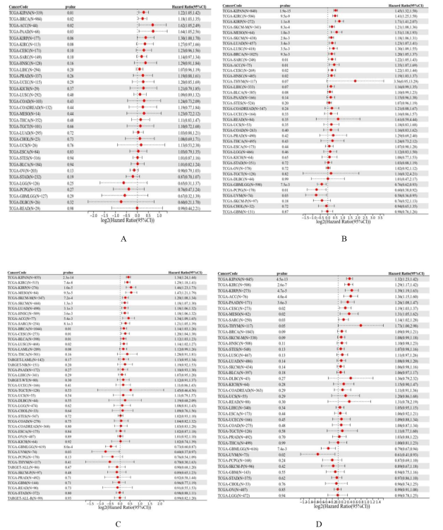
4.2. Prognostic Analysis Results of SLC7A5 Gene Expression
Figure 2. The prognosis of SLC7A5 expression in different cancers.
Note:
A. Prognostic analysis results of SLC7A5 gene (Disease -free interval)
B. Prognostic analysis results of SLC7A5 gene (Progression-free interval)
C. Prognostic analysis results of SLC7A5 gene (Overall survival)
D. Prognostic analysis results of SLC7A5 gene (Progression-free interval)
The expression data of 32 cancer species and their corresponding sample data are obtained (Disease-free interval), the expression data of 38 cancer species and their corresponding sample data are obtained (Disease-specific survival), the expression data of 44 cancer species and their corresponding sample data are obtained (Overall survival), the expression data of 38 cancer species and their corresponding sample data are obtained (Progression-free interval). Through R software the relationship between gene expression and prognosis in different cancer species are observed. High expression of SLC7A5 has poor prognosis in 4 cancer types (BRCA, KIPAN, PAAD, ACC, P<0.05)(Disease free interval), high expression in 13 cancer types (BRCA, CESC, LUAD, SARC, KIRP, KIPAN, HNSC, KIRC, LUSC, SKC-M, SKCM-M, MESO, ACC, P<0.05) and low expression in 3 cancer types (GBMLGG, UVM, PCPG, P<0.05) have poor disease specific survival (Disease- specific survival), high expression in 14 cancer types (BRCA, CESC, LUAD, SARC, KIRP, KIPAN, HNSC, KIRC, LUSC, SKCM, BLCA, SKCM-M, MESO, ACC, P<0.05) and low expression in 2 cancer types (GBMLGG, UVM, P<0.05) have poor overall survival (Overall survival), high expression in 8 cancer types (CESC, SARC, KIRP, KIPAN, KIRC, MESO, PAAD, ACC, P<0.05) and low expression in 2 cancer types (GBMLGG, UVM, P<0.05) have poor prognosis (Progression-free interval) (
Figure 2).
High expression of SLC7A5 has poor prognosis in 4 cancer types (Disease free interval), high expression in 13 cancer types and low expression in 3 cancer types have poor disease specific survival (Disease- specific survival), high expression in 14 cancer types and low expression in 2 cancer types have poor overall survival (Overall survival), high expression in 8 cancer types and low expression in 2 cancer types have poor prognosis (Progression-free interval).
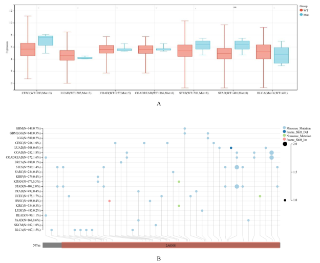
4.3. SLC7A5 Gene Mutation Analysis Results
Expression data of 7 cancer species are obtained. R software is used to calculate the expression differences of genes in different samples for each cancer. Significant difference in 1 type of cancer (STAD, P<0.05) and higher mutation rate in 4 types of cancers (STAD, STES, COAD, COADREAD, P<0.05) are observed (
Figure 3).
Figure 3. SLC7A5 gene mutation results.
Note: A. SLC7A5 gene mutation in 7 cancer species
B. SLC7A5 gene has higher mutation rate in 4 types of cancers
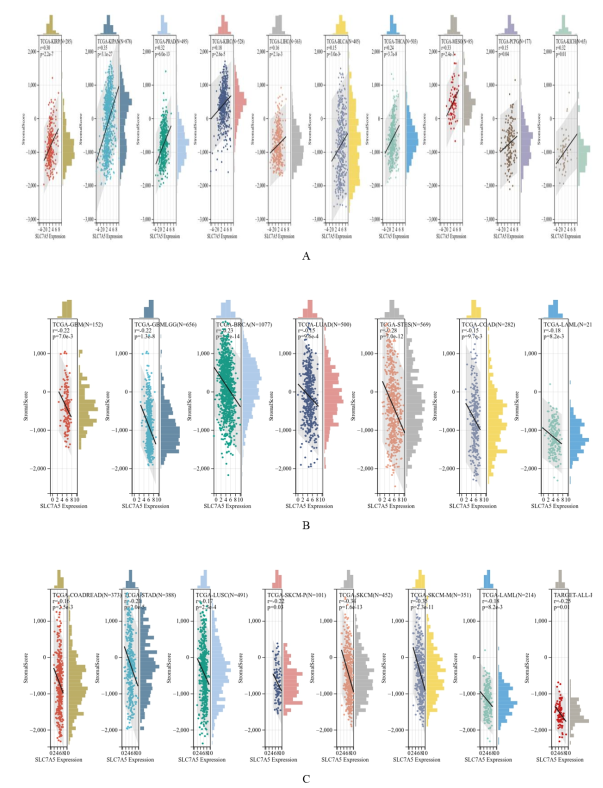
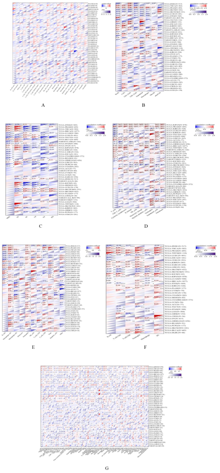
4.4. Immune Infiltration Analysis Results of SLC7A5 Gene
Figure 4. Immune infiltration results of SLC7A5.
Note: Significant correlation between gene expression and immune infiltration in 25 types of cancers
A. Ten cancer types showing significant positive correlation. As the expression of SLC7A5 gene increases, immune infiltration also increases simultaneously.
B and C. Fifteen cancer types showing a significant negative correlation. As the expression of SLC7A5 gene decreases, immune infiltration also decreases simultaneously.
Immune infiltration scores for 10180 cancer samples are obtained from a total of 44 cancer types. Through R software significant correlation between gene expression and immune infiltration are observed in 25 types of cancers, with 10 of them (KIRP, KIPAN, PRAD, KIRC, LIHC, BLCA, THCA, MESO, PCPG, KICH, P<0.05) showing significant positive correlations and 15 of them (GBM, GBMLGG, LAML, BRCA, LUAD, STES, COAD, COADREAD, STAD, LUSC, SKCM-P, SKCM, SKCM-M, LAML, ALL-R, P<0.05) showing negative correlations (
Figure 4).
4.5. Immune Cells Analysis Results of SLC7A5 Gene
Immune cell infiltration scores for 10180 cancer samples are obtained from a total of 44 cancer types. Through R software significant differences are observed in the correlation between gene expression and immune cell infiltration among 43 cancer types (CIBERSORT method: GBM, GBMLGG, LGG, UCEC, LAML, BRCA, CESC, LUAD, ESCA, STES, SARC, KIRP, KIPAN, COAD, COADREAD, PRAD, STAD, HNSC, KIRC, LUSC, THYM, LIHC, WT, SKCM-P, SKCM, BLCA, SKCM-M, THCA, NB, MESO, READ, OV, UVM, PAAD, TGCT, LAML, ALL, PCPG, ACC, ALL-R, DLBC, KICH, CHOL, 22 types of immune cells, P<0.05). The differences are observed in 40 cancer types (EPIC method: GBM, GBMLGG, LGG, UCEC, LAML, BRCA, CESC, LUAD, ESCA, STES, SARC, KIRP, KIPAN, COAD, COADREAD, PRAD, STAD, HNSC, KIRC, LUSC, THYM, LIHC, WT, SKCM-P, SKCM, BLCA, SKCM-M, THCA, NB, MESO, OV, PAAD, TGCT, LAML, ALL, PCPG, ACC, ALL-R, KICH, CHOL, 8 types of immune cells, P<0.05). The differences are observed in 38 cancer types (IPS method: GBMLGG, LGG, UCEC, LAML, BRCA, LUAD, ESCA, STES, SARC, KIRP, KIPAN, COAD, COADREAD, PRAD, STAD, HNSC, KIRC, LUSC, THYM, LIHC, WT, SKCM-P, SKCM, BLCA, SKCM-M, THCA, MESO, READ, OV, PAAD, TGCT, LAML, PCPG, ACC, ALL-R, DLBC, KICH, CHOL, 6 types of immune cells, P<0.05). The correlation are observed in 43 cancer types (MCP counter method, 10 types of immune cells, P<0.05), in 43 cancer species (QUANTISEQ method, 11 types of immune cells, P<0.05) and in 44 cancer species (xCELL method, 67 types of immune cells, P<0.05). In 9406 cancer samples of 38 cancer types, significant differences are observed in the correlation between gene expression and immune cell infiltration among 34 cancer types (Timer method: ACC, BLCA, BRCA, CESC, CHOL, DLBC, ESCA, GBM, GBMLGG, HNSC, KICH, KIPAN, KIRC, KIRP, LGG, LIHC, LUAD, LUSC, MESO, OV, PAAD, PCPG, PRAD, SARC, SKCM-M, SKCM-P, SKCM, STAD, STES, TGCT, THCA, THYM, UCEC, UVM, 6 types of immune cells, P<0.05) (
Figure 5).
Figure 5. Immune cells results of SLC7A5.
Note: A. immune cells analysis (CIBERSORT), B. immune cells analysis (EPIC)
C. immune cells analysis (IPS), D. immune cells analysis (MCP counter)
E. immune cells analysis (QUANTISEQ)
F. immune cells analysis (Timer), G. immune cells analysis (xCELL)
SLC7A5 gene expression is significantly correlated with immune cell infiltration in 43 cancer types (CIBERSORT method), in 40 cancer species (EPIC method), in 38 cancer types (IPS method), in 43 cancer types (MCP counter method), in 43 cancer types (QUANTISEQ method) and in 44 cancer types (xCELL method). In 9406 cancer samples of 38 cancer types, this gene expression is significantly correlated with immune cell infiltration in 34 cancer types (Timer method).
4.6. Genomic Heterogeneity and Clinical Staging Analysis Results of SLC7A5 Gene
Significant correlation are observed between SLC7A5 and 6 types of cancers, with a significant positive correlation in 5 types of cancers (LUAD, KIPAN, STAD, THYM, ACC, P<0.05) and a significant negative correlation in 1 type of cancer (KIRP, P<0.05) (genetic heterogeneity). Through R software the expression differences of gene in different clinical stage are observed in six types of cancers (BRCA, KIRP, KIPAN, PRAD, KIRC, BLCA, P<0.05) (clinical stage) (
Figure 6).
Figure 6. Genomic heterogeneity and clinical staging analysis Results.
Significant positive correlation in 5 types of cancers and significant negative correlation in 1 type of cancer (genetic heterogeneity) are observed. Significant differences are observed in 6 types of cancers (clinical stage).
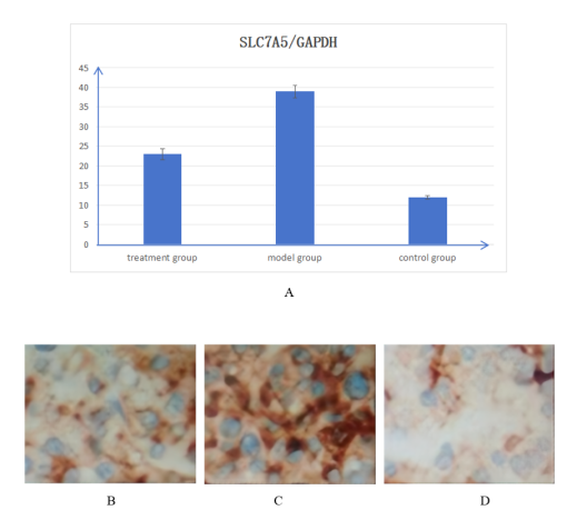
4.7. Animal Experiment Results
Through pathologic examination, we detected HCC in 16 rats (80%) in the treatment group and 17 rats (85%) in the model group after 12 weeks. In treatment group the diameters of the tumors are 1.60±0.09cm, in control group the diameters of the tumors are 1.83±0.11cm, there is significant difference between the two groups (t=2.917, P<0.05). Through RT–PCR, we found that in treatment group, model group and control group, the SLC7A5/GAPDH ratios are 23±1.4, 39±1.6 and 12±0.5, respectively. There are significant differences among the three groups (between the model group and treatment group, t=2.857, P<0.05; between the model group and control group, t=3.329, P<0.05). SLC7A5 protein reacted with anti-SLC7A5 antibody, every pathological section included 900 cells. SLC7A5 protein is expressed as follows: A total of 452 positive cells (50.2%) are in the treatment group, 791 positive cells (87.9%) are in the model group, 95 positive cells (10.6%) are in the control group, and there are significant differences among the three groups (χ
2=31.25, P<0.05, between the model group and treatment group, χ
2=38.97, P<0.05, between model group and control group) (
Figure 7).
Figure 7. RT–PCR analysis results and immunohistochemistry results of SLC7A5.
Note: A. RT–PCR analysis results of SLC7A5 in three groups
Immunohistochemistry results of SLC7A5 in treatment group
Immunohistochemistry results of SLC7A5 in model group
Immunohistochemistry results of SLC7A5 in control group
SLC7A5 is significantly elevated in HCC, but after treatment with JPH203, the expression of SLC7A5 showed a significant decrease.
5. Discussion
Amino acids are the fundamental components that make up proteins and participate in many important metabolic pathways within cells. Abnormal transport of amino acids can lead to severe amino acid absorption and metabolism disorders. Therefore, the transport of amino acids has important pathological significance. In mammals, the transmembrane transport of amino acids are mediated by various amino acid transporter proteins. As amino acids belong to small polar substances and cannot freely pass through the cell membrane
| [7] | Chen J, Wang W, Zhu L. Amino Acid Transporter as a Potential Carrier Protein for the Root-to-Shoot Translocation of Polybrominated Diphenyl Ethers in Rice. Environ Sci Technol. 2023, 57(26), 9722-9731. https://doi.org/10.1021/acs.est.3c00581 |
| [8] | Taurino G, Chiu M, Bianchi MG, et al. The SLC38A5/SNAT5 amino acid transporter: from pathophysiology to pro-cancer roles in the cancer microenvironment. Am J Physiol Cell Physiol. 2023, 325(2), C550-C562.
https://doi.org/10.1152/ajpcell.00169.2023 |
| [9] | Yahyaoui R, Pérez-Frías J. Amino Acid Transport Defects in Human Inherited Metabolic Disorders. Int J Mol Sci. 2019, 21(1), 119. https://doi.org/10.3390/ijms21010119 |
[7-9]
, they must be assisted by the corresponding amino acid transporter proteins on the cell membrane. During the proliferation, division, migration and invasion of cancer cells, a large amount of amino acids are needed to provide nutrients. Due to the faster growth of cancer cells and their lack of control by the body, the need for amino acids is more urgent. Amino acid transporters provide a carrier channel for the entry and exit of amino acids in cancer cells, so they play a crucial role in cancer occurrence and progression
| [10] | Xiao Y, Zhang H, Li Z, et al. An amino acid transporter- like protein (OsATL15) facilitates the systematic distribution of thiamethoxam in rice for controlling the brown planthopper. Plant Biotechnol J. 2022, 20(10), 1888-1901. https://doi.org/10.1111/pbi.13869 |
| [11] | Yao X, Li H, Nie J, et al. Disruption of the amino acid transporter CsAAP2 inhibits auxin-mediated root development in cucumber. New Phytol. 2023, 239(2), 639-659.
https://doi.org/10.1111/nph.18947 |
| [12] | Kukułowicz J, Pietrzak-Lichwa K, Klimończyk K, et al. The SLC6A15-SLC6A20 Neutral Amino Acid Transporter Subfamily: Functions, Diseases, and Their Therapeutic Relevance. Pharmacol Rev. 2023, 76(1), 142-193.
https://doi.org/10.1124/pharmrev.123.000886 |
[10-12]
.
Immunotherapy refers to mobilizing the body's own immune system, activating T cells and killing cancer cells. Cancer immunotherapy mainly includes four categories: cancer vaccines, immune modulators, adoptive cell immunotherapy and immune binding point blockade therapy. The basic principle of cancer vaccines is to induce a specific anti-cancer immune response in the body through cancer antigens. It can achieve the effect of treating cancers or preventing recurrence. Immunomodulators refer to non-specific biological products that enhance and regulate immune function. Such as interleukin and interferon. Adoptive cell immunotherapy refers to the induction, activation and expansion of mononuclear cells isolated from the patient's peripheral blood. Then the mononuclear cells were injected into the patient's body, inducing cancer cells death or directly killing cancer cells. Immune binding point immunotherapy is that a drug blocks immune checkpoints. Therefore, the immune system can recognize and attack cancer cells. Such as nivolumab, pembrolizumab and atezolizumab. Existing evidence suggests that amino acid transporters are closely related to immune binding point immunotherapy
| [13] | Yang L, Wang Q, He L, et al. The critical role of cancer microbiome in cancer immunotherapy. Cancer Biol The. 2024, 25(1), 2301801. https://doi.org/10.1080/15384047.2024.2301801 |
| [14] | Lucas AP, Lewis AR, Kasi PM, et al. Abscopal downstaging of intermediate stage hepatocellular via combination cryoablation and immunotherapy with complete pathologic response. Radiol Case Rep. 2023, 19(3), 910-914.
https://doi.org/10.1016/j.radcr.2023.11.062 |
| [15] | Wang J, He T, Gao Q, Chang H, et al. The dysfunctional Wnt pathway down-regulates MLH1/SET expression and promotes microsatellite instability and immunotherapy response in colorectal cancer. Genes Dis. 2023, 11(2), 542-545.
https://doi.org/10.1016/j.gendis.2023.03.026 |
[13-15]
.
SLC7A5 is an important member of the L-type amino acid transporter. It is mainly responsible for transporting large, branched, aromatic neutral amino acids, including some essential amino acids. The amino acid transporter SLC7A5 and its helper subunit SLC3A2 form heterodimers, which are necessary for effective uptake of glutamine and leucine
| [16] | Liu D, Ren H, Wen G, et al. Nicotine up-regulates SLC7A5 expression depending on TRIM29 in non-small cell lung cancer. Genes Di. 2023, 11(2), 582- 584.
https://doi.org/10.1016/j.gendis.2023.04.016 |
| [17] | Canhasi L, Tina E, Eremo AG. Hypoxia-mimetic by CoCl2 increases SLC7A5 expression in breast cancer cells in vitro. BMC Res Notes. 2023, 16(1), 366.
https://doi.org/10.1186/s13104-023-06650-2 |
| [18] | Song M, Liu J. Circ_0067717 promotes colorectal cancer cell growth, invasion and glutamine metabolism by serving as a miR-497-5p sponge to upregulate SLC7A5. Histol Histopathol. 2023, 38(1), 53-64. https://doi.org/10.14670/HH-18-494 |
[16-18]
. In addition to allowing essential amino acids to flow in and out through the plasma membrane and subcellular compartments (such as lysosomes and mitochondria), increasing evidence suggests that amino acid transporters are involved in sensing amino acid levels. So they can activate mammalian rapamycin complex 1 (mTORC1), induce the expression of the c-Myc gene and lead to cancer formation. Existing studies have shown that SLC7A5 is abnormally overexpressed in some cancer tissues (such as HCC), but there is only limited statistics and summary of its specific biological significance. Given that SLC7A5 research will have important implications in cancer treatment, nutrition and other life sciences research, we consulted the TCGA and GTE
X databases
| [19] | Hu Y, Liu F, Mi R. Glioma cells achieve malignant progression by fusion with macrophages to gain high SLC7A5 expression. J Neurooncol. 2025, 176(2), 126.
https://doi.org/10.1007/s11060-025-05382-6 |
| [20] | Onishi Y, Hiraiwa M, Kamada H, et al. Hypoxia affects Slc7a5 expression through HIF-2α in differentiated neuronal cells. FEBS Open Bio. 2019, 9(2), 241-247.
https://doi.org/10.1002/2211-5463.12559 |
| [21] | Hisada T, Kondo N, Wanifuchi-Endo Y, et al. Co-expression effect of LLGL2 and SLC7A5 to predict prognosis in ERα-positive breast cancer. Sci Rep. 2022, 12(1), 16515.
https://doi.org/10.1038/s41598-022-20225-4 |
[19-21]
. Statistical analysis of the differential expression and prognosis of SLC7A5 in different cancers, especially identifying its relationship with immune infiltration and immune cells, lay a solid foundation for its role in cancer treatment.
In statistics, by retrieving and analyzing data from the TCGA and GTEx databases, we observed many differences of SLC7A5 in different cancer tissues, including the expression, prognosis, gene mutations, relationship with immune infiltration and relationship with immune cells. Several major changes in SLC7A5 are discovered from it. Firstly, SLC7A5 shows an elevated state in most cancers and a decrease in a few cancers. It indicates that most cancers show active proliferation and high demand for amino acids, leading to the increase in SLC7A5. Due to SLC7A5 being a light chain subunit of multiple transmembrane amino acid transporters, it is important structure for exerting the function of transporters. So the phenomenon of its high expression in various cancer tissues suggests that once SLC7A5 is inhibited or disrupted, the transmembrane transporter structure collapses, the entry and exit of amino acids in the cells are immediately blocked, the growth of cancer cells will be restricted, even the possibility of cancer cell death may occur. This is a clear target that can be utilized. Secondly, cancers with different levels of SLC7A5 expression show significant differences in patient prognosis. Among cancers with high expression of SLC7A5, the vast majority have poor prognosis. Active cancers can cause significant damage to the body. That is to say, SLC7A5 is closely related to the prognosis of patients, which also prompts our thinking. The existing patient prognosis evaluation system is mainly based on TNM staging, and patients with cancer staging in stages III and IV usually have lost the opportunity for surgery. After the era of targeted therapy and immunotherapy began, some patients who are previously defined as in the middle and late stages still received treatment opportunities. So, since SLC7A5 is highly expressed in most cancer tissues and closely related to patient prognosis, can it be used as a supplement to TNM staging, making prognosis analysis more detailed and thus seeking treatment opportunities for more patients? Even for patients in the middle and late stages, if there is a phenomenon of high expression of SLC7A5, there is prospect and hope for treatment. However, it should be noted that factors such as age, stage and choice of treatment method are closely related to the patient's prognosis. Therefore, in addition to SLC7A5, a comprehensive evaluation of multiple factors is also necessary. Thirdly, the low mutation rate of SLC7A5 provides convenience for drug development and continuous treatment. Fourthly, it is particularly important that SLC7A5 is closely related to immune infiltration and immune cells. Due to the need for systematic exploration of the expression patterns and prognostic value of immune-related genes and molecules in various cancers, we adopted immune infiltration analysis and immune cells analysis. In different immune cell detection methods, it has been found that SLC7A5 is highly correlated with immune cells in various cancers, whether positive or negative, which may lead to changes in immune status. In cancers with changes in SLC7A5 expression, it can be observed that the number of immune cells, including B cells, T cells, NK cells, macrophages, etc., has increased to varying degrees, indicating an increasing trend in the body's defense ability. Especially in cancer tissues with increased expression of SLC7A5, this change is particularly evident, which provides us with great inspiration in immunotherapy. Existing immunotherapy drugs mainly achieve the effect of inhibiting or killing cancer cells by changing the recognition between T cells and cancer cells. So, can we mobilize more immune cells and enhance the effectiveness of immunotherapy by changing the expression level of SLC7A5? That is to say, SLC7A5 may become a new option for immunotherapy
| [22] | Huang X, Sun T, Wang J, et al. Metformin Reprograms Tryptophan Metabolism to Stimulate CD8+ T-cell Function in Colorectal Cancer. Cancer Res. 2023, 83(14), 2358-2371.
https://doi.org/10.1158/0008-5472.CAN-22-3042 |
| [23] | Ping S, Wang S, Zhao Y, et al. Identification and validation of a ferroptosis-related gene signature for predicting survival in skin cutaneous melanoma. Cancer Med. 2022, 11(18), 3529-3541. https://doi.org/10.1002/cam4.4706 |
| [24] | Jin C, Zhou X, Xu M, et al. Pharmacological and structural insights into nanvuranlat, a selective LAT1 (SLC7A5) inhibitor, and its N-acetyl metabolite with implications for cancer therapy. Sci Rep. 2025, 15(1), 2903.
https://doi.org/10.1038/s41598-025-87522-6 |
[22-24]
. At present, data from basic experiments and clinical treatments conducted in over 3000 laboratories and clinical centers around the world shows that the use of immunotherapy drugs alone is not very effective in the treatment of cancers. So, can SLC7A5 improve this phenomenon? Further experiments are needed for observation and determination. Fifthly, in the detection of gene heterogeneity and clinical staging, it can be seen that they have little effect on the expression of SLC7A5. This indicates that in the clinical staging of most cancers, SLC7A5 exhibits a sustained high expression phenomenon, providing a relatively stable foundation for subsequent corresponding treatments
| [25] | Zhao X, Jin L, Liu Y, et al. Bioinformatic analysis of the role of solute carrier-glutamine transporters in breast cancer. Ann Transl Med. 2022, 10(14), 777.
https://doi.org/10.21037/atm-22-2620 |
| [26] | Solvay M, Holfelder P, Klaessens S, et al. Tryptophan depletion sensitizes the AHR pathway by increasing AHR expression and GCN2/LAT1-mediated kynurenine uptake, and potentiates induction of regulatory T lymphocytes. J Immunother Cancer. 2023, 11(6), e006728.
https://doi.org/10.1136/jitc-2023-006728 |
| [27] | Sato K, Miyamoto M, Takano M, et al. Significant relationship between the LAT1 expression pattern and chemoresistance in ovarian clear cell carcinoma. Virchows Arch. 2019, 474(6), 701-710. https://doi.org/10.1007/s00428-019-02520-0 |
[25-27]
. In animal experiments, we established a rat model of HCC and treated it with JPH203 (a inhibitor of SLC7A5). In the treatment group, significant effects are observed, with a significant decrease in the expression level of SLC7A5, indicating that cancer growth is inhibited. This provides us with exciting insights. In clinical practice, if inhibitors of SLC7A5 can be used, can the immune microenvironment be improved? Can the effect of immunotherapy be improved? Can patients survive with cancers for a longer period of time, even if the cancer persists? Will the quality of life of patients be improved? These require more observation and experimentation.
Meanwhile, our research also has limitations. Firstly, in our study, various aspects of SLC7A5 are evaluated, but its expression and role at the protein level require further exploration. Secondly, we retrieved information from different databases and further observation is needed to determine if there are any biases or deviations. Thirdly, we have analyzed the above situation from the database, the function of SLC7A5 inhibitor is validated in an animal model of cancer and good results are observed, but whether it can be helpful for cancer treatment still needs to be verified through more basic researches and clinical trials. Overall, our pan-cancer analysis and animal experiments of SLC7A5 indicates that its expression is closely related to cancer prognosis, immune infiltration, immune cells, cancer heterogeneity, and clinical staging
| [28] | Menyhárt O, Fekete JT, Győrffy B. Resistance to Combined Anthracycline-Taxane Chemotherapy Is Associated with Altered Metabolism and Inflammation in Breast Carcinomas. Int J Mol Sci. 2024, 25(2), 1063.
https://doi.org/10.3390/ijms25021063 |
| [29] | Gautam S, Latif S, Kang YS. Effect of Various Pathological Conditions on Nitric Oxide Level and L-Citrulline Uptake in Motor Neuron-Like (NSC-34) Cell Lines. Biomol Ther (Seoul). 2024, 32(1), 154-161.
https://doi.org/10.4062/biomolther.2023.110 |
[28, 29]
. SLC7A5 inhibitors may inhibit cancer growth. This may provide new directions for cancer immunotherapy. At the same time, the influence of multiple factors needs to be considered, such as tumor purity, metabolic demand and stromal composition. In summary, SLC7A5 and JPH203 may be applied in practical research work.