Review Article | | Peer-Reviewed

Gastrointestinal Stromal Tumour Management at the Bouake University Hospital

Received: 20 August 2025     Accepted: 3 September 2025     Published: 25 September 2025
Views:       Downloads:
Abstract

Gastric GISTs are rare tumours that are often detected late. The aim of this study was to describe the different treatment modalities for gastric GISTs at Bouake University Hospital. It was retrospective study of 17 patients with gastric GIST between January 2013 and December 2023. GISTs accounted for 4.2% of all gastric tumours. The main reason for consultation was an abdominal mass. The average duration of progression was 10.9 months ± 2.1 months. FOGD, performed in 7 patients, suspected GIST in only one case. Abdominal CT scan, performed in 15 patients, was suggestive of GIST in 8 cases. The tumour was located in the antrum in 11 cases. There was extension to neighbouring organs in 5 cases. The tumours were classified as stage III in 9 cases. Immunohistochemistry confirmed the diagnosis of GIST with expression of C-KIT (CD117) antibodies in all cases. GIST is most often diagnosed at the stage of a large abdominal mass. Immunohistochemistry, which allows for diagnostic confirmation, is not accessible to most patients.

Published in Journal of Surgery (Volume 13, Issue 5)
DOI 10.11648/j.js.20251305.13
Page(s) 134-140
Creative Commons

This is an Open Access article, distributed under the terms of the Creative Commons Attribution 4.0 International License (http://creativecommons.org/licenses/by/4.0/), which permits unrestricted use, distribution and reproduction in any medium or format, provided the original work is properly cited.

Copyright

Copyright © The Author(s), 2025. Published by Science Publishing Group

Keywords

Stomach, GIST, Immunohistochemistry, Stromal Tumours

1. Introduction
Gastrointestinal stromal tumours (GISTs) are mesenchymal tumours that develop at the expense of Cajal cells or pacemaker cells. Gastric localisation is the most common in 60 to 70% of cases . These tumours are rare. They remain asymptomatic for a long time, so they are often discovered at a stage when they have grown into large masses . The diagnosis of GIST is suggested by clinical and radiological signs, but confirmation is provided by immunohistochemistry . The development of targeted therapies has completely revolutionised the management of aggressive recurrent gastric stromal tumours with metastases. However, surgery remains the treatment of choice for localised stromal tumours . The five-year survival rate after surgical resection varies between 20% and 95% depending on the degree of malignancy .
The aim of this study was to describe the methods used to manage gastric GISTs at Bouaké University Hospital.
2. Methods
The study involved 17 patients treated for a stromal tumour of the stomach confirmed by immunohistochemistry. There were 11 men and 6 women. The average age was 59.2 years, ranging from 47 to 76 years.
This was a descriptive cross-sectional study covering an 11-year period from January 2013 to December 2023, conducted in the digestive surgery department of Bouaké University Hospital. The diagnosis of GITS was confirmed by immunohistochemistry.
The parameters studied were diagnostic data (clinical and paraclinical signs), therapeutic data (surgery, chemotherapy) and outcome data (morbidity, mortality, survival).
3. Results
3.1. Diagnostic Characteristics
The average time to consultation was 10.9 months ± 2.1 months, with extremes ranging from 3 weeks to 24 months. Eight patients (47.1%) had consulted more than 12 months after diagnosis. Five patients (29.4%) consulted within the first six months.
In 16 patients, this was the first appearance of the tumour. In one patient, it was a recurrence of a tumour that had been operated on four years earlier.
Patients consulted for an abdominal mass in 76.5% of cases (n=13), abdominal pain in 17.6% (n=3) and for an obstruction in 5.6% (n=1).
The general condition was altered in 10 patients and unchanged in 7 patients. An abdominal mass was noted in all patients. The various clinical signs are listed in Table 1.
Table 1. Clinical signs presented by patients.

Clinical signs

Numbers

Percentages

Abdominal mass

17

100

Abdominal pain

12

70,6

Constipation

10

58,8

Weight loss

9

52,9

Clinical anaemia

7

41,2

Anorexia, asthenia

5

29,4

Vomiting

4

23,5

Oedema of the lower limbs

2

11,8

Abdominal CT scans performed in 15 patients (88.2%) led to a suspected diagnosis of GIST of the stomach in 8 cases (Figure 1). It pointed to a tumour of the mesentery in 6 cases. In one case, a diagnosis of false pancreatic cyst was suggested.
Ultrasound performed in 7 patients (41.2%) revealed a heterogeneous tissue mass (n=6) or a hypoechoic mass (n=1). It suggested a tumour of the duodenum in two cases and a tumour of the mesentery in one case. In four cases, the origin was not specified.
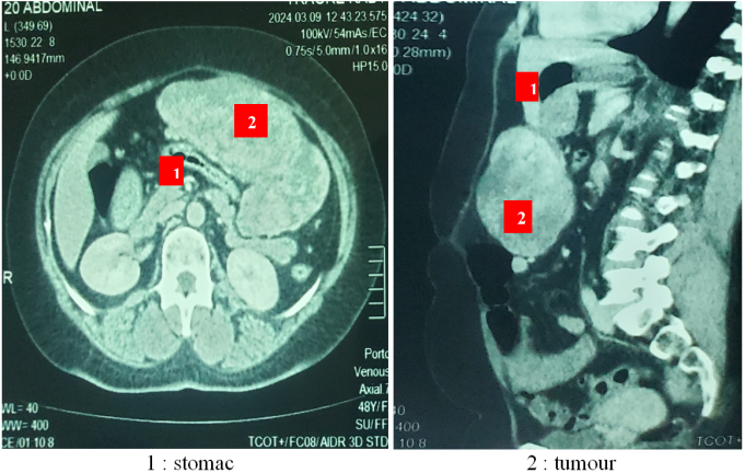
Figure 1. Transverse and sagittal CT scans showing a tumour developing at the expense of the gastric wall.
Upper endoscopy, performed in seven patients, revealed erythematous gastropathy without any objective tumour lesions in six cases. In one case, it suggested a diagnosis of gastric stromal tumour by showing erythematous gastric mucosa depressed by an extrinsic mass (Figure 2).
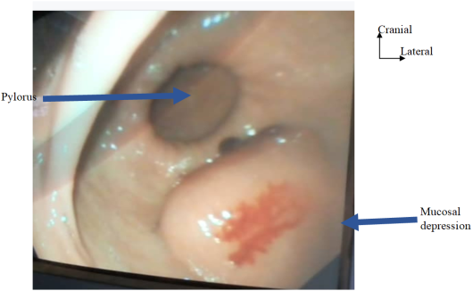
Figure 2. Endoscopic view of a depression in the gastric mucosa caused by an extraluminal mass with erosion of the mucosa, suggesting a GIST.
On pathological examination
The mass was encapsulated in 64.7% of cases (n=11). It was unencapsulated in 4 cases (23.5%). The mass was firm in 14 cases. Areas of necrosis were noted in 3 cases. The tumour was encephaloid in 3 cases (17.6%).
The majority of cases (n=9; 52.9%) were spindle cell stromal tumours. In 6 cases (35.3%), the tumours were epithelial cell tumours and in 2 cases (11.8%), they were mixed cell tumours.
According to the MIETTINIEN and LASOTA prognostic classification , the risk of recurrence was high in 8 cases (47.1%), moderate in 4 cases (23.5%) and low in 5 cases (29.4%).
Immunohistochemistry used several marker antibodies. C-KIT and Dog1+ antibodies were marked in all cases, confirming the diagnosis of gastric GISTs.
Immunohistochemistry classified the tumours as high grade in 8 patients (47.0%), low grade in 7 cases (41.2%) and moderate grade in 2 cases (11.8%).
3.2. Therapeutics Characteristics
The patients were classified as ASA II in 13 cases, ASA III in 3 cases, and ASA I in 1 case.
All patients underwent median laparotomy. The average tumour size was 22.65 cm ± 11 cm, with extremes of 5 cm and 40 cm. The tumour developed at the expense of the gastric antrum in 64.7% of cases (n=11) and the greater curvature in 35.3% of cases (n=6). (Figures 3 and 4).
The tumour was localised in 12 cases (70.6%). In 5 cases, there was extension to neighbouring organs, notably the transverse colon in 3 cases, the mesocolon in 2 cases, the liver in 1 case and the spleen in 1 case. Th e tumours were classified as T3 in 52.9% (n=17) according to the TNM classification [Table 2]. They were stage II in 9 cases (52.9%), stage I in 3 cases (17.6%) and stage III in 4 cases (23.6%).
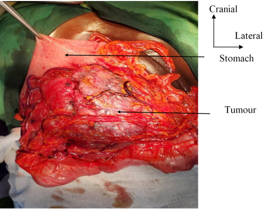
Figure 3. Tumour developed at the expense of the posterior face of the gastric antrum.
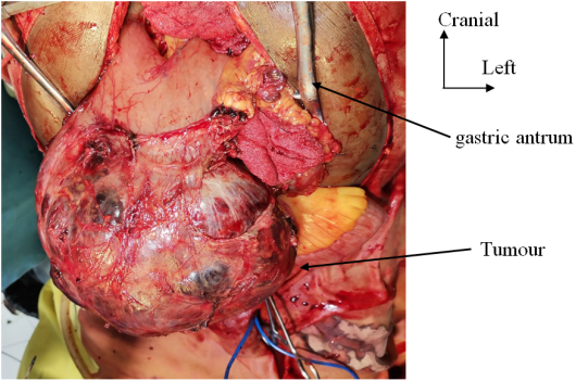
Figure 4. Necrotic-haemorrhagic tumour mass developed at the expense of the lower edge of the antrum.
Table 2. TNM classification (8th edition, 2017).

TNM grade

Numbers

Percentage

T

T1

0

0

T2

1

5,9

T3

9

52,9

T4

7

41.2

N

N0

15

88,2

N1

2

11,8

M

M0

17

100

Surgical procedures: a partial gastrectomy was performed in all cases. This involved either an atypical, sealed gastrectomy in 14 cases (Figure 5) or a lower polar gastrectomy in 3 cases. Certain procedures were associated with these gastrectomies [Table 3]. Tumour resection was complete (R0) in 15 cases and R1 in 2 cases.
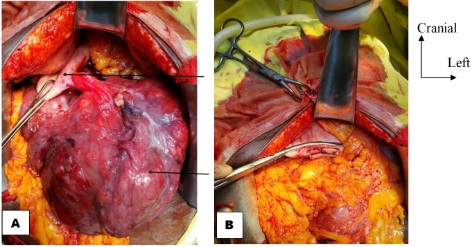
Figure 5. (A) Large pedunculated tumour on the lateral edge of the greater curvature of the stomach. (B) Stump after sealed gastrectomy.
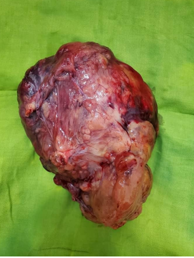
Figure 6. Tumour mass after gastrectomy.
No patients received preoperative chemotherapy. Postoperatively, only four patients were able to receive chemotherapy with imatinib.
All patients received pre-, intra- and postoperative resuscitation. Two patients were admitted to intensive care immediately after surgery.
An iso-group iso-Rhesus blood transfusion was performed in 15 patients.
A dual antibiotic therapy consisting of ceftriaxone and metronidazole was administered to all patients.
Heparin therapy, an anti-ulcer medication, and dual analgesic therapy (paracetamol + tramadol) were systematically administered.
Table 3. Distribution according to the different surgical procedures performed.

Procedures

Numbers

Perrcentages

Partial gastrectomy

17

100

Segmental colectomy of the transverse colon

03

17,6

Splenectomy

02

11,8

Partial resection of the mesocolon

01

5,9

3.3. Postoperative Complications
Seven patients experienced postoperative complications. These complications included parietal suppuration (n=3), anaemia (n=3), parietal haemorrhage (n=1) and acute urinary retention (n=1).
Three patients died postoperatively, representing a mortality rate of 17.6%. The characteristics of the deceased patients are described in Table 4.
Table 4. Characteristics of deceased patients.

Characteristics

Patient 1

Patient 2

Patients 3

Gender

Male

Male

Female

Age

59 years old

69 years old

49 years old

History

Epigastric pain

Epigastric pain

Epigastric pain

Time to progression

16 months

10 months

6 months

Mode of onset

Recurrent

Primary

Primary

General condition

Preserved

Preserved

Altered

Tumour size

25 cm

30 cm

20 cm

Tumour location

Antrum

Large curvature

Antrum

Local spread

Localised

Invasion of the transverse colon

Invasion of transverse colon, liver, spleen

Procedure

Sealed gastrectomy

Sealed gastrectomy, colectomy

Sealed gastrectomy + colectomy + splenectomy

Type of resection

R0

R0

R1

Stage of progression

pT4

pT4

Cause of death

Haemorrhagic shock

PPO

Cachexia

Time to death

H8

Day 7

Day 10

The average length of hospital stay was 9.35 days, ranging from 1 to 17 days.
The average follow-up period was 15.6 ± 8.7 months, ranging from 0 to 38 months. All 14 patients who were discharged were reviewed at 1 month and 3 months. After 36 months, 9 patients were lost to follow-up. No recurrences were noted.
4. Discussion
GISTs are asymptomatic for a long time, until they become large or cause complications. They may be discovered incidentally when they are small during an upper gastrointestinal endoscopy, or more rarely during a CT scan. This method of detection accounts for approximately 20% of cases . The most common symptoms of gastric stromal tumours are gastrointestinal bleeding (20 to 50%) or a palpable mass (25 to 40%) . In our study, the main reason for consultation or circumstance of discovery was an abdominal mass. In our series, the tumour was mobile in all cases. The mobile nature of the tumour is well described in gastric GISTs . The average size of the mass was 22.65 cm. The large size of the tumours in our series reflects the insidious and latent evolution of these tumours.
At the paraclinical level, upper gastrointestinal endoscopy may be normal in 20% of cases when tumours develop extra-luminally or extra-gastrically. The wall appears normal or rigid or shows extra-luminal bulging suggestive of extrinsic compression . It allows for lesion and peripheral biopsies to be performed, but the sensitivity of biopsies for the diagnosis of gastric GISTs is unreliable (15 to 30%) because biopsies are often superficial . In our study, OGDE (esophago-gastroduodenal endoscopy) led to suspicion of gastric GIST in only one case, showing a parietal depression with erythematous mucosa, but the biopsies performed were negative.
Echoendoscopy remains the best examination for characterising gastric submucosal tumours and differentiating them from extrinsic compression . It also helps guide biopsies. However, we did not perform any biopsies as this technique was not available.
Abdominal computed tomography is a useful tool for detecting large gastric stromal tumours, assessing local and general spread, and guiding tissue biopsy. In our series, abdominal CT scans led to a diagnosis of gastric GIST in 8 out of 15 patients (53.3%), most often revealing a multilobulated mass with areas of necrosis developing at the expense of the gastric wall. In other cases, the CT scan did not confirm a gastric GIST but rather a mesenteric tumour. This could be due to the large volume of the mass, often estimated at more than 30 cm. Hence the interest of MRI, which is more sensitive and specific than CT in the diagnosis of large gastric GISTs.
In our study, the mass was located in the majority of cases in the antrum, which is the preferred site described in the literature . In most cases, it was a tissue mass, usually encapsulated; non-encapsulated tumours have a high recurrence rate .
There are several histological types, but the main ones are spindle cell stromal tumours, epithelioid cell stromal tumours, and mixed cell stromal tumours. The spindle cell variant accounts for 70% of cases . This variant was found in 52.9% of cases in our study.
The diagnosis of GIST is suggested by histology, but diagnostic confirmation is provided by immunohistochemistry, which also allows the grade of malignancy of the tumour to be determined . In our study, 47% of tumours had a high grade of malignancy.
The development of targeted therapies, particularly Imatinib, has completely revolutionised the management of locally advanced or metastatic gastric stromal tumours. However, surgery remains the treatment of choice for localised stromal tumours.
When surgery is feasible, complete resection of the tumour should be the first-line treatment, as it remains the standard treatment for non-metastatic gastric stromal tumours and is a factor in a good prognosis for the patient. The tumour must therefore be removed en bloc with its pseudocapsule, if possible with healthy margins to prevent recurrence . There is no consensus on the optimal width of surgical margins: a margin of 2 cm is considered reasonable. On the other hand, wider excision has not shown any additional benefit . In our study, we performed complete resection extending into healthy tissue (R0) in 88.2% of cases. Pre- or intraoperative tumour rupture is a factor associated with poor prognosis. The tumour should therefore be handled with care during surgery to avoid the spread of tumour cells throughout the body . Lymph node dissection is not necessary, as stromal tumours of the stomach very rarely metastasise to the lymph nodes . However, it is still important to remain vigilant and remove any lymph nodes that appear suspicious. Locally advanced tumours that spread to neighbouring organs; complete surgery is only possible if one or more adjacent viscera are removed .
We performed two splenectomies and two segmental colectomies of the transverse colon when these organs were invaded by the tumour. These extensive resections are sometimes mutilating and must be adjusted according to the organs involved. This ‘classical’ approach is currently being questioned since the discovery of Imatinib. The use of this new molecule in the treatment of gastric stromal tumours would make it possible to limit the initial surgical procedure and subsequently increase the chances of complete resection . We did not perform this preoperative chemotherapy with Imatinib for two main reasons. Firstly, we did not have diagnostic certainty in the majority of cases (58.8%). Secondly, the majority of patients were farmers and did not have the financial means to undergo chemotherapy.
The majority of post-operative recurrences occur within 5 years . The risk is highest in the first 2 years, but late relapses, more than 10 years after surgery, have been reported . In our series, we noted one case of tumour recurrence that had been operated on two years earlier. The risk of recurrence is mainly correlated with two parameters: the size and mitotic index of these tumours.
The 5-year survival rate is approximately 20% for tumours larger than 10 cm in diameter, 40% for tumours between 5 and 10 cm in diameter, and 60% for tumours smaller than 5 cm in diameter . We were unable to estimate the actual survival rate of our patients due to the large number who were lost to follow-up after 36 months. Only 5 patients were seen again at 3 years, representing a survival rate of 29.4%.
5. Conclusion
The management of gastric GISTs is based on surgery, which remains the treatment of choice with a high resectability rate. Recurrences are common. For locally advanced tumours (greater than 10 cm or invading a neighbouring organ), targeted therapy with Imatinib makes it possible to limit the initial surgical procedure, subsequently increase the chances of complete resection and reduce the risk of recurrence. Early diagnosis and greater access to targeted therapy before and after surgery would improve the prognosis for gastric GISTs.
Abbreviations

GIST

Gastro Intestinal Stroma Tumour

OGDE

Esophago-Gastroduodenal Endoscopy

TNM

Tumour, Node, Metastasis

Conflicts of Interest
The authors declare no conflicts of interest.
References
[1] Brun-stang C, Bedossa P, Blay JY, JMC, Le Cesne A, Monges G. Les tumeurs stromales gastro-intestinales: prévalences et incidences en France. Gastroenterol Clin Biol 2004; 28: 102-5.
[2] Saïd HRT, Nawal Y. Advances and therapeutic indications in gastrointestinal stromal tumours. Alg J med Heal res 2022; 1(3): 6.
[3] Bucher P, Egger J, Gervaz P. An audit of surgical management of gastrointestinal stromal tumors (GIST). Eur J Surg Oncol 2006; 32: 310-4.
[4] Bucher P, Villiger P, Egger J. Management of gastrointestinal stromal tumors: From diagnosis to treatment. swiss Med Wkly 2004; 134: 145-53.
[5] Yan H, Marchettini P, Acherman YI. Prognostic assessment of gastrointestinal stromal tumour. Am J Clin Oncol. 2003; 26: 221-228.
[6] Miettinen M and Lasota J. Gastrointestinal stromal tumors: pathology and prognosis at different sites. Semin Diagn Pathol 2006; 23: 70-83.
[7] Joensuu H, Fletcher C, Dimitrijevic S, Silberman S, Roberts P, Demetri G. Management of malignant gastrointestinal stromal tumors. Lancet Oncol 2002; 3: 655-64.
[8] Miettinen M, Sobin LH, Sarlomo-Rikala M. Immunohistochemical spectrum of GISTs at different sites and their differential diagnosis with a référencez to CD117 (KIT). Mod Pathol 2000; 13: 1134-42.
[9] Nguyen VU, Taylor A. Gastrointestinal stromale tumors leiomyoma/leiomysarcoma. E Med Gastrointest 2006; 11 p.
[10] Bettini G, Morini M, Marcato PS. Gastrointestinal spindle cell tumours of the dog: histological and immunohistochemical study. J. Comp. Pathol 2003; 129: 283-93.
[11] Dematteo RP, Heinrich MC, El-Rifai WM, Demetri G. Clinical management of gastrointestinal stromal tumors: before and after STI 571. Hum Pathol 2002; 33: 466-77.
[12] Demetri GD, Van Oosterom AT, Garrett CR, Blackstein ME, Shah MH, Verweij J et al. Efficacy and safety of sunitinib in patients with advanced gastrointestinal stromal tumour after failure of imatinib: a randomised controlled trial. Lancet. 2006; 368: 1329-38.
[13] Kreiker J, Daou R, Aftimos G. Tumeurs stromales gastriques Présentations de deux cas avec étude immunohistochimique. Revue de la littérature. J Méd Lib 2002; 50(5-6): 226-36.
[14] Clère F, Carola E, Halimi C, Gramont AD, Y Panis. Actualités sur les tumeurs stromales gastro-intestinales: à partir de sept observations de tumeurs malignes. Rev Méd Int 2002; 23: 499- 507.
[15] Barrier A, M. Huguier M, Levard H, Montario T, Fagniez PL, Sauvanet A et les Associations françaises de recherche en chirurgie. Tumeurs gastriques conjonctives. Résultats d’une étude multicentrique. Chirurgie 1999; 124: 494-502.
[16] N’Dah KJ, Troh E, Doukouré B; Koffi KE, Abouna AD, Traoré B et Al. Aspects anatomo-cliniques des tumeurs stromales gastro-intestinales à Abibjan. Rev Afr Pathol 2013; 12(1-2): 21-30.
[17] Ian Judson, Ramesh Bulusu, Beatrice Seddon, Adam Dangoor, Newton Wong, Satvinder Mudan. UK clinical practice guidelines for the management of gastrointestinal stromal tumours (GIST). Clinical Sarcoma Research 2017; 7: 6.
[18] Mazur MT and Clark HB. Gastric stromal tumors. Reappraisal of histogenesis. Am J Surg pathol. 1983; 7: 507-519.
Cite This Article
  • APA Style

    Mamadou, T., Ismael, L. B. K., Bernadette, N. A., Amos, E. S., Amos, K. B., et al. (2025). Gastrointestinal Stromal Tumour Management at the Bouake University Hospital. Journal of Surgery, 13(5), 134-140. https://doi.org/10.11648/j.js.20251305.13

    Copy | Download

    ACS Style

    Mamadou, T.; Ismael, L. B. K.; Bernadette, N. A.; Amos, E. S.; Amos, K. B., et al. Gastrointestinal Stromal Tumour Management at the Bouake University Hospital. J. Surg. 2025, 13(5), 134-140. doi: 10.11648/j.js.20251305.13

    Copy | Download

    AMA Style

    Mamadou T, Ismael LBK, Bernadette NA, Amos ES, Amos KB, et al. Gastrointestinal Stromal Tumour Management at the Bouake University Hospital. J Surg. 2025;13(5):134-140. doi: 10.11648/j.js.20251305.13

    Copy | Download

  • @article{10.11648/j.js.20251305.13,
      author = {Traore Mamadou and Leh Bi Kalou Ismael and N’Dri Ahou Bernadette and Ekra Serge Amos and Kouakou Blaise Amos and Bamba Inza and Akowendo Djahou Eczechiel and Anzoua Kouakou Ibrahim and Kouakou Kouame Bernadin},
      title = {Gastrointestinal Stromal Tumour Management at the Bouake University Hospital
    },
      journal = {Journal of Surgery},
      volume = {13},
      number = {5},
      pages = {134-140},
      doi = {10.11648/j.js.20251305.13},
      url = {https://doi.org/10.11648/j.js.20251305.13},
      eprint = {https://article.sciencepublishinggroup.com/pdf/10.11648.j.js.20251305.13},
      abstract = {Gastric GISTs are rare tumours that are often detected late. The aim of this study was to describe the different treatment modalities for gastric GISTs at Bouake University Hospital. It was retrospective study of 17 patients with gastric GIST between January 2013 and December 2023. GISTs accounted for 4.2% of all gastric tumours. The main reason for consultation was an abdominal mass. The average duration of progression was 10.9 months ± 2.1 months. FOGD, performed in 7 patients, suspected GIST in only one case. Abdominal CT scan, performed in 15 patients, was suggestive of GIST in 8 cases. The tumour was located in the antrum in 11 cases. There was extension to neighbouring organs in 5 cases. The tumours were classified as stage III in 9 cases. Immunohistochemistry confirmed the diagnosis of GIST with expression of C-KIT (CD117) antibodies in all cases. GIST is most often diagnosed at the stage of a large abdominal mass. Immunohistochemistry, which allows for diagnostic confirmation, is not accessible to most patients.
    },
     year = {2025}
    }
    

    Copy | Download

  • TY  - JOUR
    T1  - Gastrointestinal Stromal Tumour Management at the Bouake University Hospital
    
    AU  - Traore Mamadou
    AU  - Leh Bi Kalou Ismael
    AU  - N’Dri Ahou Bernadette
    AU  - Ekra Serge Amos
    AU  - Kouakou Blaise Amos
    AU  - Bamba Inza
    AU  - Akowendo Djahou Eczechiel
    AU  - Anzoua Kouakou Ibrahim
    AU  - Kouakou Kouame Bernadin
    Y1  - 2025/09/25
    PY  - 2025
    N1  - https://doi.org/10.11648/j.js.20251305.13
    DO  - 10.11648/j.js.20251305.13
    T2  - Journal of Surgery
    JF  - Journal of Surgery
    JO  - Journal of Surgery
    SP  - 134
    EP  - 140
    PB  - Science Publishing Group
    SN  - 2330-0930
    UR  - https://doi.org/10.11648/j.js.20251305.13
    AB  - Gastric GISTs are rare tumours that are often detected late. The aim of this study was to describe the different treatment modalities for gastric GISTs at Bouake University Hospital. It was retrospective study of 17 patients with gastric GIST between January 2013 and December 2023. GISTs accounted for 4.2% of all gastric tumours. The main reason for consultation was an abdominal mass. The average duration of progression was 10.9 months ± 2.1 months. FOGD, performed in 7 patients, suspected GIST in only one case. Abdominal CT scan, performed in 15 patients, was suggestive of GIST in 8 cases. The tumour was located in the antrum in 11 cases. There was extension to neighbouring organs in 5 cases. The tumours were classified as stage III in 9 cases. Immunohistochemistry confirmed the diagnosis of GIST with expression of C-KIT (CD117) antibodies in all cases. GIST is most often diagnosed at the stage of a large abdominal mass. Immunohistochemistry, which allows for diagnostic confirmation, is not accessible to most patients.
    
    VL  - 13
    IS  - 5
    ER  - 

    Copy | Download

Author Information