Abstract
Canine parvovirus enteritis (CPVE) remains a highly contagious and frequently fatal viral disease affecting young dogs globally. This case report describes the clinical progression and postmortem findings of CPVE in a 7-week-old male German Shepherd puppy presented to the D.I.K Osori Veterinary Teaching Hospital, Sokoto, Nigeria. The patient presented with a two-day history of anorexia, vomiting, and foul-smelling diarrhea after being housed with a dog that had recently recovered from CPVE. Clinical examination revealed 5% dehydration, tachycardia, and tachypnea. A rapid fecal antigen test confirmed CPVE. Hematological analysis revealed normocytic normochromic anemia with moderate neutrophilia, suggesting an acute inflammatory response associated with severe intestinal injury and highlighting an atypical neutrophilic hematological response in CPVE. Despite aggressive fluid resuscitation with Ringer's Lactate and dual antimicrobial intervention, the patient succumbed to the infection three days post-admission. Gross necropsy revealed severe, diffuse mucohemorrhagic enteritis with a characteristic "ground-glass" serosal appearance, mesenteric lymphadenomegaly, and diffuse pulmonary congestion and edema. Histopathological evaluation confirmed severe lymphoid depletion in the mesenteric lymph nodes and proteinaceous alveolar exudates in the lungs. The combined clinical and pathological findings were consistent with severe systemic circulatory and inflammatory disturbances preceding death, highlighting the importance of recognizing atypical hematological presentations in CPVE.
Keywords
Canine Parvovirus, Clinicopathology, Neutrophilia, Necropsy, Histopathology, Puppy
1. Introduction
Canine parvovirus type 2 (CPV-2) is a highly contagious and frequently fatal viral disease of young dogs, particularly puppies between six weeks and six months of age
| [1] | Behera, S. K., Singh, Y. D., Roychoudhury, P. Clinico-pathological and necropsy findings in a 4-month old mixed-breed pup with canine parvovirus-2 infection and its genetic characterization. Journal of Entomology and Zoology Studies. 2020, 8(5), 573-577. |
| [11] | Decaro, N., Buonavoglia, C. Canine parvovirus—A review of epidemiological and diagnostic aspects. The Veterinary Journal. 2012, 192(1), 21-25. https://doi.org/10.1016/j.tvjl.2011.08.025 |
| [15] | Miranda, C., Thompson, G. Canine parvovirus: the constant evolution of a point mutant virus. Veterinary Research. 2016, 47(1), 110. https://doi.org/10.1186/s13567-016-0394-4 |
[1, 11, 15]
. The virus belongs to the family Parvoviridae and primarily affects rapidly dividing cells, especially intestinal crypt epithelial cells, lymphoid tissues, and bone marrow progenitor cells
| [2] | Nivy, R., Hahn, S., Perl, S., et al. A Fatal Outbreak of Parvovirus Infection: First Detection of Canine Parvovirus Type 2c in Israel with Secondary Escherichia coli Septicemia and Meningoencephalitis. Israel Journal of Veterinary Medicine. 2011, 66(3), 96-101. |
| [3] | Odueko, F. D. Literature review on canine parvoviral enteritis variants in Nigeria. Journal of Dairy, Veterinary & Animal Research. 2020, 9(1), 26-32.
https://doi.org/10.15406/jdvar.2020.09.00274 |
| [14] | Stuetzer, B., Hartmann, K. Feline parvovirus infection and associated diseases. The Veterinary Journal. 2014, 201(2), 150-155. https://doi.org/10.1016/j.tvjl.2014.05.027 |
[2, 3, 14]
. Infection commonly results in severe hemorrhagic enteritis, dehydration, immunosuppression, and, in severe cases, death.
Transmission occurs mainly through the fecal-oral route either by direct contact with infected dogs or indirectly through contaminated fomites and environments
. The virus is highly resistant in the environment and may remain infectious for prolonged periods
| [2] | Nivy, R., Hahn, S., Perl, S., et al. A Fatal Outbreak of Parvovirus Infection: First Detection of Canine Parvovirus Type 2c in Israel with Secondary Escherichia coli Septicemia and Meningoencephalitis. Israel Journal of Veterinary Medicine. 2011, 66(3), 96-101. |
[2]
. Following infection, viral replication within intestinal crypts results in destruction of the intestinal mucosa, villous collapse, and loss of mucosal integrity, thereby predisposing affected animals to secondary bacterial invasion and systemic inflammatory complications.
Clinical manifestations commonly include anorexia, lethargy, vomiting, foul-smelling hemorrhagic diarrhea, dehydration, and varying degrees of leukopenia
| [1] | Behera, S. K., Singh, Y. D., Roychoudhury, P. Clinico-pathological and necropsy findings in a 4-month old mixed-breed pup with canine parvovirus-2 infection and its genetic characterization. Journal of Entomology and Zoology Studies. 2020, 8(5), 573-577. |
[1]
. Hematological findings classically include panleukopenia, particularly neutropenia and lymphopenia, resulting from viral destruction of bone marrow progenitor cells
| [4] | Thilahayswari, J., Nagarajan, K., Hemalatha, S., Chandrasekar, M. Canine Parvovirus-2a infection in a juvenile pup: Bone marrow findings and clinical implications. Indian Journal of Veterinary Pathology. 2026, 50(1), 36-39.
https://doi.org/10.56093/cr49qj17 |
[4]
. However, atypical hematological presentations characterized by normal or elevated leukocyte counts have occasionally been reported in severe inflammatory CPVE cases.
This report describes the clinical presentation, hematological findings, necropsy lesions, and histopathological changes observed in a fatal case of CPVE in a juvenile German Shepherd puppy presented to the D.I.K Osori Veterinary Teaching Hospital, Sokoto, Nigeria.
2. Case Presentation
2.1. History and Signalment
On Wednesday 25th of February 2026, a client from the Arkilla area of Sokoto, Sokoto State of Nigeria, presented a 7-week-old male German Shepherd dog weighing 6.2 kg at the Small Animal unit of the D.I.K Osori Veterinary Teaching Hospital, Usmanu Danfodiyo University, Sokoto. The patient presented with complaints of being off feed, vomiting, and diarrhea, all noticed two days prior to presentation. Epidemiological history revealed that the dog was recently purchased and transported from Jos, Plateau State, Nigeria. Upon arrival in Sokoto State, it was housed in the same confinement as another resident dog that had recovered from Canine Parvovirus Enteritis (CPVE) two weeks earlier. The patient was fed a diet of fish, meat, and table remnants. The client claimed the dog received its first shot of the DHLPP vaccine while in Jos; however, the vaccination record (green book) was misplaced by the client. There was no history of prior medication before clinical presentation.
2.2. Physical Examination and Clinical Findings
Upon initial physical examination, the patient was dull, lethargic, and actively passing profuse foul-smelling diarrhea. The ocular and oral mucous membranes were slightly pale, with a capillary refill time (CRT) of less than 2 seconds. The patient weighed 6.2 kg and was assessed to have a 5% dehydration deficit. The body condition score was determined to be 3 on a scale of 1 to 5. Vital parameters revealed marked tachycardia and tachypnea, while the rectal core body temperature remained within normal reference limits. The exact vital parameter configurations compared against established standards are structured in
Table 1.
Table 1. Vital Parameters of the Patient vs. Canine Reference Ranges.
Parameter | Patient Value | Reference Range |
Temperature (°C) | 38.9 | 38.3 - 39.2 |
Pulse Rate (beats/min) | 139 | 70 – 120 |
Respiratory Rate (cycles/min) | 80 | 18 – 34 |
Reference values sourced from Kahn et al.,
| [7] | Kahn, C. M., Line, S., Aiello, S. E. The Merck Veterinary Manual. 9th ed. Whitehouse Station, NJ: Merck & Co., Inc.; 2005, pp. 1-2300. |
[7]
.
Based on these combined clinical inputs, a comprehensive problem list was compiled: dullness, dehydration, emesis, anorexia, foul-smelling diarrhea, tachycardia, and tachypnea. Differential diagnoses focused on Canine Parvovirus Enteritis and Canine Distemper, with a strong tentative diagnosis directed toward CPVE based on exposure history.
2.3. Diagnostic Plan and Laboratory Profiling
The diagnostic plan included hospitalization, fluid stabilization, rapid fecal antigen testing, complete blood count (CBC), and routine hemoparasite screening.
The fecal sample tested positive for Canine Parvovirus using a rapid ELISA-based antigen test kit. Blood collected via jugular venipuncture into EDTA tubes was submitted for complete blood count (CBC). Hematological evaluation revealed moderate neutrophilia (14.3x10
9/L) with a total leukocyte count near the upper reference limit (17.0 x10
9/L). Manual peripheral blood smear evaluation for band neutrophils or toxic morphologic changes was not performed. The definitive hematological profile is outlined in
Table 2.
Table 2. Hematological Profile of the CPV-Infected Puppy.
PARAMETERS | OBTAINED VALUES | REFERENCE VALUES | CLINICAL INTERPRETATION |
PCV (L/L) | 0.33* | 0.35-0.57 | Decreased (Anemia) |
HB (g/L) | 110* | 120-200 | Decreased |
RBC (x1012/L) | 4.38* | 5.4-8.5 | Decreased |
MCV (fL) | 70 | 60-77 | Normocytic |
MCH (pg) | 23.4 | 19-24 | Normochromic |
MCHC (g/dl) | 333 | 330-360 | Normochromic |
WBC (X109/L) | 17.0 | 6-18 | Normal (Upper Limit) |
Neutrophils (x109/L) | 14.3* | 3-12 | Increased (Neutrophilia) |
Lymphocytes (x109/L) | 2.4 | 1-5 | Normal |
Monocyte (x109/L) | 0.17* | 0.2-1.5 | Decreased (Monocytopenia) |
Eosinophils (x109/L) | 0.17 | 0.1-0.8 | Normal |
Basophils (X 109/L) | 0 | Rare | Normal |
Source: Adapted from MSD Veterinary Manual, Hematology (Complete Blood Count
| [8] | Fielder, S. E. Hematology (Complete Blood Count) Reference Ranges. In MSD Veterinary Manual. Rahway, NJ: Merck & Co., Inc.; 2024. Available from:
https://www.msdvetmanual.com/ [Accessed 29 May 2026]. |
[8]
.
Following confirmation of CPVE by rapid antigen testing, hematological evaluation revealed moderate neutrophilia despite the confirmed viral infection, representing an atypical leukogram compared with the classical leukopenic presentation. No hemoparasites were detected on Giemsa-stained blood smears.
2.4. Therapeutic Management and Clinical Progression
Therapy was aimed at correcting dehydration, controlling vomiting, and minimizing secondary bacterial complications associated with intestinal mucosal damage.
Fluid Therapy: Intravenous Ringer’s Lactate solution was administered to correct dehydration and fluid loss. Based on a body weight of 6.2 kg and 5% dehydration, the fluid deficit was calculated as:
An initial replacement volume of 310 mL was administered intravenously.
Antimicrobial Therapy: Long-acting 15% Amoxicillin at 15 mg/kg IM and Ceftriaxone 10% at 30 mg/kg IV were administered to minimize the risk of secondary bacterial complications associated with intestinal mucosal disruption.
Antiemetic Therapy: Metoclopramide 0.5% was administered IV at 0.5 mg/kg to control vomiting.
Supportive Therapy: injectable Vitamin B-complex was administered IM at 1 mL/10 kg for three days.
Despite supportive and antimicrobial therapy, the patient’s clinical condition progressively deteriorated, and death was recorded on February 28, 2026. The carcass was subsequently submitted for necropsy and histopathological evaluation, as detailed in the clinical timeline in
Table 3.
Table 3. Patient Clinical Progress.
Date | Temperature (°C) | Pulse rate (beats/min) | Respiratory rate (cycles/min) | Remarks |
26/02/2026 | 39.2 | 134 | 84 | The patient was weak, vomiting and passing foul smelling diarrhoea. Fluid and Medications were administered. |
27/02/2026 | 39.9 | 139 | 100 | The patient condition had not improved vomiting and diarrhoea had continued. Fluid and Medications were administered. |
28/02/2026 | | | | The patient died and the carcass was submitted to Necropsy/Histopathology unit for Postmortem examination. |
3. Pathological Findings
3.1. Gross Pathology
Postmortem examination was conducted immediately after death on February 28, 2026. External evaluation revealed a carcass in poor-to-moderately poor body condition with sunken eyes and a rough hair coat, consistent with severe dehydration. Foul-smelling, dark red mucohemorrhagic feces heavily stained the perineal region and hindquarters.
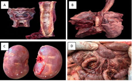
Upon opening the abdominal cavity, the small intestinal segments particularly the duodenum and jejunum were severely congested, dark red, and dilated by abundant liquid hemorrhagic contents. The intestinal serosal surface exhibited a classic, dull "ground-glass" appearance. The underlying intestinal mucosa was diffusely thickened, hyperemic, and displayed multifocal regions of severe hemorrhage. The mesenteric lymph nodes were markedly enlarged and deeply congested. The gallbladder was severely distended with stagnant bile. Upon exposing the thoracic cavity, the lungs were diffusely congested and edematous, exhibiting a patchy dark-red mottling across all lobes. The tracheal lumen was filled with abundant, white frothy fluid exudate. The kidneys were moderately congested with rounded margins.
Figure 1. Gross pathological findings of the abdominal and thoracic cavities. Label A (Trachea): Arrows point to the trachea lumen containing abundant fluid exudate. Label B (Lungs): Demonstrates diffuse pulmonary congestion, dark-red patchy hepatization, and frothy exudate at the bronchial bifurcations. Label C (Kidney): Moderately congested kidneys with rounded margins. Label D (Small Intestine): Demonstrates the intense diffuse mucosal hyperemia and the classic granular "ground-glass" texture of the serosal surface.
3.2. Histopathology
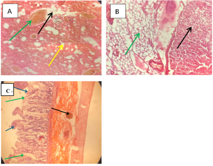
Histopathological examination of selected tissues further characterized the systemic lesions observed during necropsy. Microscopically, the lungs showed severe diffuse pulmonary edema characterized by thickening of the alveolar septa, accumulation of proteinaceous fluid within alveolar spaces, vascular congestion, fibrin deposition, inflammatory cellular infiltration, and multifocal hemorrhages (
Figure 2A). Sections of the mesenteric lymph nodes revealed marked lymphoid depletion with congestion of blood vessels and replacement of depleted lymphoid tissue by adipose tissue, consistent with severe lymphoid depletion (
Figure 2B). Histopathological evaluation of the small intestine also demonstrated acute, severe mucosal disruption accompanied by marked submucosal vascular changes. Within the mucosal layer, there was extensive epithelial necrosis, prominent villous blunting, and widespread sloughing of necrotic cells into the intestinal lumen. Furthermore, the submucosa was markedly expanded by severe edema and contained engorged, hyperemic blood vessels densely packed with erythrocytes (
Figure 2C).
Figure 2. Histopathological changes in the lungs and lymphoid organs (H&E Stain, X100 magnification). Label A (Lungs): Arrows point to dense sheets of proteinaceous fluid completely obliterating the alveolar airspaces, paired with marked alveolar septal thickening, vascular congestion, and multifocal hemorrhages. Label B (Mesenteric Lymph Node): Highlights advanced lymphoid depletion within the germinal centers and the replacement of normal architecture by infiltrating adipose tissue cells. Lebel C (Intestine): Shows mucosal layer demonstrating epithelial necrosis, villous blunting, and sloughing into the lumen, alongside marked expansion of the submucosa secondary to severe edema and severely engorged, congested submucosal blood vessels densely packed with erythrocytes.
3.3. Morphological Diagnosis
1) Severe, acute, diffuse necrotic enteritis with villous blunting, sloughing, severe submucosal edema, and marked submucosal vascular congestion.
2) Marked mesenteric lymphoid depletion with congestion.
3) Severe, diffuse pulmonary congestion and alveolar edema.
4) Moderate acute renal congestion with mild tubular degeneration.
5) Marked gallbladder distension consistent with prolonged anorexia.
4. Discussion
The clinical history, laboratory findings, gross lesions, and histopathological changes observed in this case were consistent with fatal Canine Parvovirus Enteritis (CPVE). The recent transportation of the puppy and exposure to an environment previously occupied by a recovering CPVE case likely increased the risk of infection, as CPV-2 is highly resistant in contaminated environments and may remain infectious for prolonged periods
| [2] | Nivy, R., Hahn, S., Perl, S., et al. A Fatal Outbreak of Parvovirus Infection: First Detection of Canine Parvovirus Type 2c in Israel with Secondary Escherichia coli Septicemia and Meningoencephalitis. Israel Journal of Veterinary Medicine. 2011, 66(3), 96-101. |
| [3] | Odueko, F. D. Literature review on canine parvoviral enteritis variants in Nigeria. Journal of Dairy, Veterinary & Animal Research. 2020, 9(1), 26-32.
https://doi.org/10.15406/jdvar.2020.09.00274 |
| [13] | Kalli, I., Leontides, L. S., Mylonakis, M. E., Billinis, C., Kontos, V., Koutinas, A. F. Factors associated with survival in dogs with natural canine parvovirus type 2 infection. Journal of Veterinary Internal Medicine. 2010, 24(4), 885-891.
https://doi.org/10.1111/j.1939-1676.2010.0538.x |
[2, 3, 13]
.
A notable feature of this case was the absence of classical leukopenia. Instead, the patient exhibited moderate neutrophilia with a total leukocyte count near the upper reference limit. Classical hematological findings in CPVE commonly include leukopenia, neutropenia, and lymphopenia due to viral destruction of rapidly dividing hematopoietic precursor cells within the bone marrow
| [4] | Thilahayswari, J., Nagarajan, K., Hemalatha, S., Chandrasekar, M. Canine Parvovirus-2a infection in a juvenile pup: Bone marrow findings and clinical implications. Indian Journal of Veterinary Pathology. 2026, 50(1), 36-39.
https://doi.org/10.56093/cr49qj17 |
| [5] | Li, H., Li, S., Pan, Y., Shi, Q. Decoding canine parvovirus: biomarkers for diagnosis and advances in vaccine development to address emerging challenges. Frontiers in Veterinary Science. 2025, 12, 1624275.
https://doi.org/10.3389/fvets.2025.1624275 |
| [10] | Goddard, A., Leisewitz, A. L., Christopher, M. M., Duncan, N. M., Becker, P. J. Prognostic usefulness of blood leukocyte changes in canine parvoviral enteritis. Journal of Veterinary Internal Medicine. 2008, 22(2), 309-316.
https://doi.org/10.1111/j.1939-1676.2008.0062.x |
| [12] | Schoeman, J. P., Goddard, A., Leisewitz, A. L. Biomarkers in canine parvovirus enteritis. New Zealand Veterinary Journal. 2013, 61(4), 217-222.
https://doi.org/10.1080/00480169.2013.776451 |
[4, 5, 10, 12]
. In contrast, the neutrophilic leukogram observed in this case may reflect an intense inflammatory response associated with extensive intestinal mucosal injury Although manual blood smear evaluation for band neutrophils or toxic morphologic changes was not performed, the clinical presentation; characterized by pyrexia, severe necrohemorrhagic diarrhea, tachycardia, and rapid systemic collapse strongly indicates an acute systemic inflammatory response syndrome (SIRS) and endotoxemia rather than a stress leukogram
. Translocation of enteric bacteria across the disrupted intestinal mucosa triggers systemic cytokine release, driving acute neutrophilic release from the bone marrow storage pool
| [10] | Goddard, A., Leisewitz, A. L., Christopher, M. M., Duncan, N. M., Becker, P. J. Prognostic usefulness of blood leukocyte changes in canine parvoviral enteritis. Journal of Veterinary Internal Medicine. 2008, 22(2), 309-316.
https://doi.org/10.1111/j.1939-1676.2008.0062.x |
[10]
. This atypical hematological presentation highlights the variability that may occur in advanced CPVE cases and suggests that the absence of leukopenia should not exclude CPVE from differential diagnosis in clinically compatible cases.
The severe mucohemorrhagic enteritis observed grossly and microscopically reflected destruction of intestinal crypt epithelial cells by the virus, resulting in villous collapse, mucosal hemorrhage, and loss of intestinal integrity
| [2] | Nivy, R., Hahn, S., Perl, S., et al. A Fatal Outbreak of Parvovirus Infection: First Detection of Canine Parvovirus Type 2c in Israel with Secondary Escherichia coli Septicemia and Meningoencephalitis. Israel Journal of Veterinary Medicine. 2011, 66(3), 96-101. |
| [6] | Tangolli, A. A., Kumar, G. K. C., Shankar, B. P. Post mortem and histopathological changes in puppies died with canine parvoviral infection. International Journal of Veterinary Sciences and Animal Husbandry. 2024, 9(6), 354-359.
https://doi.org/10.22271/chemi.2024.v9.i6e.354 |
| [9] | Sinani, A., Kusi, I. Pathologic Findings in Dogs Died of CPV-2 in Kosovo. Albanian Journal of Agricultural Sciences. 2016, 15(3), 154-159. |
| [16] | Mazzaferro, E. M. Update on Canine Parvovirus. Veterinary Clinics of North America: Small Animal Practice. 2020, 50(6), 1339-1349. https://doi.org/10.1016/j.cvsm.2020.07.008 |
[2, 6, 9, 16]
. These lesions likely contributed to the profuse hemorrhagic diarrhea, dehydration, and rapid clinical deterioration observed before death.
Marked lymphoid depletion within the mesenteric lymph nodes supports the tropism of CPV-2 for rapidly dividing lymphoid cells and is consistent with severe immunosuppression
| [4] | Thilahayswari, J., Nagarajan, K., Hemalatha, S., Chandrasekar, M. Canine Parvovirus-2a infection in a juvenile pup: Bone marrow findings and clinical implications. Indian Journal of Veterinary Pathology. 2026, 50(1), 36-39.
https://doi.org/10.56093/cr49qj17 |
[4]
. The pulmonary congestion and edema observed in this case may have been associated with severe systemic inflammatory and circulatory disturbances secondary to intestinal injury.
The distended gallbladder observed at necropsy was likely associated with prolonged anorexia and reduced gastrointestinal motility, which have been reported in severe systemic illnesses associated with CPVE
| [1] | Behera, S. K., Singh, Y. D., Roychoudhury, P. Clinico-pathological and necropsy findings in a 4-month old mixed-breed pup with canine parvovirus-2 infection and its genetic characterization. Journal of Entomology and Zoology Studies. 2020, 8(5), 573-577. |
[1]
. Taken together, the clinical progression, hematological abnormalities, necropsy findings, and histopathological lesions support the possibility that severe inflammatory responses in advanced CPVE may occasionally mask the classical leukopenic presentation.
5. Conclusion
This report describes a fatal case of Canine Parvovirus Enteritis (CPVE) in a juvenile German Shepherd puppy characterized by severe mucohemorrhagic enteritis, marked lymphoid depletion, pulmonary edema, and an atypical hematological presentation marked by neutrophilia rather than the classical leukopenia commonly associated with the disease.
The case highlights that CPVE may occasionally present with atypical hematological findings characterized by neutrophilia and near-normal leukocyte counts rather than classical leukopenia, particularly in severe inflammatory states associated with extensive intestinal damage. It underscores the importance of integrating clinical, hematological, necropsy, and histopathological findings during evaluation, particularly when blood parameters deviate from the typical presentation. Furthermore, this case illustrates that an atypical leukogram should not exclude CPVE as a differential diagnosis. Limitations of this report include the absence of a manual peripheral blood smear evaluation to quantify band neutrophils and toxic leukocyte changes, as well as the lack of microbiological culture to identify specific secondary translocating bacterial pathogens.
Abbreviations
CBC | Complete Blood Count |
CPV-2 | Canine Parvovirus Type 2 |
CPVE | Canine Parvovirus Enteritis |
CRT | Capillary Refill Time |
H&E | Hematoxylin and Eosin |
PCV | Packed Cell Volume |
Acknowledgments
The authors acknowledge the clinical and technical staff of the D. I. K Osori Veterinary Teaching Hospital and the Department of Veterinary Pathology, Usmanu Danfodiyo University, Sokoto, for their technical assistance during clinical management and postmortem procedures.
Author Contributions
Abubakar Abdulrauf Hassan: Conceptualization, Formal Analysis, Investigation, Writing – original draft
Muhammad Jimoh: Investigation
Shamsuddeen Muhammad: Data curation, Investigation
Peter Charles Mshelia: Project administration, Supervision
Ahmad Muhammad Bako: Investigation, Resources
Ibrahim Abubakar Ali: Investigation, Resources
Nathaniel Jude Govo: Investigation, Resources
Muhammad Salisu Abubakar: Supervision, Validation, Writing – review & editing
Data Availability Statement
The data supporting the outcome of this research work has been reported in this manuscript.
Conflicts of Interest
The authors declare no conflicts of interest.
References
| [1] |
Behera, S. K., Singh, Y. D., Roychoudhury, P. Clinico-pathological and necropsy findings in a 4-month old mixed-breed pup with canine parvovirus-2 infection and its genetic characterization. Journal of Entomology and Zoology Studies. 2020, 8(5), 573-577.
|
| [2] |
Nivy, R., Hahn, S., Perl, S., et al. A Fatal Outbreak of Parvovirus Infection: First Detection of Canine Parvovirus Type 2c in Israel with Secondary Escherichia coli Septicemia and Meningoencephalitis. Israel Journal of Veterinary Medicine. 2011, 66(3), 96-101.
|
| [3] |
Odueko, F. D. Literature review on canine parvoviral enteritis variants in Nigeria. Journal of Dairy, Veterinary & Animal Research. 2020, 9(1), 26-32.
https://doi.org/10.15406/jdvar.2020.09.00274
|
| [4] |
Thilahayswari, J., Nagarajan, K., Hemalatha, S., Chandrasekar, M. Canine Parvovirus-2a infection in a juvenile pup: Bone marrow findings and clinical implications. Indian Journal of Veterinary Pathology. 2026, 50(1), 36-39.
https://doi.org/10.56093/cr49qj17
|
| [5] |
Li, H., Li, S., Pan, Y., Shi, Q. Decoding canine parvovirus: biomarkers for diagnosis and advances in vaccine development to address emerging challenges. Frontiers in Veterinary Science. 2025, 12, 1624275.
https://doi.org/10.3389/fvets.2025.1624275
|
| [6] |
Tangolli, A. A., Kumar, G. K. C., Shankar, B. P. Post mortem and histopathological changes in puppies died with canine parvoviral infection. International Journal of Veterinary Sciences and Animal Husbandry. 2024, 9(6), 354-359.
https://doi.org/10.22271/chemi.2024.v9.i6e.354
|
| [7] |
Kahn, C. M., Line, S., Aiello, S. E. The Merck Veterinary Manual. 9th ed. Whitehouse Station, NJ: Merck & Co., Inc.; 2005, pp. 1-2300.
|
| [8] |
Fielder, S. E. Hematology (Complete Blood Count) Reference Ranges. In MSD Veterinary Manual. Rahway, NJ: Merck & Co., Inc.; 2024. Available from:
https://www.msdvetmanual.com/
[Accessed 29 May 2026].
|
| [9] |
Sinani, A., Kusi, I. Pathologic Findings in Dogs Died of CPV-2 in Kosovo. Albanian Journal of Agricultural Sciences. 2016, 15(3), 154-159.
|
| [10] |
Goddard, A., Leisewitz, A. L., Christopher, M. M., Duncan, N. M., Becker, P. J. Prognostic usefulness of blood leukocyte changes in canine parvoviral enteritis. Journal of Veterinary Internal Medicine. 2008, 22(2), 309-316.
https://doi.org/10.1111/j.1939-1676.2008.0062.x
|
| [11] |
Decaro, N., Buonavoglia, C. Canine parvovirus—A review of epidemiological and diagnostic aspects. The Veterinary Journal. 2012, 192(1), 21-25.
https://doi.org/10.1016/j.tvjl.2011.08.025
|
| [12] |
Schoeman, J. P., Goddard, A., Leisewitz, A. L. Biomarkers in canine parvovirus enteritis. New Zealand Veterinary Journal. 2013, 61(4), 217-222.
https://doi.org/10.1080/00480169.2013.776451
|
| [13] |
Kalli, I., Leontides, L. S., Mylonakis, M. E., Billinis, C., Kontos, V., Koutinas, A. F. Factors associated with survival in dogs with natural canine parvovirus type 2 infection. Journal of Veterinary Internal Medicine. 2010, 24(4), 885-891.
https://doi.org/10.1111/j.1939-1676.2010.0538.x
|
| [14] |
Stuetzer, B., Hartmann, K. Feline parvovirus infection and associated diseases. The Veterinary Journal. 2014, 201(2), 150-155.
https://doi.org/10.1016/j.tvjl.2014.05.027
|
| [15] |
Miranda, C., Thompson, G. Canine parvovirus: the constant evolution of a point mutant virus. Veterinary Research. 2016, 47(1), 110.
https://doi.org/10.1186/s13567-016-0394-4
|
| [16] |
Mazzaferro, E. M. Update on Canine Parvovirus. Veterinary Clinics of North America: Small Animal Practice. 2020, 50(6), 1339-1349.
https://doi.org/10.1016/j.cvsm.2020.07.008
|
Cite This Article
-
APA Style
Hassan, A. A., Jimoh, M., Muhammad, S., Mshelia, P. C., Bako, A. M., et al. (2026). Fatal Canine Parvoviral Enteritis Associated with Neutrophilia Rather Than Classical Leukopenia in a Juvenile German Shepherd Puppy. Animal and Veterinary Sciences, 14(5), 137-143. https://doi.org/10.11648/j.avs.20261405.12
 Copy
|
Copy
|
 Download
ACS Style
Hassan, A. A.; Jimoh, M.; Muhammad, S.; Mshelia, P. C.; Bako, A. M., et al. Fatal Canine Parvoviral Enteritis Associated with Neutrophilia Rather Than Classical Leukopenia in a Juvenile German Shepherd Puppy. Anim. Vet. Sci. 2026, 14(5), 137-143. doi: 10.11648/j.avs.20261405.12
Copy
|
Download
AMA Style
Hassan AA, Jimoh M, Muhammad S, Mshelia PC, Bako AM, et al. Fatal Canine Parvoviral Enteritis Associated with Neutrophilia Rather Than Classical Leukopenia in a Juvenile German Shepherd Puppy. Anim Vet Sci. 2026;14(5):137-143. doi: 10.11648/j.avs.20261405.12
Copy
|
Download
-
@article{10.11648/j.avs.20261405.12,
  author = {Abubakar Abdulrauf Hassan and Muhammad Jimoh and Shamsuddeen Muhammad and Peter Charles Mshelia and Ahmad Muhammad Bako and Ibrahim Abubakar Ali and Nathaniel Jude Govo and Muhammad Salisu Abubakar},
  title = {Fatal Canine Parvoviral Enteritis Associated with Neutrophilia Rather Than Classical Leukopenia in a Juvenile German Shepherd Puppy},
  journal = {Animal and Veterinary Sciences},
  volume = {14},
  number = {5},
  pages = {137-143},
  doi = {10.11648/j.avs.20261405.12},
  url = {https://doi.org/10.11648/j.avs.20261405.12},
  eprint = {https://article.sciencepublishinggroup.com/pdf/10.11648.j.avs.20261405.12},
  abstract = {Canine parvovirus enteritis (CPVE) remains a highly contagious and frequently fatal viral disease affecting young dogs globally. This case report describes the clinical progression and postmortem findings of CPVE in a 7-week-old male German Shepherd puppy presented to the D.I.K Osori Veterinary Teaching Hospital, Sokoto, Nigeria. The patient presented with a two-day history of anorexia, vomiting, and foul-smelling diarrhea after being housed with a dog that had recently recovered from CPVE. Clinical examination revealed 5% dehydration, tachycardia, and tachypnea. A rapid fecal antigen test confirmed CPVE. Hematological analysis revealed normocytic normochromic anemia with moderate neutrophilia, suggesting an acute inflammatory response associated with severe intestinal injury and highlighting an atypical neutrophilic hematological response in CPVE. Despite aggressive fluid resuscitation with Ringer's Lactate and dual antimicrobial intervention, the patient succumbed to the infection three days post-admission. Gross necropsy revealed severe, diffuse mucohemorrhagic enteritis with a characteristic "ground-glass" serosal appearance, mesenteric lymphadenomegaly, and diffuse pulmonary congestion and edema. Histopathological evaluation confirmed severe lymphoid depletion in the mesenteric lymph nodes and proteinaceous alveolar exudates in the lungs. The combined clinical and pathological findings were consistent with severe systemic circulatory and inflammatory disturbances preceding death, highlighting the importance of recognizing atypical hematological presentations in CPVE.},
 year = {2026}
}
Copy
|
Download
-
TY  - JOUR
T1  - Fatal Canine Parvoviral Enteritis Associated with Neutrophilia Rather Than Classical Leukopenia in a Juvenile German Shepherd Puppy
AU  - Abubakar Abdulrauf Hassan
AU  - Muhammad Jimoh
AU  - Shamsuddeen Muhammad
AU  - Peter Charles Mshelia
AU  - Ahmad Muhammad Bako
AU  - Ibrahim Abubakar Ali
AU  - Nathaniel Jude Govo
AU  - Muhammad Salisu Abubakar
Y1  - 2026/09/24
PY  - 2026
N1  - https://doi.org/10.11648/j.avs.20261405.12
DO  - 10.11648/j.avs.20261405.12
T2  - Animal and Veterinary Sciences
JF  - Animal and Veterinary Sciences
JO  - Animal and Veterinary Sciences
SP  - 137
EP  - 143
PB  - Science Publishing Group
SN  - 2328-5850
UR  - https://doi.org/10.11648/j.avs.20261405.12
AB  - Canine parvovirus enteritis (CPVE) remains a highly contagious and frequently fatal viral disease affecting young dogs globally. This case report describes the clinical progression and postmortem findings of CPVE in a 7-week-old male German Shepherd puppy presented to the D.I.K Osori Veterinary Teaching Hospital, Sokoto, Nigeria. The patient presented with a two-day history of anorexia, vomiting, and foul-smelling diarrhea after being housed with a dog that had recently recovered from CPVE. Clinical examination revealed 5% dehydration, tachycardia, and tachypnea. A rapid fecal antigen test confirmed CPVE. Hematological analysis revealed normocytic normochromic anemia with moderate neutrophilia, suggesting an acute inflammatory response associated with severe intestinal injury and highlighting an atypical neutrophilic hematological response in CPVE. Despite aggressive fluid resuscitation with Ringer's Lactate and dual antimicrobial intervention, the patient succumbed to the infection three days post-admission. Gross necropsy revealed severe, diffuse mucohemorrhagic enteritis with a characteristic "ground-glass" serosal appearance, mesenteric lymphadenomegaly, and diffuse pulmonary congestion and edema. Histopathological evaluation confirmed severe lymphoid depletion in the mesenteric lymph nodes and proteinaceous alveolar exudates in the lungs. The combined clinical and pathological findings were consistent with severe systemic circulatory and inflammatory disturbances preceding death, highlighting the importance of recognizing atypical hematological presentations in CPVE.
VL  - 14
IS  - 5
ER  -
Copy
|
Download






 Share This Article
 Twitter
 Linked In
 Facebook