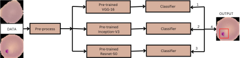
Malaria is a potentially lethal infectious disease caused by the Plasmodium parasite. The transmission of this disease to humans occurs via the bites of Anopheles mosquitoes that are infected with the pathogen. The impact of this disease on the health systems of vulnerable nations, especially in sub-Saharan Africa, is profound and catastrophic. Malaria infiltrates and reproduces within red blood cells, leading to their destruction and the release of harmful substances into the circulation. The parasite’s capacity to adhere to and alter the surface of red blood cells might induce their adhesiveness, impeding blood circulation in crucial organs including the brain and spleen. Hence, it is crucial to employ effective methods for promptly identifying malaria in order to preserve patients’ lives. The primary objective of this project is to establish a very effective model for the early detection of malaria. For the study trials, we utilized malaria pictures depicting both parasitized and uninfected red blood cells. We employed a transfer learning ensemble model, utilizing three distinct pretrained models: VGG16, Resnet-50, and Inception-V3. The models were trained with softmax activation, Adam optimizer with a learning rate of 0.002, categorical-crossentropy loss function, and accuracy matrices. Ultimately, in order to get an improved outcome, we combine all three models and obtain an accuracy rate of 98.6%. We evaluate our model using data that was not used throughout the training and validation procedure.
| Published in | Computational Biology and Bioinformatics (Volume 13, Issue 1) |
| DOI | 10.11648/j.cbb.20251301.12 |
| Page(s) | 17-21 |
| Creative Commons |
This is an Open Access article, distributed under the terms of the Creative Commons Attribution 4.0 International License (http://creativecommons.org/licenses/by/4.0/), which permits unrestricted use, distribution and reproduction in any medium or format, provided the original work is properly cited. |
| Copyright |
Copyright © The Author(s), 2025. Published by Science Publishing Group |
Transfer Learning, Pre-tained Model, VGG, Resnet, Inceptin, Malaria Classification, Ensemble Model
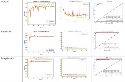
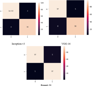
Model | ACC | AUC | Precision | Recall | F1 |
|---|---|---|---|---|---|
Inception-V3 | 97% | 99% | 97% | 97% | 97% |
VGG-16 | 0.93% | 98% | 93% | 93% | 93% |
Resnet-50 | 97% | 99% | 96.8% | 97% | 97% |
Proposed | 98.6% | 99% | 98% | 98% | 98% |
ACC | Accuracy |
AUC | Area Under the Curve |
ROC | Receiver Operating Characteristic |
| [1] | Vijayalakshmi, A., 2020. Deep learning approach to detect malaria from microscopic images. Multimedia Tools and Applications, 79, pp. 15297-15317. |
| [2] | White, N. J., 2017. Malaria parasite clearance. Malaria journal, 16(1), p. 88. |
| [3] | Vandana, T. and Fidock, D. A., 2021. Malaria parasite beats the heat. Nature Microbiology, 6(9), pp. 1105-1107. |
| [4] | Malaria, R. B., 2005. World malaria report 2005. World Health Organization and UNICEF. |
| [5] | Wilson, M. L., 2012. Malaria rapid diagnostic tests. Clinical infectious diseases, 54(11), pp. 1637-1641. |
| [6] | Nadjm, B. and Behrens, R. H., 2012. Malaria: An update for physicians. Infectious Disease Clinics, 26(2), pp. 243-259. |
| [7] | Gollin, D. and Zimmermann, C., 2007. Malaria: Disease impacts and long-run income differences. |
| [8] | Raza, A., Qadri, A. M., Akhtar, I., Samee, N. A. and Alabdulhafith, M., 2023. LogRF: An approach to human pose estimation using skeleton landmarks for physiotherapy fitness exercise correction. IEEE Access. |
| [9] | Raza, A., Munir, K., Almutairi, M., Younas, F., Fareed, M. M. S. and Ahmed, G., 2022. A Novel Approach to Classify Telescopic Sensors Data Using Bidirectional-Gated Recurrent Neural Networks. Applied Sciences, 12(20), p. 10268. |
| [10] | Suraksha, S., Santhosh, C. and Vishwa, B., 2023, January. Classification of Malaria cell images using Deep Learning Approach. In 2023 Third International Conference on Advances in Electrical, Computing, Communication and Sustainable Technologies (ICAECT) (pp. 1-5). IEEE. |
| [11] | Raza, A., Rustam, F., Mallampati, B., Gali, P. and Ashraf, I., 2023. Preventing crimes through gunshots recog- nition using novel feature engineering and meta-learning ap- proach. IEEE Access. |
| [12] | Salamah, U., Sarno, R., Arifin, A. Z., Nugroho, A. S., Rozi, I. E. and Asih, P. B. S., 2019. A robust segmentation for malaria parasite detection of thick blood smear microscopic images. Int. J. Adv. Sci. Eng. Inf. Technol., 9(4), pp. 1450-1459. |
| [13] | Alassaf, A. and Sikkandar, M. Y., 2022. Intelligent Deep Transfer Learning Based Malaria Parasite Detection and Classification Model Using Biomedical Image. Computers, Materials Continua, 72(3). |
| [14] | Sriporn, K., Tsai, C. F., Tsai, C. E. and Wang, P., 2020. Analyzing malaria disease using effective deep learning approach. Diagnostics, 10(10), p. 744. |
| [15] |
JUNEL SOLIS, BioImage informatics II malaria dataset — kaggle, 2023, Available at
https://www.kaggle.com/datasets/junelsolis/bioimage-informatics-ii-malariadataset |
APA Style
Karim, Z., Mahmud, K. B. O., Mahmud, A. A., Al-Amin, A., Chowdhury, T. T. (2025). Classification and Detection of Malaria from Parasitized and Uninfected Red Blood Cell Images Using Transfer Learning Based Ensemble Model. Computational Biology and Bioinformatics, 13(1), 17-21. https://doi.org/10.11648/j.cbb.20251301.12
ACS Style
Karim, Z.; Mahmud, K. B. O.; Mahmud, A. A.; Al-Amin, A.; Chowdhury, T. T. Classification and Detection of Malaria from Parasitized and Uninfected Red Blood Cell Images Using Transfer Learning Based Ensemble Model. Comput. Biol. Bioinform. 2025, 13(1), 17-21. doi: 10.11648/j.cbb.20251301.12
@article{10.11648/j.cbb.20251301.12,
  author = {Zadidul Karim and Kazi Bil Oual Mahmud and Abdullah Al Mahmud and Abdullah Al-Amin and Tanima Tasmin Chowdhury},
  title = {Classification and Detection of Malaria from Parasitized and Uninfected Red Blood Cell Images Using Transfer Learning Based Ensemble Model
},
  journal = {Computational Biology and Bioinformatics},
  volume = {13},
  number = {1},
  pages = {17-21},
  doi = {10.11648/j.cbb.20251301.12},
  url = {https://doi.org/10.11648/j.cbb.20251301.12},
  eprint = {https://article.sciencepublishinggroup.com/pdf/10.11648.j.cbb.20251301.12},
  abstract = {Malaria is a potentially lethal infectious disease caused by the Plasmodium parasite. The transmission of this disease to humans occurs via the bites of Anopheles mosquitoes that are infected with the pathogen. The impact of this disease on the health systems of vulnerable nations, especially in sub-Saharan Africa, is profound and catastrophic. Malaria infiltrates and reproduces within red blood cells, leading to their destruction and the release of harmful substances into the circulation. The parasite’s capacity to adhere to and alter the surface of red blood cells might induce their adhesiveness, impeding blood circulation in crucial organs including the brain and spleen. Hence, it is crucial to employ effective methods for promptly identifying malaria in order to preserve patients’ lives. The primary objective of this project is to establish a very effective model for the early detection of malaria. For the study trials, we utilized malaria pictures depicting both parasitized and uninfected red blood cells. We employed a transfer learning ensemble model, utilizing three distinct pretrained models: VGG16, Resnet-50, and Inception-V3. The models were trained with softmax activation, Adam optimizer with a learning rate of 0.002, categorical-crossentropy loss function, and accuracy matrices. Ultimately, in order to get an improved outcome, we combine all three models and obtain an accuracy rate of 98.6%. We evaluate our model using data that was not used throughout the training and validation procedure. },
 year = {2025}
}
TY  - JOUR
T1  - Classification and Detection of Malaria from Parasitized and Uninfected Red Blood Cell Images Using Transfer Learning Based Ensemble Model

AU  - Zadidul Karim
AU  - Kazi Bil Oual Mahmud
AU  - Abdullah Al Mahmud
AU  - Abdullah Al-Amin
AU  - Tanima Tasmin Chowdhury
Y1  - 2025/07/21
PY  - 2025
N1  - https://doi.org/10.11648/j.cbb.20251301.12
DO  - 10.11648/j.cbb.20251301.12
T2  - Computational Biology and Bioinformatics
JF  - Computational Biology and Bioinformatics
JO  - Computational Biology and Bioinformatics
SP  - 17
EP  - 21
PB  - Science Publishing Group
SN  - 2330-8281
UR  - https://doi.org/10.11648/j.cbb.20251301.12
AB  - Malaria is a potentially lethal infectious disease caused by the Plasmodium parasite. The transmission of this disease to humans occurs via the bites of Anopheles mosquitoes that are infected with the pathogen. The impact of this disease on the health systems of vulnerable nations, especially in sub-Saharan Africa, is profound and catastrophic. Malaria infiltrates and reproduces within red blood cells, leading to their destruction and the release of harmful substances into the circulation. The parasite’s capacity to adhere to and alter the surface of red blood cells might induce their adhesiveness, impeding blood circulation in crucial organs including the brain and spleen. Hence, it is crucial to employ effective methods for promptly identifying malaria in order to preserve patients’ lives. The primary objective of this project is to establish a very effective model for the early detection of malaria. For the study trials, we utilized malaria pictures depicting both parasitized and uninfected red blood cells. We employed a transfer learning ensemble model, utilizing three distinct pretrained models: VGG16, Resnet-50, and Inception-V3. The models were trained with softmax activation, Adam optimizer with a learning rate of 0.002, categorical-crossentropy loss function, and accuracy matrices. Ultimately, in order to get an improved outcome, we combine all three models and obtain an accuracy rate of 98.6%. We evaluate our model using data that was not used throughout the training and validation procedure.
VL  - 13
IS  - 1
ER  -
Electrical & Electronic Engineering, University of Asia Pacific, Dhaka, Bangladesh
Electrical & Electronic Engineering, University of Asia Pacific, Dhaka, Bangladesh
Electrical & Electronic Engineering, University of Asia Pacific, Dhaka, Bangladesh
Electrical & Electronic Engineering, University of Asia Pacific, Dhaka, Bangladesh
Electrical & Electronic Engineering, University of Asia Pacific, Dhaka, Bangladesh
Information