
 ,
Rachad Kondo Moussa,
Borelle Padonou,
Félicité Agbayi,
Fustel Sossa,
Tibaud Ebessa,
David Mawu-Kolo Bokodaho,
Francis Soummonni,
David Dossou,
Xavier Fadonougbo,
Philippe Agbalika,
Murielle Hounkponou,
Djidjoho Joël Arnaud Sonou,
Léopold Houétondji Codjo
Left ventricular (LV) pseudoaneurysm is a contained rupture of the myocardial wall sealed off by the pericardium. It usually complicates myocardial infarction or cardiac surgery, whereas non-coronary forms are exceptional, particularly in young patients, and their etiology is often difficult to establish. We report the case of a 27-year-old woman with an unspecified childhood heart disease, admitted for prolonged pericarditic chest pain associated with fever, cough, weight loss and New York Heart Association (NYHA) class III dyspnea. Examination showed sinus tachycardia at 100 beats per minute, a body mass index of 18.97 kg/m2 and isolated jugular venous distension. The electrocardiogram showed PR segment depression, and the chest radiograph showed cardiomegaly (cardiothoracic ratio 0.60) with a bulge of the left middle cardiac border. Transthoracic echocardiography, performed for the work-up of this chest pain, revealed a communicating LV pseudoaneurysm of the basal anterolateral wall (55 mm in diameter, 28 mm neck, no thrombus) and a moderate fibrinous pericardial effusion. Cardiac magnetic resonance imaging confirmed the diagnosis (sac of 62 × 49 mm) and showed no myocardial enhancement suggestive of myocarditis. Laboratory tests showed multifactorial anemia (hemoglobin 8.3 g/dL) with signs suggestive of hemolysis and a C-reactive protein level of 96 mg/L; blood cultures and the tuberculosis work-up were negative. The patient received high-dose aspirin and weight-adjusted colchicine, as first-line therapy for acute pericarditis, and a blood transfusion, with a favorable course after two weeks of hospitalization. Surgical repair was indicated, but cardiac surgery was not available in the country at that time. While awaiting a state-organized cardiac surgery mission, the patient was lost to follow-up, reportedly had four episodes of decompensated heart failure, and died of sudden circulatory collapse in October 2024, approximately nine months after diagnosis. No autopsy was performed; rupture of the pseudoaneurysm is suspected but remains a hypothesis. The etiology remains undetermined, and a congenital and/or infectious (pericardial) origin is possible but unproven. Echocardiography is the first-line examination, and because of the risk of rupture, surgical repair is recommended even in asymptomatic patients and should be made accessible in low-resource settings.
| Published in | Cardiology and Cardiovascular Research (Volume 10, Issue 4) |
| DOI | 10.11648/j.ccr.20261004.12 |
| Page(s) | 112-117 |
| Creative Commons |
This is an Open Access article, distributed under the terms of the Creative Commons Attribution 4.0 International License (http://creativecommons.org/licenses/by/4.0/), which permits unrestricted use, distribution and reproduction in any medium or format, provided the original work is properly cited. |
| Copyright |
Copyright © The Author(s), 2026. Published by Science Publishing Group |
Pseudoaneurysm, Left Ventricle, Young Adult, Echocardiography, Cardiac Magnetic Resonance, Benin, Pericarditis
CMR | Cardiac Magnetic Resonance |
LV | Left Ventricular |
LVEF | Left Ventricular Ejection Fraction |
NYHA | New York Heart Association |
PsA | Pseudoaneurysm |
| [1] | Frances, C., Romero, A., Grady, D. Left ventricular pseudoaneurysm. J Am Coll Cardiol. 1998, 32(3), 557-561. |
| [2] | Karimanasseri, C., Rahimov, D., Sankisa, D., Nasher, N., Mustafa, M., Allimulla, A., et al. Management and outcomes of post-myocardial infarction left ventricular pseudoaneurysm: a case-level systematic review. Crit Pathw Cardiol. 2026. |
| [3] | Ndiaye, M. B., Ba, F. G., Bodian, M., Diao, M., Kane, A. D., Sarr, S. A., et al. Faux anévrisme du ventricule gauche du sujet jeune: à propos de trois cas [Pseudoaneurysm of the left ventricle in young patients: a propos of three cases]. Ann Cardiol Angeiol (Paris). 2015, 64(4), 300-304. |
| [4] | Alexander, G., Ashwath, M. L. Cardiac pseudoaneurysms: a clinical case series. Eur Heart J Case Rep. 2024, 8(1), ytad636. |
| [5] | Antunes, P. E., Silva, R., Ferrão de Oliveira, J., Antunes, M. J. Left ventricular aneurysms: early and long-term results of two types of repair. Eur J Cardiothorac Surg. 2005, 27(2), 210-215. |
| [6] | Konen, E., Merchant, N., Gutierrez, C., Provost, Y., Mickleborough, L., Paul, N. S., et al. True versus false left ventricular aneurysm: differentiation with MR imaging—initial experience. Radiology. 2005, 236(1), 65-70. |
| [7] | Jafari Afshar, E., Tayebi, A., Samimisedeh, P., Shahnavaz, V., Madady, A., Rastad, H., et al. Clinical and imaging findings in 504 cases with left ventricular pseudoaneurysm following myocardial infarction: a comprehensive systematic review and meta-analysis. Cardiol Res Pract. 2026, 9208154. |
| [8] | Schulz-Menger, J., Collini, V., Gröschel, J., Adler, Y., Brucato, A., Christian, V., et al. 2025 ESC Guidelines for the management of myocarditis and pericarditis. Eur Heart J. 2025, 46(40), 3952-4041. |
| [9] | Effiom, V. B., Alassiri, A. K., Femi-Lawal, V., Genda, E., Ibekwe, J. L., Anyinkeng, A. B. S., et al. Challenges to open-heart surgery in sub-Saharan Africa: a narrative review. Braz J Cardiovasc Surg. 2026, 41(1), e20240351. |
| [10] | Mackenzie, J. W., Lemole, G. M. Pseudoaneurysm of the left ventricle. Tex Heart Inst J. 1994, 21(4), 296-301. |
| [11] | Prêtre, R., Linka, A., Jenni, R., Turina, M. I. Surgical treatment of acquired left ventricular pseudoaneurysms. Ann Thorac Surg. 2000, 70(2), 553-557. |
| [12] | Brown, S. L., Gropler, R. J., Harris, K. M. Distinguishing left ventricular aneurysm from pseudoaneurysm: a review of the literature. Chest. 1997, 111(5), 1403-1409. |
| [13] | Benouaich, V., Marcheix, B., Grunenwald, E., et al. Complications tardives d’un faux anévrisme du ventricule gauche: infection du thrombus et péricardite purulente [Late complications of a left ventricular false aneurysm: thrombus infection and purulent pericarditis]. Ann Cardiol Angeiol (Paris). 2007, 56(6), 316-318. |
| [14] | Banar, S., Tolouietabar, Y., Shojaei, M. A., Khesali, H., Pouraliakbar, H., Hosseini, S. A simplified approach to managing a complex infected left ventricular pseudoaneurysm. J Cardiothorac Surg. 2024, 19, 652. |
| [15] | Chraibi, S., Beghetti, M., El Hattaoui, M., Fadouach, S., Kalangos, A., Mehadji, B. A., et al. Faux anévrisme après ligature d'un canal artériel opéré avec succès [False aneurysm after ligation of a successfully operated ductus arteriosus]. Ann Cardiol Angeiol (Paris). 2003, 52(3), 188-190. |
| [16] | Haine, S. E., Paelinck, B. P., Vrints, C. J. Post-traumatic focal true left ventricular aneurysm. Heart. 2004, 90(9), 1009. |
| [17] | Dada, M., Noyez, L., Verheugt, F. W. A., Brouwer, R. M. H. J. Delayed diagnosis of a post-traumatic ventricular aneurysm? Interact Cardiovasc Thorac Surg. 2003, 2(1), 84-86. |
| [18] | Zouhri, O., Hajji, Y., Tazi Mezalek, A., Cheikhaoui, Y. Traumatic left ventricular apical pseudoaneurysm in a young woman: a rare and life-threatening condition. Eur Heart J Case Rep. 2026, 10(6), ytag401. |
APA Style
Abatti, C. G., Moussa, R. K., Padonou, B., Agbayi, F., Sossa, F., et al. (2026). Non-Coronary Left Ventricular Pseudoaneurysm Diagnosed During the Evaluation of Pericarditic Chest Pain in a Young Woman in Benin: A Case Report. Cardiology and Cardiovascular Research, 10(4), 112-117. https://doi.org/10.11648/j.ccr.20261004.12
ACS Style
Abatti, C. G.; Moussa, R. K.; Padonou, B.; Agbayi, F.; Sossa, F., et al. Non-Coronary Left Ventricular Pseudoaneurysm Diagnosed During the Evaluation of Pericarditic Chest Pain in a Young Woman in Benin: A Case Report. Cardiol. Cardiovasc. Res. 2026, 10(4), 112-117. doi: 10.11648/j.ccr.20261004.12
AMA Style
Abatti CG, Moussa RK, Padonou B, Agbayi F, Sossa F, et al. Non-Coronary Left Ventricular Pseudoaneurysm Diagnosed During the Evaluation of Pericarditic Chest Pain in a Young Woman in Benin: A Case Report. Cardiol Cardiovasc Res. 2026;10(4):112-117. doi: 10.11648/j.ccr.20261004.12
@article{10.11648/j.ccr.20261004.12,
  author = {Carmel Gloria Abatti and Rachad Kondo Moussa and Borelle Padonou and Félicité Agbayi and Fustel Sossa and Tibaud Ebessa and David Mawu-Kolo Bokodaho and Francis Soummonni and David Dossou and Xavier Fadonougbo and Philippe Agbalika and Murielle Hounkponou and Djidjoho Joël Arnaud Sonou and Léopold Houétondji Codjo},
  title = {Non-Coronary Left Ventricular Pseudoaneurysm Diagnosed During the Evaluation of Pericarditic Chest Pain in a Young Woman in Benin: A Case Report},
  journal = {Cardiology and Cardiovascular Research},
  volume = {10},
  number = {4},
  pages = {112-117},
  doi = {10.11648/j.ccr.20261004.12},
  url = {https://doi.org/10.11648/j.ccr.20261004.12},
  eprint = {https://article.sciencepublishinggroup.com/pdf/10.11648.j.ccr.20261004.12},
  abstract = {Left ventricular (LV) pseudoaneurysm is a contained rupture of the myocardial wall sealed off by the pericardium. It usually complicates myocardial infarction or cardiac surgery, whereas non-coronary forms are exceptional, particularly in young patients, and their etiology is often difficult to establish. We report the case of a 27-year-old woman with an unspecified childhood heart disease, admitted for prolonged pericarditic chest pain associated with fever, cough, weight loss and New York Heart Association (NYHA) class III dyspnea. Examination showed sinus tachycardia at 100 beats per minute, a body mass index of 18.97 kg/m2 and isolated jugular venous distension. The electrocardiogram showed PR segment depression, and the chest radiograph showed cardiomegaly (cardiothoracic ratio 0.60) with a bulge of the left middle cardiac border. Transthoracic echocardiography, performed for the work-up of this chest pain, revealed a communicating LV pseudoaneurysm of the basal anterolateral wall (55 mm in diameter, 28 mm neck, no thrombus) and a moderate fibrinous pericardial effusion. Cardiac magnetic resonance imaging confirmed the diagnosis (sac of 62 × 49 mm) and showed no myocardial enhancement suggestive of myocarditis. Laboratory tests showed multifactorial anemia (hemoglobin 8.3 g/dL) with signs suggestive of hemolysis and a C-reactive protein level of 96 mg/L; blood cultures and the tuberculosis work-up were negative. The patient received high-dose aspirin and weight-adjusted colchicine, as first-line therapy for acute pericarditis, and a blood transfusion, with a favorable course after two weeks of hospitalization. Surgical repair was indicated, but cardiac surgery was not available in the country at that time. While awaiting a state-organized cardiac surgery mission, the patient was lost to follow-up, reportedly had four episodes of decompensated heart failure, and died of sudden circulatory collapse in October 2024, approximately nine months after diagnosis. No autopsy was performed; rupture of the pseudoaneurysm is suspected but remains a hypothesis. The etiology remains undetermined, and a congenital and/or infectious (pericardial) origin is possible but unproven. Echocardiography is the first-line examination, and because of the risk of rupture, surgical repair is recommended even in asymptomatic patients and should be made accessible in low-resource settings.},
 year = {2026}
}
TY  - JOUR
T1  - Non-Coronary Left Ventricular Pseudoaneurysm Diagnosed During the Evaluation of Pericarditic Chest Pain in a Young Woman in Benin: A Case Report
AU  - Carmel Gloria Abatti
AU  - Rachad Kondo Moussa
AU  - Borelle Padonou
AU  - Félicité Agbayi
AU  - Fustel Sossa
AU  - Tibaud Ebessa
AU  - David Mawu-Kolo Bokodaho
AU  - Francis Soummonni
AU  - David Dossou
AU  - Xavier Fadonougbo
AU  - Philippe Agbalika
AU  - Murielle Hounkponou
AU  - Djidjoho Joël Arnaud Sonou
AU  - Léopold Houétondji Codjo
Y1  - 2026/11/12
PY  - 2026
N1  - https://doi.org/10.11648/j.ccr.20261004.12
DO  - 10.11648/j.ccr.20261004.12
T2  - Cardiology and Cardiovascular Research
JF  - Cardiology and Cardiovascular Research
JO  - Cardiology and Cardiovascular Research
SP  - 112
EP  - 117
PB  - Science Publishing Group
SN  - 2578-8914
UR  - https://doi.org/10.11648/j.ccr.20261004.12
AB  - Left ventricular (LV) pseudoaneurysm is a contained rupture of the myocardial wall sealed off by the pericardium. It usually complicates myocardial infarction or cardiac surgery, whereas non-coronary forms are exceptional, particularly in young patients, and their etiology is often difficult to establish. We report the case of a 27-year-old woman with an unspecified childhood heart disease, admitted for prolonged pericarditic chest pain associated with fever, cough, weight loss and New York Heart Association (NYHA) class III dyspnea. Examination showed sinus tachycardia at 100 beats per minute, a body mass index of 18.97 kg/m2 and isolated jugular venous distension. The electrocardiogram showed PR segment depression, and the chest radiograph showed cardiomegaly (cardiothoracic ratio 0.60) with a bulge of the left middle cardiac border. Transthoracic echocardiography, performed for the work-up of this chest pain, revealed a communicating LV pseudoaneurysm of the basal anterolateral wall (55 mm in diameter, 28 mm neck, no thrombus) and a moderate fibrinous pericardial effusion. Cardiac magnetic resonance imaging confirmed the diagnosis (sac of 62 × 49 mm) and showed no myocardial enhancement suggestive of myocarditis. Laboratory tests showed multifactorial anemia (hemoglobin 8.3 g/dL) with signs suggestive of hemolysis and a C-reactive protein level of 96 mg/L; blood cultures and the tuberculosis work-up were negative. The patient received high-dose aspirin and weight-adjusted colchicine, as first-line therapy for acute pericarditis, and a blood transfusion, with a favorable course after two weeks of hospitalization. Surgical repair was indicated, but cardiac surgery was not available in the country at that time. While awaiting a state-organized cardiac surgery mission, the patient was lost to follow-up, reportedly had four episodes of decompensated heart failure, and died of sudden circulatory collapse in October 2024, approximately nine months after diagnosis. No autopsy was performed; rupture of the pseudoaneurysm is suspected but remains a hypothesis. The etiology remains undetermined, and a congenital and/or infectious (pericardial) origin is possible but unproven. Echocardiography is the first-line examination, and because of the risk of rupture, surgical repair is recommended even in asymptomatic patients and should be made accessible in low-resource settings.
VL  - 10
IS  - 4
ER  -
University Cardiology Clinic, National Teaching Hospital Hubert Koutoukou Maga of Cotonou, Cotonou, Benin
University Cardiology Clinic, National Teaching Hospital Hubert Koutoukou Maga of Cotonou, Cotonou, Benin
University Cardiology Clinic, National Teaching Hospital Hubert Koutoukou Maga of Cotonou, Cotonou, Benin
University Cardiology Clinic, National Teaching Hospital Hubert Koutoukou Maga of Cotonou, Cotonou, Benin
University Cardiology Clinic, National Teaching Hospital Hubert Koutoukou Maga of Cotonou, Cotonou, Benin
University Cardiology Clinic, National Teaching Hospital Hubert Koutoukou Maga of Cotonou, Cotonou, Benin
University Cardiology Clinic, National Teaching Hospital Hubert Koutoukou Maga of Cotonou, Cotonou, Benin
Cardiology Department, Calavi International Hospital, Abomey-Calavi, Benin
University Cardiology Clinic, National Teaching Hospital Hubert Koutoukou Maga of Cotonou, Cotonou, Benin
University Cardiology Clinic, National Teaching Hospital Hubert Koutoukou Maga of Cotonou, Cotonou, Benin
University Cardiology Clinic, National Teaching Hospital Hubert Koutoukou Maga of Cotonou, Cotonou, Benin
University Cardiology Clinic, National Teaching Hospital Hubert Koutoukou Maga of Cotonou, Cotonou, Benin
University Cardiology Clinic, National Teaching Hospital Hubert Koutoukou Maga of Cotonou, Cotonou, Benin
Cardiology Department, Calavi International Hospital, Abomey-Calavi, Benin
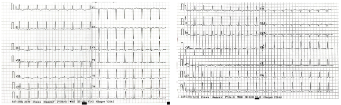
Figure 1. Twelve-lead electrocardiogram showing regular sinus tachycardia at 100 beats per minute and PR segment depression (A and B: two tracings).
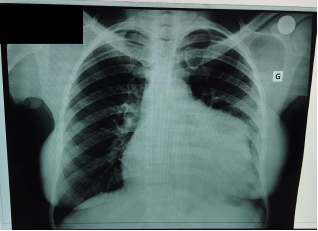
Figure 2. Posteroanterior chest radiograph showing cardiomegaly (cardiothoracic ratio 0.60) and a bulge of the left middle cardiac border, consistent with the pseudoaneurysm sac. G: left side.
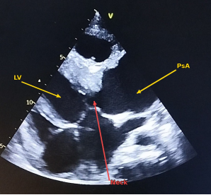
Figure 3. Transthoracic echocardiography, apical four-chamber view, showing the left ventricular pseudoaneurysm (PsA, arrow) communicating with the left ventricle (LV) through a 28-mm neck (Neck, red arrow); maximal sac diameter 55 mm. LV: left ventricle; PsA: pseudoaneurysm.
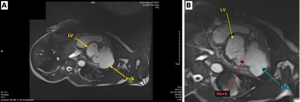
Figure 4. Cardiac magnetic resonance axial cine images showing the left ventricular pseudoaneurysm. (A) Pseudoaneurysm (PsA, arrow; 62 × 49 mm) attached to the left ventricle (LV). (B) Magnified view showing the neck (red arrow) connecting the left ventricle (yellow arrow) to the pseudoaneurysm sac (blue arrow). LV: left ventricle; PsA: pseudoaneurysm.

Information


 Share This Article
 Twitter
 Linked In
 Facebook

 Copy
 Download