Research Article | | Peer-Reviewed

A Diagnostic Challenge: Giant Cell Arteritis Presenting as Fever of Unknown Origin

Received: 25 August 2026     Accepted: 19 October 2026     Published: 16 November 2026
Views:       Downloads:
Abstract

Background: Fever of unknown origin (FUO) in elderly patients presents a major diagnostic challenge due to its broad differential diagnosis and frequently nonspecific clinical manifestations. Although infections, malignancies, and connective tissue diseases are common etiologies, atypical presentations of vasculitis remain an underrecognized cause of persistent fever. Giant cell arteritis classically presents with headache, jaw claudication, and visual symptoms; however, a subset of patients may present solely with constitutional symptoms, leading to delayed diagnosis and increased risk of irreversible complications. We report a rare case of biopsy-confirmed non-giant-cell temporal arteritis presenting as prolonged FUO without cranial symptoms despite markedly elevated inflammatory markers and an extensive negative infectious and malignancy evaluation. Case Report: An 85-year-old female presented with several weeks of low-grade fever and fatigue, without associated headache, jaw claudication, or visual deficits. Initial laboratory workup was pertinent for leukocytosis and markedly elevated inflammatory markers (ESR/CRP). Extensive workup, including blood cultures, urinalysis, chest and abdominal CT imaging, transthoracic echocardiography, tuberculosis testing, and autoimmune serologies, was unrevealing. Multiple courses of empiric broad-spectrum antibiotics provided no symptom relief. Due to persistent elevation of ESR and CRP, and in the absence of an identifiable source, an autoimmune etiology such as temporal arteritis was higher on the differential. A temporal artery biopsy was subsequently performed and showed features consistent with non-giant cell arteritis. High-dose corticosteroid therapy was initiated, leading to rapid resolution of her fever and constitutional symptoms, as well as normalization of white blood cell count. Conclusion: This case highlights the importance of considering temporal arteritis in elderly patients with persistent fever of unknown origin, markedly elevated ESR and CRP, and unrevealing infectious or malignant workup, even in the absence of classic cranial manifestations. It also emphasizes the diagnostic value of temporal artery biopsy in atypical presentations and expands the limited literature describing biopsy-confirmed non-giant-cell arteritis presenting primarily as FUO. Key clinical message: Non-giant cell temporal arteritis should be considered in elderly patients with fever of unknown origin and markedly elevated inflammatory markers even in the absence of classic cranial symptoms. Early recognition and prompt corticosteroid therapy can lead to rapid clinical improvement and prevent irreversible complications such as vision loss and vascular ischemia.

Published in Clinical Medicine Research (Volume 15, Issue 5)
DOI 10.11648/j.cmr.20261505.12
Page(s) 75-80
Creative Commons

This is an Open Access article, distributed under the terms of the Creative Commons Attribution 4.0 International License (http://creativecommons.org/licenses/by/4.0/), which permits unrestricted use, distribution and reproduction in any medium or format, provided the original work is properly cited.

Copyright

Copyright © The Author(s), 2026. Published by Science Publishing Group

Keywords

Fever of Unknown Origin, Non-Giant Cell Arteritis, Arteritis, Temporal Arteritis, Autoimmune Conditions

1. Introduction
Fever of unknown encompasses a broad differential diagnosis that includes infections, malignancies, autoimmune disorders, connective tissue diseases, and vasculitides . In older adults, infectious and neoplastic causes are frequently prioritized during evaluation; however, atypical inflammatory disorders may present with isolated constitutional symptoms and remain diagnostically elusive. Among these, giant cell arteritis (GCA) is an important but often underrecognized cause of FUO, particularly when classic cranial manifestations are absent.
Temporal arteritis is a subtype of giant cell arteritis, which is an autoimmune inflammation of medium and large vessels, resulting in vascular wall thickening and intima proliferation, narrowing of the lumen, and ultimately tissue ischemia. Temporal arteritis specifically refers to inflammation of the temporal artery and other cranial arteries. The classic symptoms include headache, scalp tenderness, jaw discomfort and/or pain, weight loss, and fever . However, this condition can also present non-specifically, such as fevers, lethargy, and general malaise. Around 30% of cases can present with neurological symptoms including peripheral neuropathies and 15% can present with fever as the sole hallmark manifestation .
Temporal arteritis is an ophthalmologic emergency due to its high risk of rapidly progressive vision loss; 14-70% of patients with giant cell arteritis had eye involvement and 20% had partial or total permanent vision loss in one or both eyes . The most common cause of this vision loss is due to the involvement of the arteritic anterior ischemic optic neuropathy, and this accounts for 81.2% of permanent vision loss cases due to giant cell arteritis . Along with vision loss, temporal arteritis can also cause damage to blood vessels, leading to higher risk of aneurysms or stroke/transient ischemic attacks.
Atypical presentations of autoimmune vasculitis have been increasingly recognized in the literature as a significant source of diagnostic uncertainty. Several reports describe patients presenting primarily with constitutional symptoms without the hallmark cranial features traditionally associated with arteritis . In a large cohort study, Stagnaro et al. found that 45 of 180 GCA patients presented with FUO and elevated ESR/CRP without otherwise justified inflammation. After exclusion of infection, malignancy, and hematologic disease, temporal artery biopsy was positive in 52%, and diagnosis was delayed longer than in patients with classic cranial symptoms . Temporal artery biopsy should be considered in patients older than 50 with persistent fevers and unexplained elevation of ESR or CRP after reasonable exclusion of infection and malignancy, even when headache, jaw claudication, scalp tenderness, visual symptoms, or temporal artery abnormalities are absent. Biopsy remains valuable in this setting because abnormal specimens may show mononuclear or granulomatous inflammation, with or without giant cells, and negative cranial symptoms should not preclude evaluation when the inflammatory phenotype remains otherwise unexplained.
Prior literature shows that up to one-third of patients may initially lack classic symptoms, resulting in delays in diagnosis and treatment that increase the risk of irreversible complications. By aligning with this body of evidence, the present case contributes to a broader understanding of the variable clinical spectrum of autoimmune vasculitis and reinforces the need for clinicians to consider temporal arteritis in the absence of typical clinical findings.
2. Case Presentation
An 85-year-old female with a significant medical history of congestive heart failure with preserved ejection fraction, chronic kidney disease, and chronic obstructive pulmonary disease presented to the hospital with five days of progressive altered mentation. Her son, who was present on admission, reported that she had appeared more confused, as well as increased generalized weakness and anorexia over this period. The patient had been largely minimally interactive throughout this five day period prior to/during admission and thus the son was the primary historian. The patient was febrile with a temperature of 103.1F. Initial laboratory tests, as seen in Table 1, were pertinent for a white blood cell count of 20,000 and mildly elevated liver enzymes.
Table 1. Laboratory Values.

Lab

Value

Normal Range

Units

Glucose

109

70-100

mg/dL

Sodium

135

136-145

meq/L

Potassium

4.9

3.5-5.3

meq/L

BUN

19

8-27

mg/dL

Creatinine

1.27

0.60-1.60

mg/dL

Alkaline phosphatase

152

34-130

U/L

Aspartate aminotransferase

49

0-40

U/L

Alanine aminotransferase

27

0-68

U/L

Bilirubin, total

1

0-1.1

mg/dL

Hemoglobin

12

12-16

g/dL

White blood cell count

20

4.5-11

103/μL

Table shows initial laboratory values for the patient on admission and normal ranges.
Given concern for severe infection, including possible bacteremia or meningitis, broad-spectrum antibiotics were initiated empirically upon admission. A lumbar puncture was performed in the emergency department on hospital day 1, and cerebrospinal fluid studies, blood cultures, and meningitis panels were ultimately negative. Additional early infectious evaluation, including urinalysis, rapid streptococcal testing, respiratory viral panel, and hepatitis serologies, was unrevealing. Because of the elevated liver enzymes, a right upper quadrant ultrasound was obtained but showed no acute hepatobiliary pathology CT imaging of the abdomen, pelvis, and head, including evaluation of the paranasal sinuses, did not identify an infectious source.
Due to persistent fevers and leukocytosis despite antimicrobial therapy, infectious disease specialists were consulted early during the hospitalization, prompting expansion of the infectious workup over the following several days. Serologic and microbiologic testing for HIV, Cryptococcus, Typhus, Bartonella, Brucella, Coxiella, and Histoplasma were all negative. Treponemal testing was not performed due to absence of hallmark neurosyphilis signs including chronic dorsalis tabes and the acute nature of the febrile state of the patient. CT abd/pelvis and chest with contrast also did not demonstrate lesions suspicious for gummas. A transthoracic echocardiogram demonstrated no evidence of infective endocarditis. No findings concerning for severe acute aortitis were observed. Aortic root diameter was under 4cm . Despite broad-spectrum antibiotics, antipyretics, and extensive infectious evaluation, the patient continued to have recurrent fevers and worsening inflammatory markers without an identifiable source.
Given the persistent constitutional symptoms and leukocytosis, hematologic malignancy also remained high on the differential diagnosis. Peripheral smear showed no evidence of blasts or other malignant features. On hospitalization day 9 a tagged white blood cell scan was also obtained to evaluate for occult infectious or inflammatory foci but yielded inconclusive findings. A bone marrow biopsy was subsequently performed on hospitalization day 10 to evaluate for lymphoma and leukemia, which were ultimately excluded. Hemophagocytic lymphohistiocytosis was briefly considered because of a ferritin level greater than 1,000 ng/mL (Table 2), but the overall clinical picture and additional laboratory findings were not supportive.
Table 2. Laboratory Values.

Lab

Value

Normal Range

Units

Erythrocyte sedimentation rate

>120

0-30

mm/h

C-reactive protein

466.3

0-3.2

mg/L

Ferritin

1202.86

8-252

ng/mL

Table shows inflammatory marker lab values obtained later during the hospitalization along with the normal ranges for the lab values.
Despite these extensive investigations, the patient continued to have recurrent fevers and elevated white blood cell counts. Although she did not report headache, scalp tenderness, jaw claudication, visual disturbances, or other classic cranial manifestations of temporal arteritis, an autoimmune or vasculitic process became increasingly concerning because of the markedly elevated inflammatory markers, with ESR greater than 120 mm/hr and CRP of 466.3 mg/L on admission (Table 2). Given the patient’s age, persistent fever of unknown origin, generalized malaise, and profound inflammatory marker elevation disproportionate to the otherwise negative workup, temporal arteritis emerged as a leading diagnostic consideration despite the absence of cranial symptoms. Additionally the absence of marked eosinophilia, history of asthma, acute epistaxis/nasal mucosal findings and associated pre-renal azotemia all precluded from considering c-anca/p-anca or complement mediated serologies before proceeding with a temporal biopsy on hospitalization day 14 .
Empiric high-dose Prednisone therapy at 60 mg daily was started due to concern for possible vasculitis and the risk of delayed treatment. Patient then underwent temporal artery biopsy. Within days of corticosteroid initiation, the patient demonstrated rapid clinical improvement with resolution of fevers, normalization of the white blood cell count, and improvement in mentation and generalized weakness. The temporal artery biopsy later revealed findings consistent with active giant-cell arteritis, confirming the diagnosis.
3. Discussion
3.1. Diagnosis
According to the American College of Rheumatology (ACR), the diagnostic criteria for giant cell arteritis involves three of the five criteria listed as follows : I. Onset of symptoms occur at age > 50 years. II. Onset of new headache. III. Temporal artery tenderness or decreased pulsation. IV. Elevated erythrocyte sedimentation rate (ESR) (> 50 mm/h). V. Histologic evidence of necrotizing arteritis in temporal artery with mononuclear cell infiltration or granulomatous inflammation usually with multinucleated giant cells. The EULAR 2022 criteria for diagnosis of GCA requires a score of six with the following features/symptoms having a score of at least two points: morning stiffness of neck, scalp tenderness, sudden vision loss, new onset temporal headache, jaw/tongue claudication, physically obvious temporal artery tenderness/firmness/pulse weakness, ESR max > 50 mm/hr or CRP > 10mg/L (prior to treatment), positive biopsy/halo sign on ultrasound, bilateral axillary artery inflammation present on imaging, and FDG uptake throughout aorta. Of these our patient would have had a pre-test score of 3 given the ESR and CRP alone. The positive histopathology finding would give the patient a score of 8 all together greater than the 5 that is needed to classify pathology clinically as Giant Cell Arteritis .
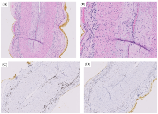
The temporal artery biopsy criterion itself is highly specific for giant cell arteritis, and thus is the gold standard for diagnosis. Temporal artery biopsies have a specificity of 100% and a sensitivity of 77%, indicating that positive pathology results can accurately confirm the presence of arteritis but negative results cannot exclude this diagnosis. The false negative rates of the biopsy, which range from 9% to 61%, can be attributed to timing of biopsy, the length of the specimen obtained, and “skip lesions” within the artery. This can be minimized by obtaining the biopsy within one week of starting steroids and attaining a specimen that is 20-30 mm in length . Histologically, temporal artery biopsy showing infiltration of inflammatory cells, including lymphocytes and macrophages, in the three layers of the artery is confirmatory for arteritis. Giant cells can be seen in the histology for arteritis but they are not required to make a diagnosis of temporal arteritis . In our patient, multiple tissue sections were examined histologically, all of which were 20 mm in length, to reduce the risk of sampling error associated with skip lesions.
Although most cases of temporal arteritis fall under the spectrum of classically presenting giant cell arteritis, a subset of patients present with the characteristic multinucleated giant cells absent on histopathology. These non-classic variants often present with similar clinical symptoms, elevations in inflammatory markers, and risks of ischemic complications as classic GCA, but may demonstrate more subtle granulomatous changes with lymphocytic and macrophage-predominant infiltration. Because these cases rely on recognizing the clinical picture and overall pattern of inflammatory changes, they can pose a greater diagnostic challenge.
Importantly, non-classically presenting GCA must be distinguished histologically from other vasculitides and infectious arteritides. Polyarteritis nodosa and ANCA-associated vasculitides may demonstrate necrotizing inflammation of medium-sized vessels but more commonly exhibit fibrinoid necrosis, neutrophilic infiltration, and systemic multiorgan manifestations, findings that were not present in this case . Infectious arteritides, including syphilitic, fungal, or bacterial arteritis, may also produce vascular inflammation but are typically associated with identifiable organisms, suppurative inflammation, or positive microbiologic studies. In our patient, extensive infectious evaluation was unrevealing, and histopathologic examination did not demonstrate features suggestive of infectious arteritis. Thus, specific microbiology studies were not pursued on tissue biopsy.
Figure 1. Histology of Temporal Artery Biopsy Specimen.
Immunostains, such as CD3 and CD68, can be used to support the diagnosis of temporal arteritis by highlighting inflammatory cells within the arterial wall. CD3 is a T-cell marker that identifies T lymphocytes, which are the predominant inflammatory cells in temporal arteritis. The immunostain in Figure 1 shows marked infiltration of CD3 T lymphocytes in the intima, media, and adventitia of the affected artery. CD68 is a macrophage marker that identifies macrophages involved in granuloma formation and tissue destruction. The immunostain often shows macrophages concentrated near the internal elastic lamina as shown in Figure 1D. Macrophages can also be seen within giant cells when they are present .
Slide shows focal inflammation in intima, media, and adventitia of the temporal artery (A) (B) that is consistent with active arteritis. No granulomatous inflammation, fibrinoid necrosis, intimal hyperplasia were observed. No giant cells were identified. Immunostains of CD3 highlight lymphocytes (C) and CD68 highlight macrophages (D).
3.2. Treatment
High-dose corticosteroids are the primary treatment for patients with suspected temporal arteritis and should be started immediately, even before a biopsy confirms the diagnosis. Prompt initiation of steroids is crucial to prevent complications, such as vision loss. However, it is important to recognize that steroids can mask symptoms like rashes and fever, potentially delaying the diagnosis of other conditions. Therefore, thorough clinical evaluation is necessary before starting steroid therapy to ensure appropriate management .
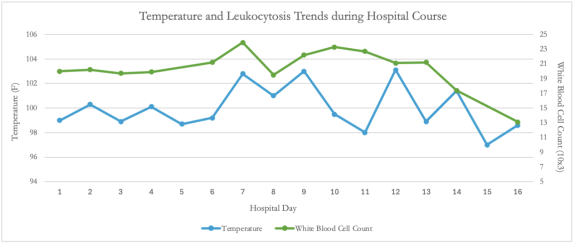
Steroid dosing for temporal arteritis typically starts at 1 mg/kg of body weight daily . Clinically, most patients experience rapid improvement upon the initiation of steroids, as seen in Figure 2, likely due to the suppression of IL-6 and the acute phase response. Steroid tapering can generally begin two to four weeks after treatment initiation, once the patient shows clinical improvement and laboratory markers, such as ESR and CRP, normalize . During the first year of treatment, close monitoring by a Rheumatologist is essential, with monthly checks of inflammatory markers. As the patient improves, the frequency of monitoring decreases, with bimonthly checks in the second year and three- to six-month intervals for long-term follow up. The standard tapering protocol involves reducing the steroid taper dose by 10-20% each month, with a slower taper (1 mg/month) once the dose drops below 10 mg/day . Patients may experience flares, characterized by symptom relapse and elevated inflammatory markers. In cases of severe flare, re-induction with the initial dose of 1 mg/kg/day of steroids is recommended. For mild flares, a 10-20% increase in the steroid dose is typically sufficient to control symptoms . Newer biologic therapies such as the IL-6 inhibitor tocilizumab have become more popularly used as adjunctive therapies to steroids with some evidence in industry trials of decreased remission rates. Dosing was initially approved at 6mg/kg weekly subcutaneous injection administration however additional studies have recommended up to 8mg/kg. Around 42% of patients in the GiACTA trial experienced two-year drug free remission after combined therapy with tocilizumab. No clear standard of tocilizumab duration is recommended; cessation is dependent on provider-patient individualized monitoring. However a subset of patients under remission still experienced flares and diagnosing remission is a difficult multimodal process with no clear singular biomarker panel .
Figure 2. Fever and White Blood Cell Count Trends.
High dose steroids were initiated on hospital day thirteen, at which point, the patient stopped spiking fevers and white blood cell count started to decline. Fevers were unresponsive to antibiotics as seen below, thereby suggesting that the fevers did not occur due to infectious sources.
Long-term steroid use exposes patients to a range of potential adverse effects, including hypertension, hyperglycemia, hypercholesterolemia, osteoporosis, avascular necrosis, glaucoma, peptic ulcer disease, and depression, among others . As such, patients require close monitoring and proactive strategies to mitigate these risks. To reduce the likelihood of gastroduodenal irritations associated with prolonged steroid therapy, starting proton pump inhibitors can be considered based on patient history and concomitant use of tocilizumab which has its own GI perforation risk . To counteract bone loss, supplementation with Vitamin D and Calcium should be initiated, and patients should be encouraged to start weight-bearing exercises or even physical therapy to maintain muscle strength. Additionally, prophylaxis against Pneumocystis Jiroveci pneumonia can be considered based on patient risk factors and clinical symptoms . Ulcer prophylaxis with PPI in the setting of steroid therapy alone is not recommended but concomitant non-selective NSAID use is an indication. . Careful and comprehensive management of patients on long-term high-dose steroid therapy is essential to ensuring effective treatment while minimizing the risk of complications.
4. Conclusion
This case highlights the importance of systematic workup in patients with fever of unknown origin, along with the necessity of maintaining broad diagnosis differentials despite atypical symptoms. Temporal arteritis should be strongly considered in patients over 50 years of age presenting with unexplained fever, elevated inflammatory markers, and non-specific symptoms. Infectious and hematologic/neoplastic etiologies should be ruled out early on, however, autoimmune processes must remain in the differentials when treating patients with similarly presenting symptoms. Early recognition and treatment with high-dose steroids can result in significant clinical improvement while mitigating the risk of severe complications such as irreversible vision loss, aneurysms, and strokes.
Abbreviations

FUO

Fever of Uknown Origin

GCA

Giant Cell Arteritis

Author Contributions
Vishnu Vandana Dubakula: Conceptualization, Formal Analysis, Investigation, Methodology, Visualization, Writing – original draft, Writing – review & editing
Sai Deepak Muthyala: Writing – review & editing
Megan Kudlak: Visualization, Writing – original draft, Writing – review & editing
Sameera Vohra: Writing – review & editing
Tauqeer Yousuf: Writing – review & editing
Conflicts of Interest
The authors declare no conflicts of interest.
References
[1] Jiang Z, Ji H, Dong J. Temporal artery biopsy for suspected giant cell arteritis: a mini review. Indian J Ophthalmol. 2023; 71(10): 3299–3304.
[2] Kumar V, Abbas AK, Aster JC. Robbins and Cotran Pathologic Basis of Disease. 10th ed. Philadelphia: Elsevier; 2020. p. 511–513.
[3] Weyand CM, Goronzy JJ. Giant-cell arteritis and polymyalgia rheumatica. N Engl J Med. 2014; 371(1): 52–63.
[4] Williams J, Bellamy R. Fever of unknown origin. Clin Med (Lond). 2008; 8(5): 526–530.
[5] Zhou L, Luneau K, Weyand CM, Biousse V, Newman NJ, Grossniklaus HE. Clinicopathologic correlations in giant cell arteritis: a retrospective study of 107 cases. Ophthalmology. 2009; 116(8): 1574–1580.
[6] Lipsky BA, Weitz DC. Corticosteroid-induced adverse events in adults: frequency, screening, and prevention. Drugs. 2010; 70(10): 1329–1345.
[7] Salvarani C, Cantini F, Hunder GG. Polymyalgia rheumatica and giant-cell arteritis. Lancet. 2008; 372(9634): 234–245.
[8] Stagnaro, Chiara, et al. “Misdiagnosis of Giant Cell Arteritis Presenting as Fever of Unknown Origin.” ACR Meeting Abstracts, American College of Rheumatology, 2014.
[9] Hansen ME, Springer J, Byram K. Diagnostic and Therapeutic Challenges of Vasculitis. Can J Cardiol. 2026; 38(5): 623-633.
[10] Kronbichler A, Bajema IM, Bruchfeld A, Kirsztajn GM, Stone JH. Review Diagnosis and management of ANCA-associated vasculitis. Lancet. 2023; 403(10427): 683-698.
[11] Ponte C, Grayson PC, Robson JC, et al. 2022 American College of Rheumatology / EULAR classification criteria for giant cell arteritis. 2022: 1647-1653.
[12] Jennette, J. Charles, et al. “2012 Revised International Chapel Hill Consensus Conference Nomenclature of Vasculitides.” Arthritis & Rheumatism, vol. 65, no. 1, 2013, pp. 1–11. Wiley Online Library.
[13] Ford JA, Gewurz D, Gewurz-singer O. Tocilizumab in giant cell arteritis: an update for the clinician. 2023: 135-140.
[14] Caplan AS, Mecoli CA, Micheletti RG. Prophylaxis Against Pneumocystis Pneumonia. JAMA. 2023; 330(19): 1908–1909.
[15] Ford JA, Gewurz D, Gewurz-singer O. Tocilizumab in giant cell arteritis: an update for the clinician. 2023: 135-140.
Cite This Article
  • APA Style

    Dubakula, V. V., Muthyala, S. D., Kudlak, M., Vohra, S., Yousuf, T. (2026). A Diagnostic Challenge: Giant Cell Arteritis Presenting as Fever of Unknown Origin. Clinical Medicine Research, 15(5), 75-80. https://doi.org/10.11648/j.cmr.20261505.12

    Copy | Download

    ACS Style

    Dubakula, V. V.; Muthyala, S. D.; Kudlak, M.; Vohra, S.; Yousuf, T. A Diagnostic Challenge: Giant Cell Arteritis Presenting as Fever of Unknown Origin. Clin. Med. Res. 2026, 15(5), 75-80. doi: 10.11648/j.cmr.20261505.12

    Copy | Download

    AMA Style

    Dubakula VV, Muthyala SD, Kudlak M, Vohra S, Yousuf T. A Diagnostic Challenge: Giant Cell Arteritis Presenting as Fever of Unknown Origin. Clin Med Res. 2026;15(5):75-80. doi: 10.11648/j.cmr.20261505.12

    Copy | Download

  • @article{10.11648/j.cmr.20261505.12,
      author = {Vishnu Vandana Dubakula and Sai Deepak Muthyala and Megan Kudlak and Sameera Vohra and Tauqeer Yousuf},
      title = {A Diagnostic Challenge: Giant Cell Arteritis Presenting as Fever of Unknown Origin},
      journal = {Clinical Medicine Research},
      volume = {15},
      number = {5},
      pages = {75-80},
      doi = {10.11648/j.cmr.20261505.12},
      url = {https://doi.org/10.11648/j.cmr.20261505.12},
      eprint = {https://article.sciencepublishinggroup.com/pdf/10.11648.j.cmr.20261505.12},
      abstract = {Background: Fever of unknown origin (FUO) in elderly patients presents a major diagnostic challenge due to its broad differential diagnosis and frequently nonspecific clinical manifestations. Although infections, malignancies, and connective tissue diseases are common etiologies, atypical presentations of vasculitis remain an underrecognized cause of persistent fever. Giant cell arteritis classically presents with headache, jaw claudication, and visual symptoms; however, a subset of patients may present solely with constitutional symptoms, leading to delayed diagnosis and increased risk of irreversible complications. We report a rare case of biopsy-confirmed non-giant-cell temporal arteritis presenting as prolonged FUO without cranial symptoms despite markedly elevated inflammatory markers and an extensive negative infectious and malignancy evaluation. Case Report: An 85-year-old female presented with several weeks of low-grade fever and fatigue, without associated headache, jaw claudication, or visual deficits. Initial laboratory workup was pertinent for leukocytosis and markedly elevated inflammatory markers (ESR/CRP). Extensive workup, including blood cultures, urinalysis, chest and abdominal CT imaging, transthoracic echocardiography, tuberculosis testing, and autoimmune serologies, was unrevealing. Multiple courses of empiric broad-spectrum antibiotics provided no symptom relief. Due to persistent elevation of ESR and CRP, and in the absence of an identifiable source, an autoimmune etiology such as temporal arteritis was higher on the differential. A temporal artery biopsy was subsequently performed and showed features consistent with non-giant cell arteritis. High-dose corticosteroid therapy was initiated, leading to rapid resolution of her fever and constitutional symptoms, as well as normalization of white blood cell count. Conclusion: This case highlights the importance of considering temporal arteritis in elderly patients with persistent fever of unknown origin, markedly elevated ESR and CRP, and unrevealing infectious or malignant workup, even in the absence of classic cranial manifestations. It also emphasizes the diagnostic value of temporal artery biopsy in atypical presentations and expands the limited literature describing biopsy-confirmed non-giant-cell arteritis presenting primarily as FUO. Key clinical message: Non-giant cell temporal arteritis should be considered in elderly patients with fever of unknown origin and markedly elevated inflammatory markers even in the absence of classic cranial symptoms. Early recognition and prompt corticosteroid therapy can lead to rapid clinical improvement and prevent irreversible complications such as vision loss and vascular ischemia.},
     year = {2026}
    }
    

    Copy | Download

  • TY  - JOUR
    T1  - A Diagnostic Challenge: Giant Cell Arteritis Presenting as Fever of Unknown Origin
    AU  - Vishnu Vandana Dubakula
    AU  - Sai Deepak Muthyala
    AU  - Megan Kudlak
    AU  - Sameera Vohra
    AU  - Tauqeer Yousuf
    Y1  - 2026/11/16
    PY  - 2026
    N1  - https://doi.org/10.11648/j.cmr.20261505.12
    DO  - 10.11648/j.cmr.20261505.12
    T2  - Clinical Medicine Research
    JF  - Clinical Medicine Research
    JO  - Clinical Medicine Research
    SP  - 75
    EP  - 80
    PB  - Science Publishing Group
    SN  - 2326-9057
    UR  - https://doi.org/10.11648/j.cmr.20261505.12
    AB  - Background: Fever of unknown origin (FUO) in elderly patients presents a major diagnostic challenge due to its broad differential diagnosis and frequently nonspecific clinical manifestations. Although infections, malignancies, and connective tissue diseases are common etiologies, atypical presentations of vasculitis remain an underrecognized cause of persistent fever. Giant cell arteritis classically presents with headache, jaw claudication, and visual symptoms; however, a subset of patients may present solely with constitutional symptoms, leading to delayed diagnosis and increased risk of irreversible complications. We report a rare case of biopsy-confirmed non-giant-cell temporal arteritis presenting as prolonged FUO without cranial symptoms despite markedly elevated inflammatory markers and an extensive negative infectious and malignancy evaluation. Case Report: An 85-year-old female presented with several weeks of low-grade fever and fatigue, without associated headache, jaw claudication, or visual deficits. Initial laboratory workup was pertinent for leukocytosis and markedly elevated inflammatory markers (ESR/CRP). Extensive workup, including blood cultures, urinalysis, chest and abdominal CT imaging, transthoracic echocardiography, tuberculosis testing, and autoimmune serologies, was unrevealing. Multiple courses of empiric broad-spectrum antibiotics provided no symptom relief. Due to persistent elevation of ESR and CRP, and in the absence of an identifiable source, an autoimmune etiology such as temporal arteritis was higher on the differential. A temporal artery biopsy was subsequently performed and showed features consistent with non-giant cell arteritis. High-dose corticosteroid therapy was initiated, leading to rapid resolution of her fever and constitutional symptoms, as well as normalization of white blood cell count. Conclusion: This case highlights the importance of considering temporal arteritis in elderly patients with persistent fever of unknown origin, markedly elevated ESR and CRP, and unrevealing infectious or malignant workup, even in the absence of classic cranial manifestations. It also emphasizes the diagnostic value of temporal artery biopsy in atypical presentations and expands the limited literature describing biopsy-confirmed non-giant-cell arteritis presenting primarily as FUO. Key clinical message: Non-giant cell temporal arteritis should be considered in elderly patients with fever of unknown origin and markedly elevated inflammatory markers even in the absence of classic cranial symptoms. Early recognition and prompt corticosteroid therapy can lead to rapid clinical improvement and prevent irreversible complications such as vision loss and vascular ischemia.
    VL  - 15
    IS  - 5
    ER  - 

    Copy | Download

Author Information
  • Baylor Scott & White Health Round Rock Internal Medicine Residency, BSW Round Rock, Round Rock, USA

  • Texas A&M Naresh K. Vashisht College of Medicine, Texas A&M University, Round Rock, USA

  • Baylor Scott & White Health Round Rock Internal Medicine Residency, BSW Round Rock, Round Rock, USA

  • Baylor Scott & White Health Round Rock Internal Medicine Department, BSW Round Rock, Round Rock, USA

  • Baylor Scott & White Health Round Rock Internal Medicine Department, BSW Round Rock, Round Rock, USA