Introduction: Harlequin ichthyosis is the most severe form of autosomal recessive congenital ichthyosis. It is caused by mutations in the ABCA12 gene, leading to a major defect in epidermal lipid transport and a profound impairment of the skin barrier. The estimated incidence ranges from 1 in 300,000 to 1 in 1,000,000 live births. Clinically, affected neonates present at birth with thick hyperkeratotic plates separated by deep fissures, associated with bilateral ectropion, eclabium, and limb contractures. Despite advances in neonatal intensive care and the introduction of systemic retinoids, mortality remains high. We report here a case of harlequin ichthyosis observed in Madagascar. Case presentation: We report the case of a full-term newborn, the first child of a 15-year-old mother, delivered vaginally with a birth weight of 2540 g. A first-trimester prenatal ultrasound was reported as normal. No parental consanguinity was known. At day 0 of life, physical examination revealed massive generalized hyperkeratosis with large thick plates separated by deep erythematous fissures, giving the skin an “armor-like” appearance. Marked bilateral ectropion, eclabium, nasal flattening, dysmorphic auricles, and limb contractures were also observed. The newborn was admitted to the neonatal intensive care unit. Supportive care including topical emollients, correction of hydro-electrolytic disturbances, and infection prevention was initiated. Systemic retinoids could not be introduced. By day 4 of life, increased skin rigidity, widening of the fissures, and distal dark discoloration of the extremities suggestive of ischemic compromise were observed. The clinical course was marked by death at day 6 of life. Discussion: Harlequin ichthyosis results from impaired lipid transport caused by ABCA12 mutations, leading to severe disruption of the stratum corneum and skin barrier function. Prenatal diagnosis by ultrasound is possible but remains difficult and is often made late in pregnancy. The unfavorable outcome in our case highlights the challenges in managing this condition in resource-limited settings. Conclusion: Harlequin ichthyosis remains a severe neonatal emergency. Early recognition, prompt supportive care, and specialized multidisciplinary management are essential to improve prognosis.
This is an Open Access article, distributed under the terms of the Creative Commons Attribution 4.0 International License (http://creativecommons.org/licenses/by/4.0/), which permits unrestricted use, distribution and reproduction in any medium or format, provided the original work is properly cited.
. It is caused by mutations in the ABCA12 gene, which encodes a lipid transporter essential for the proper function of lamellar bodies in keratinocytes and for maintaining the integrity of the skin barrier
[2]
Kelsell DP, Norgett EE, Unsworth H, Teh MT, Cullup T, Mein CA, et al. Mutations in ABCA12 underlie the severe congenital skin disease harlequin ichthyosis. Am J Hum Genet. 2005; 76(5): 794–803.
. Dysfunction of this mechanism leads to a major disruption of the stratum corneum, resulting in massive hyperkeratosis and increased transepidermal water loss
[3]
Akiyama M. ABCA12 mutations and autosomal recessive congenital ichthyosis. Hum Mutat. 2010; 31(10): 1090–1096.
. The incidence of this rare condition is estimated to range from 1 in 300,000 to 1 in 1,000,000 live births according to published series
[4]
Rajpopat S, Moss C, Mellerio J, Vahlquist A, Ganemo A, Hellström-Pigg M, et al. Harlequin ichthyosis: a review of clinical and molecular findings in 45 cases. Arch Dermatol. 2011; 147(6): 681–686.
Clinically, affected neonates present with thick hyperkeratotic plates arranged in large polygonal scales separated by deep erythematous fissures, associated with bilateral ectropion, eclabium, and characteristic facial abnormalities
[3]
Akiyama M. ABCA12 mutations and autosomal recessive congenital ichthyosis. Hum Mutat. 2010; 31(10): 1090–1096.
Tsivilika M, Kavvadas D, Karachrysafi S, Sioga A, Papamitsou T. Management of harlequin ichthyosis: a brief review of the recent literature. Children (Basel). 2022; 9(6): 893.
. Skin rigidity may lead to limb contractures as well as systemic complications related to the disruption of the skin barrier, including severe dehydration, hydro-electrolytic imbalance, infections, and respiratory distress
[5]
Tsivilika M, Kavvadas D, Karachrysafi S, Sioga A, Papamitsou T. Management of harlequin ichthyosis: a brief review of the recent literature. Children (Basel). 2022; 9(6): 893.
Advances in neonatal intensive care and the early use of systemic retinoids have contributed to improved survival over recent decades
[6]
Lilly E, Biesbroek L, et al. Congenital ichthyosis: a practical clinical guide on current treatments and future perspectives. Clin Cosmet Investig Dermatol. 2023; 16: 1071–1085.
We report here a case of harlequin ichthyosis in a Malagasy newborn seen at the University Hospital of Place Kabary, Antsiranana.
2. Case Report
The patient was a newborn from northern Madagascar, born to a 15-year-old mother in her first pregnancy and a 19-year-old father. The delivery occurred vaginally in a primary healthcare center. Birth weight was 2540 g. A first-trimester prenatal ultrasound had been performed and was reported as normal. No similar family history or previous neonatal deaths were reported. There was no known parental consanguinity, although both parents originated from the same village in southern Madagascar.
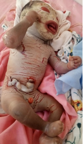
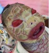
At birth, clinical examination revealed massive generalized hyperkeratosis with thick yellowish plates arranged in large polygonal lamellae, associated with deep erythematous fissures involving the trunk and thighs, giving the skin a rigid armor-like appearance. Marked bilateral ectropion with conjunctival exposure, eclabium with persistent mouth opening, flattening of the nasal pyramid due to cutaneous tension, hypoplastic and dysmorphic auricles, and flexion contractures of the limbs were also observed (Figures 1 and 2). This presentation corresponds to the typical neonatal phenotype described in the literature
[5]
Tsivilika M, Kavvadas D, Karachrysafi S, Sioga A, Papamitsou T. Management of harlequin ichthyosis: a brief review of the recent literature. Children (Basel). 2022; 9(6): 893.
Figure 2. Bilateral ectropion and eclabium at birth.
Management consisted of repeated application of emollients, nasogastric feeding and enteral hydration due to feeding difficulties related to eclabium, correction of hydro-electrolytic disturbances, and infection prevention. Due to the impossibility of obtaining peripheral or umbilical venous access, intravenous fluid therapy could not be initiated. Oral amoxicillin-clavulanic acid was administered empirically according to body weight because parenteral antibiotic therapy was not feasible. Lubrication with artificial tears was also initiated, as recommended in the literature
[6]
Lilly E, Biesbroek L, et al. Congenital ichthyosis: a practical clinical guide on current treatments and future perspectives. Clin Cosmet Investig Dermatol. 2023; 16: 1071–1085.
. Supplemental oxygen via nasal cannula was provided to support respiratory function. Systemic retinoid therapy could not be introduced due to limited resources.
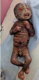
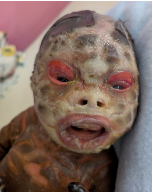
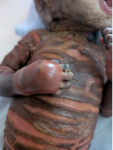
By day 4 of life, worsening of the skin fissures and increased rigidity of the integument were noted, with persistence of the marked bilateral ectropion and eclabium (Figures 3 and 4), as well as distal dark discoloration of the extremities suggestive of ischemic compromise (Figure 5).
Figure 5. Distal extremity discoloration suggestive of ischemia at day 4 of life.
The clinical course was marked by death on day 6 of life due to multifactorial complications, including severe transepidermal fluid loss, suspected sepsis, and progressive respiratory compromise related to thoracic skin rigidity.
3. Discussion
Harlequin ichthyosis is a rare genodermatosis caused by mutations in the ABCA12 gene, leading to a major defect in lipid transport within keratinocytes
[2]
Kelsell DP, Norgett EE, Unsworth H, Teh MT, Cullup T, Mein CA, et al. Mutations in ABCA12 underlie the severe congenital skin disease harlequin ichthyosis. Am J Hum Genet. 2005; 76(5): 794–803.
The diagnosis is usually established at birth based on the typical clinical presentation characterized by thick hyperkeratotic plates, deep fissures, ectropion, and eclabium
[5]
Tsivilika M, Kavvadas D, Karachrysafi S, Sioga A, Papamitsou T. Management of harlequin ichthyosis: a brief review of the recent literature. Children (Basel). 2022; 9(6): 893.
. Several case series have reported similar clinical findings in newborns affected by harlequin ichthyosis
[4]
Rajpopat S, Moss C, Mellerio J, Vahlquist A, Ganemo A, Hellström-Pigg M, et al. Harlequin ichthyosis: a review of clinical and molecular findings in 45 cases. Arch Dermatol. 2011; 147(6): 681–686.
Prenatal diagnosis is possible but remains challenging. Ultrasound examination may reveal suggestive abnormalities such as ectropion, persistent open mouth, or abnormal ears, although these signs often appear late during pregnancy
[10]
Zhou XJ, Wang H, et al. Prenatal diagnosis of harlequin ichthyosis by ultrasonography. Ann Transl Med. 2021; 9(3): 266.
. Diagnostic confirmation relies on the identification of ABCA12 mutations through genetic analysis
[2]
Kelsell DP, Norgett EE, Unsworth H, Teh MT, Cullup T, Mein CA, et al. Mutations in ABCA12 underlie the severe congenital skin disease harlequin ichthyosis. Am J Hum Genet. 2005; 76(5): 794–803.
Hotz A, Bourrat E, Kopp J, Oji V, Süßmuth K, Komlosi K, et al. Mutational spectrum of the ABCA12 gene and genotype–phenotype correlation in autosomal recessive congenital ichthyosis. Genes (Basel). 2023; 14(3): 717.
. This molecular diagnosis, based on DNA analysis, can be performed using chorionic villus sampling or amniocentesis. Fetal skin biopsy may also contribute to the diagnosis from the 18th week of gestation
[2]
Kelsell DP, Norgett EE, Unsworth H, Teh MT, Cullup T, Mein CA, et al. Mutations in ABCA12 underlie the severe congenital skin disease harlequin ichthyosis. Am J Hum Genet. 2005; 76(5): 794–803.
Hotz A, Bourrat E, Kopp J, Oji V, Süßmuth K, Komlosi K, et al. Mutational spectrum of the ABCA12 gene and genotype–phenotype correlation in autosomal recessive congenital ichthyosis. Genes (Basel). 2023; 14(3): 717.
. In subsequent pregnancies at risk, when the familial mutation is known, targeted prenatal diagnosis can be performed by chorionic villus sampling between 11 and 13 weeks of gestation or by amniocentesis from 15 weeks of gestation to detect the specific mutation
[2]
Kelsell DP, Norgett EE, Unsworth H, Teh MT, Cullup T, Mein CA, et al. Mutations in ABCA12 underlie the severe congenital skin disease harlequin ichthyosis. Am J Hum Genet. 2005; 76(5): 794–803.
Several studies have shown that management in neonatal intensive care units combined with early administration of systemic retinoids, particularly acitretin, may improve survival in patients with harlequin ichthyosis
[6]
Lilly E, Biesbroek L, et al. Congenital ichthyosis: a practical clinical guide on current treatments and future perspectives. Clin Cosmet Investig Dermatol. 2023; 16: 1071–1085.
Glick JB, Craiglow BG, Choate KA, Kato H, Fleming RE, Siegfried E, et al. Improved management of harlequin ichthyosis with advances in neonatal intensive care. Pediatrics. 2017; 139(1): e20161003.
. Early initiation of systemic retinoids has also been associated with faster desquamation of hyperkeratotic plaques and improved skin flexibility, which may reduce the risk of respiratory restriction and distal ischemic complications
[6]
Lilly E, Biesbroek L, et al. Congenital ichthyosis: a practical clinical guide on current treatments and future perspectives. Clin Cosmet Investig Dermatol. 2023; 16: 1071–1085.
Systemic complications observed in these patients are mainly related to disruption of the skin barrier, leading to excessive fluid loss, hydro-electrolytic imbalance, and increased susceptibility to infections
[5]
Tsivilika M, Kavvadas D, Karachrysafi S, Sioga A, Papamitsou T. Management of harlequin ichthyosis: a brief review of the recent literature. Children (Basel). 2022; 9(6): 893.
Similar fatal outcomes have been reported in low-resource settings, where limited access to intensive care and invasive management significantly affects neonatal survival
[13]
Vella V, Maulida M, Earlia N, et al. A fatal case of harlequin ichthyosis: Experience from low-resource setting. Clin Case Rep. 2024; 12(3): e8084.
In our case, the absence of peripheral or umbilical venous access significantly limited optimal fluid and electrolyte management. Although enteral hydration, oral antibiotic therapy, and supplemental oxygen were provided, these measures were insufficient to prevent clinical deterioration. This highlights the challenges of managing harlequin ichthyosis in resource-limited settings, where access to neonatal intensive care, vascular access, laboratory monitoring, and systemic retinoids remains restricted
[5]
Tsivilika M, Kavvadas D, Karachrysafi S, Sioga A, Papamitsou T. Management of harlequin ichthyosis: a brief review of the recent literature. Children (Basel). 2022; 9(6): 893.
Lilly E, Biesbroek L, et al. Congenital ichthyosis: a practical clinical guide on current treatments and future perspectives. Clin Cosmet Investig Dermatol. 2023; 16: 1071–1085.
Harlequin ichthyosis is a rare and severe genetic disorder of the newborn, characterized by massive hyperkeratosis and severe impairment of the skin barrier secondary to mutations in the ABCA12 gene. The diagnosis is usually clinical at birth, although confirmation relies on genetic testing. Early management in neonatal intensive care units and the use of systemic retinoids may improve prognosis
[6]
Lilly E, Biesbroek L, et al. Congenital ichthyosis: a practical clinical guide on current treatments and future perspectives. Clin Cosmet Investig Dermatol. 2023; 16: 1071–1085.
Glick JB, Craiglow BG, Choate KA, Kato H, Fleming RE, Siegfried E, et al. Improved management of harlequin ichthyosis with advances in neonatal intensive care. Pediatrics. 2017; 139(1): e20161003.
Kelsell DP, Norgett EE, Unsworth H, Teh MT, Cullup T, Mein CA, et al. Mutations in ABCA12 underlie the severe congenital skin disease harlequin ichthyosis. Am J Hum Genet. 2005; 76(5): 794–803.
Rajpopat S, Moss C, Mellerio J, Vahlquist A, Ganemo A, Hellström-Pigg M, et al. Harlequin ichthyosis: a review of clinical and molecular findings in 45 cases. Arch Dermatol. 2011; 147(6): 681–686.
Tsivilika M, Kavvadas D, Karachrysafi S, Sioga A, Papamitsou T. Management of harlequin ichthyosis: a brief review of the recent literature. Children (Basel). 2022; 9(6): 893.
Lilly E, Biesbroek L, et al. Congenital ichthyosis: a practical clinical guide on current treatments and future perspectives. Clin Cosmet Investig Dermatol. 2023; 16: 1071–1085.
Hotz A, Bourrat E, Kopp J, Oji V, Süßmuth K, Komlosi K, et al. Mutational spectrum of the ABCA12 gene and genotype–phenotype correlation in autosomal recessive congenital ichthyosis. Genes (Basel). 2023; 14(3): 717.
Glick JB, Craiglow BG, Choate KA, Kato H, Fleming RE, Siegfried E, et al. Improved management of harlequin ichthyosis with advances in neonatal intensive care. Pediatrics. 2017; 139(1): e20161003.
Andriatahina, H. F. P., Jarison, L. V., Rakotonandrasana, F., Jaofeno, D. A., Ramarozatovo, L. S., et al. (2026). A Malagasy Case Report of Harlequin Ichthyosis. International Journal of Clinical Dermatology, 9(1), 72-75. https://doi.org/10.11648/j.ijcd.20260901.19
Andriatahina, H. F. P.; Jarison, L. V.; Rakotonandrasana, F.; Jaofeno, D. A.; Ramarozatovo, L. S., et al. A Malagasy Case Report of Harlequin Ichthyosis. Int. J. Clin. Dermatol.2026, 9(1), 72-75. doi: 10.11648/j.ijcd.20260901.19
Andriatahina HFP, Jarison LV, Rakotonandrasana F, Jaofeno DA, Ramarozatovo LS, et al. A Malagasy Case Report of Harlequin Ichthyosis. Int J Clin Dermatol. 2026;9(1):72-75. doi: 10.11648/j.ijcd.20260901.19
@article{10.11648/j.ijcd.20260901.19,
  author = {Herin’Ny Fitiavana Princia Andriatahina and Louisebine Volasoa Jarison and Fenohasina Rakotonandrasana and Diane Angela Jaofeno and Lala Soavina Ramarozatovo and Fahafahantsoa Rapelanoro Rabenja and Irina Mamisoa Ranaivo},
  title = {A Malagasy Case Report of Harlequin Ichthyosis},
  journal = {International Journal of Clinical Dermatology},
  volume = {9},
  number = {1},
  pages = {72-75},
  doi = {10.11648/j.ijcd.20260901.19},
  url = {https://doi.org/10.11648/j.ijcd.20260901.19},
  eprint = {https://article.sciencepublishinggroup.com/pdf/10.11648.j.ijcd.20260901.19},
  abstract = {Introduction: Harlequin ichthyosis is the most severe form of autosomal recessive congenital ichthyosis. It is caused by mutations in the ABCA12 gene, leading to a major defect in epidermal lipid transport and a profound impairment of the skin barrier. The estimated incidence ranges from 1 in 300,000 to 1 in 1,000,000 live births. Clinically, affected neonates present at birth with thick hyperkeratotic plates separated by deep fissures, associated with bilateral ectropion, eclabium, and limb contractures. Despite advances in neonatal intensive care and the introduction of systemic retinoids, mortality remains high. We report here a case of harlequin ichthyosis observed in Madagascar. Case presentation: We report the case of a full-term newborn, the first child of a 15-year-old mother, delivered vaginally with a birth weight of 2540 g. A first-trimester prenatal ultrasound was reported as normal. No parental consanguinity was known. At day 0 of life, physical examination revealed massive generalized hyperkeratosis with large thick plates separated by deep erythematous fissures, giving the skin an “armor-like” appearance. Marked bilateral ectropion, eclabium, nasal flattening, dysmorphic auricles, and limb contractures were also observed. The newborn was admitted to the neonatal intensive care unit. Supportive care including topical emollients, correction of hydro-electrolytic disturbances, and infection prevention was initiated. Systemic retinoids could not be introduced. By day 4 of life, increased skin rigidity, widening of the fissures, and distal dark discoloration of the extremities suggestive of ischemic compromise were observed. The clinical course was marked by death at day 6 of life. Discussion: Harlequin ichthyosis results from impaired lipid transport caused by ABCA12 mutations, leading to severe disruption of the stratum corneum and skin barrier function. Prenatal diagnosis by ultrasound is possible but remains difficult and is often made late in pregnancy. The unfavorable outcome in our case highlights the challenges in managing this condition in resource-limited settings. Conclusion: Harlequin ichthyosis remains a severe neonatal emergency. Early recognition, prompt supportive care, and specialized multidisciplinary management are essential to improve prognosis.},
 year = {2026}
}
TY  - JOUR
T1  - A Malagasy Case Report of Harlequin Ichthyosis
AU  - Herin’Ny Fitiavana Princia Andriatahina
AU  - Louisebine Volasoa Jarison
AU  - Fenohasina Rakotonandrasana
AU  - Diane Angela Jaofeno
AU  - Lala Soavina Ramarozatovo
AU  - Fahafahantsoa Rapelanoro Rabenja
AU  - Irina Mamisoa Ranaivo
Y1  - 2026/04/23
PY  - 2026
N1  - https://doi.org/10.11648/j.ijcd.20260901.19
DO  - 10.11648/j.ijcd.20260901.19
T2  - International Journal of Clinical Dermatology
JF  - International Journal of Clinical Dermatology
JO  - International Journal of Clinical Dermatology
SP  - 72
EP  - 75
PB  - Science Publishing Group
SN  - 2995-1305
UR  - https://doi.org/10.11648/j.ijcd.20260901.19
AB  - Introduction: Harlequin ichthyosis is the most severe form of autosomal recessive congenital ichthyosis. It is caused by mutations in the ABCA12 gene, leading to a major defect in epidermal lipid transport and a profound impairment of the skin barrier. The estimated incidence ranges from 1 in 300,000 to 1 in 1,000,000 live births. Clinically, affected neonates present at birth with thick hyperkeratotic plates separated by deep fissures, associated with bilateral ectropion, eclabium, and limb contractures. Despite advances in neonatal intensive care and the introduction of systemic retinoids, mortality remains high. We report here a case of harlequin ichthyosis observed in Madagascar. Case presentation: We report the case of a full-term newborn, the first child of a 15-year-old mother, delivered vaginally with a birth weight of 2540 g. A first-trimester prenatal ultrasound was reported as normal. No parental consanguinity was known. At day 0 of life, physical examination revealed massive generalized hyperkeratosis with large thick plates separated by deep erythematous fissures, giving the skin an “armor-like” appearance. Marked bilateral ectropion, eclabium, nasal flattening, dysmorphic auricles, and limb contractures were also observed. The newborn was admitted to the neonatal intensive care unit. Supportive care including topical emollients, correction of hydro-electrolytic disturbances, and infection prevention was initiated. Systemic retinoids could not be introduced. By day 4 of life, increased skin rigidity, widening of the fissures, and distal dark discoloration of the extremities suggestive of ischemic compromise were observed. The clinical course was marked by death at day 6 of life. Discussion: Harlequin ichthyosis results from impaired lipid transport caused by ABCA12 mutations, leading to severe disruption of the stratum corneum and skin barrier function. Prenatal diagnosis by ultrasound is possible but remains difficult and is often made late in pregnancy. The unfavorable outcome in our case highlights the challenges in managing this condition in resource-limited settings. Conclusion: Harlequin ichthyosis remains a severe neonatal emergency. Early recognition, prompt supportive care, and specialized multidisciplinary management are essential to improve prognosis.
VL  - 9
IS  - 1
ER  -