Research Article | | Peer-Reviewed

Prognostic Role of Nucleosomes Containing Histones of the H3.1 Type in Septic Shock: Results of a Prospective Study

Received: 31 May 2026     Accepted: 13 July 2026     Published: 23 October 2026
Views:       Downloads:
Abstract

Cell-free DNA (cfDNA) and neutrophil extracellular traps (NETs), specifically marked by histone type 3.1, play a dual pathogenic role in sepsis by reflecting cellular damage and directly exacerbating tissue injury, highlighting their clinical relevance for extracorporeal elimination therapies. To evaluate their prognostic value, we conducted a prospective, single-center observational study quantifying nucleosomes containing histones of the H3.1 type (Nu.H3.1) in 52 adult patients with septic shock admitted to an intensive care unit between August 2023 and May 2024. The cohort (64% male) had a mean SOFA score of 10±4 and an APACHE II score of 22±7. The H3.1-nuc concentration was 4658 ± 3962 ng/mL, and the median cfDNA level was 3041 (876–7815) ng/mL. Statistical analysis revealed that Nu.H3.1 levels significantly and positively correlated with cfDNA (r=0.67; p=0.0001), serum lactate (r=0.4; p=0.004), and SOFA scores (r=0.41; p=0.002). Furthermore, Nu.H3.1 demonstrated significant predictive performance for both 28-day mortality (AUC=0.68; 95% CI: 0.53–0.84; p=0.037) and the development of acute kidney injury (AKI) (AUC=0.74; 95% CI: 0.57–0.90; p=0.014). In conclusion, Nu.H3.1 serves as an independent predictor of adverse outcomes in septic shock, with established threshold values identifying patients at high risk for mortality and AKI. These findings suggest that Nu.H3.1 is a promising biomarker for clinical risk stratification and for substantiating indications for the extracorporeal elimination of toxic DNA-associated structures from the systemic circulation.

Published in International Journal of Clinical and Experimental Medical Sciences (Volume 12, Issue 5)
DOI 10.11648/j.ijcems.20261205.11
Page(s) 82-95
Creative Commons

This is an Open Access article, distributed under the terms of the Creative Commons Attribution 4.0 International License (http://creativecommons.org/licenses/by/4.0/), which permits unrestricted use, distribution and reproduction in any medium or format, provided the original work is properly cited.

Copyright

Copyright © The Author(s), 2026. Published by Science Publishing Group

Keywords

Cell-free DNA, Nucleosomes h3.1, Sepsis, Septic Shock

1. Introduction
The concept of personalized intensive care requires the development of fundamentally new prognostic tools in the practice of treating septic shock. Various biomarkers are of interest as predictors of septic shock course. Some of them may serve not only as markers but also as significant factors in the pathogenesis of septic shock, which allows them to be considered as target molecules for elimination during extracorporeal therapy . Cell-free DNA (cfDNA) possesses such potential, representing a promising marker and therapeutic target in sepsis .
cfDNA represents double-stranded extracellular fragments of chromosomes and associated structural proteins and enzymes released into the bloodstream as a result of apoptosis, necrosis, or active secretion by immune system cells. The latter process represents the formation of neutrophil extracellular traps (NETs). This is an anti-infective innate protective mechanism whereby decondensed chromatin and nuclear proteins are released from neutrophils and some other immune cells, forming mesh-like structures that trap microorganisms and lyse their cell walls .
The structural unit of NETs is the nucleosome, which consists of a DNA strand wrapped around a complex of specialized nuclear proteins—histones. Histones are necessary for compact spatial organization of DNA. Histone type H3.1 is characteristic only of neutrophils and serves as a marker of NETs levels in blood plasma .
In sepsis and septic shock, "overproduction" of NETs occurs, leading to additional enhancement of the hyperinflammatory response, endothelial damage, thrombosis of microcirculation vessels, which directly contributes to the progression of organ dysfunction . Prolonged elevation of cfDNA levels in the bloodstream "overloads" the macrophage system involved in its natural elimination, reducing their phagocytic activity, which may be one of the causes of secondary immunodepression development in sepsis .
Several studies have demonstrated significant elevation of cfDNA and NETs levels in patients with sepsis, with association between the degree of this elevation and severity of organ dysfunction and increased mortality . Thus, in sepsis, cfDNA plays a dual role: it characterizes the degree of cellular damage and intensity of inflammatory response while simultaneously serving as an etiological agent of both these processes . Research on cfDNA and NETs in sepsis has acquired additional practical significance due to the emergence of extracorporeal therapy techniques aimed at their selective elimination from the bloodstream .
2. Objective
The objective of this study was to evaluate the role of NETs and cfDNA as independent predictors of mortality in patients with septic shock and their correlation with clinical and laboratory data and risk of acute kidney injury (AKI) development, as well as to study the potential of NETs as a target for blood purification techniques.
2.1. Materials and Methods
2.1.1. Study Design
Prospective single-center observational study conducted from August 2023 to May 2024 in the intensive care unit of Sergey Yudin City Clinical Hospital.
2.1.2. Inclusion Criteria
1) Age over 18 years
2) Established diagnosis of sepsis according to Sepsis-3 criteria
3) Hypotension persisting after infusion therapy at 30 mL/kg, requiring norepinephrine vasopressor therapy at a dosage exceeding 0.3 µg/kg/min
2.1.3. Exclusion Criteria
1) Uncontrolled bleeding or high risk of its occurrence
2) Presence of venous or arterial thrombosis
3) Cardiovascular events within the last 2 months: acute myocardial infarction, acute cerebrovascular accident, pulmonary embolism
4) Severe congestive chronic heart failure
5) End-stage chronic kidney disease
6) Decompensated liver cirrhosis
7) Severe combined or positional trauma
8) Presence of human immunodeficiency virus infection
9) 6Severe granulocytopenia (peripheral blood neutrophil count <500 cells/mm³)
10) 7Oncological pathology at the stage of neo- or adjuvant therapy
11) 8Continuous immunosuppressive therapy
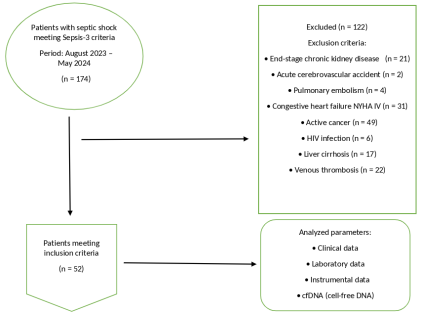
According to the specified criteria, 52 patients were included in the study (Figure 1).
Figure 1. The study's patient selection scheme.
The study was approved by the local ethics committee of Sergey Yudin City Clinical Hospital (protocol No. 5 dated 01/11/2023).
Sample size calculation for two-sided alternative hypothesis was performed based on 80% power, type I error rate of 5%, using MedCalc Software Ltd. Sample size for Single mean. https://www.medcalc.org/calc/sample-size-single-mean.php (Version 23.3.5; accessed August 16, 2025). Based on anticipated effect sizes (mean differences) and variability (standard deviation), the minimum sample size for each group was 8 patients.
2.2. Data Collection
Demographic, clinical, laboratory, and instrumental data of patients were collected and analyzed within the first 24 hours from septic shock manifestation (Table 1).
Clinical assessment: Included severity assessment using APACHE II (Acute Physiology and Chronic Health Evaluation II) and SOFA (Sequential Organ Failure Assessment) scales. Hospital treatment outcomes, duration of ICU stay, mechanical ventilation (MV), and vasopressor therapy were recorded.
Hemodynamic monitoring: Along with standard monitoring (electrocardiography, pulse oximetry), blood pressure control and echocardiography were performed.
Laboratory monitoring: Included complete blood count, biochemical blood analysis, acid-base status analysis, coagulogram, water-electrolyte parameters. Additionally, interleukin-6 (IL-6), procalcitonin (electrochemiluminescence method, Lifortonic eCL8000), C-reactive protein (CRP), ferritin, complement system proteins C3 and C4 (immunoturbidimetry, Mindray BS 800), C1 inhibitor of complement factor (fluorimetric method, Qubit4 Thermo FS), von Willebrand factor level, D-dimer (immunoturbidimetric method, Sismexc SC 5100), protein S, protein C, antithrombin III (chromogenic, photometric method, Destiny Max) were determined.
In addition to standard laboratory parameters, Nu.Q H3.1® methodology (Belgian Volition SRL, Belgium) was used to measure H3.1 nucleosome (Nu.H3.1) concentration according to manufacturer's instructions, and cfDNA was determined using the "NucleoTest" method (NPF POKARD, Russia).
"NucleoTest" is a chromogenic method for determining the concentration of DNA-containing structures, based on the ability of DNA to interact with human histone H1.3. Patient plasma test samples were added to wells of an enzyme immunoassay plate with pre-immobilized histone H1.3, followed by histone H1.3-peroxidase conjugate. DNA concentration in samples was determined spectrophotometrically by color development at 450 nm. Calibration dependence was constructed using purified DNA preparation. The method allows quantitative determination of DNA-containing structure levels in human blood plasma.
Intensive care was conducted in accordance with "Septic Shock in Adults" recommendations (FAR clinical guidelines, Surviving Sepsis Campaign, 2021) .
2.3. Statistical Analysis
Standard SPSS v.26 software packages (SPSS Inc., USA) were used for statistical processing. Shapiro-Wilk and Kolmogorov-Smirnov tests, kurtosis and skewness assessment were used to determine normality of quantitative variable distribution. If variables followed normal distribution, their values were presented as mean and standard deviation (M ± SD). If quantitative variable distribution did not follow normal, values were presented as median (Me) and interquartile range (25–75%). Nominal (qualitative) values are indicated in absolute numbers (n) and percentages (%).
Spearman's coefficient (r) was used to assess correlations between quantitative (with non-normal distribution) and ordinal variables, while Pearson's coefficient was applied for normal distribution.
To identify independent predictors of high cfDNA levels, which included all statistically significant correlations (p < 0.1), paired and multiple linear regression analyses were used. Multivariate linear regression was performed using stepwise exclusion method.
To identify dependence of Nu.H3.1 level on hospital lethal outcome and complications in patients with septic shock, receiver operating characteristic (ROC) curve analysis was used. Cut-off point was selected considering Youden index values and comparison of odds ratios for high and low indicator values.
Sensitivity, specificity, positive and negative predictive values were calculated using contingency tables.
Statistically significant differences were considered at two-sided p < 0.05.
3. Results
The final number of patients included in the study was 52 (64% men, 36% women) aged 52.1 ± 17.3 years. Body mass index (BMI) of subjects approached first-degree obesity (29.0 ± 6.2 kg/m2). All patients met septic shock criteria (organ dysfunction assessment and mortality prediction by SOFA and APACHE II scales were 10 ± 4 and 22 ± 7 points respectively, median lactate and procalcitonin levels — 2.4 [1.7–4.0] mmol/L and 15.7 [5.0–60.5] ng/mL respectively, median norepinephrine dosage — 0.52 [0.33–0.87] µg/kg/min). Main demographic, clinical-laboratory, and instrumental data are presented in Table 1.
Table 1. The main traits of patients included in the study.

General Cohort (N=52)

Demographic Characteristics

Age, years

52,1±17,3

Sex (male), %

64,0%

BMI, kg/m2

29,0±6,2

Etiology of Septic Shock

Abdominal, % (N)

35,9% (18)

Pulmonary, % (N)

37,5% (19)

Urological, % (N)

12,5% (7)

Skin and soft tissue infection, % (N)

9,0% (5)

Other, % (N)

5,1% (3)

Clinical Data

SOFA, score

10,0±4,0

Apache II, score

22,0±7,0

Heart rate, bpm

103,0±23,0

Mean arterial pressure (MAP), mmHg

61,7±8,0

Norepinephrine, µg/kg/min

0,52 (0,33-0,87)

Hospital length of stay, days

9,0 (6,0-16,0)

ICU length of stay, days

7,0(4,0-11,0)

Duration of mechanical ventilation, days

5,0 (3,0-10,0)

Duration of vasopressor support, days

5,0 (3,0-10,0)

Instrumental Data

LVEF, %

59,0 (47,0-63,0)

LVEDV, mL

95,0 (78,0-118,0)

sPAP, mmHg

35,0±11,0

Laboratory Data

PaO₂/FiO₂ ratio

300,0±138,0

Lactate, mmol/L

2,4 (1,7-4,0)

Leukocytes, ×10⁹/L

17,3 (10,6-24,4)

Neutrophils, ×10/L

14,9 (7,9-25,5)

Band neutrophils, %

8,0 (4,0-19,0)

Lymphocytes, ×10⁹/L

8,0 (5,0-12,5)

Platelets, ×10⁹/L

137,0 (89,0-220,0)

IL-6, pg/mL

434,0±387,2

CRP, mg/L

212,5±88,1

PCT, ng/mL

15,7 (5,0-60,5)

Ferritin, ng/mL

974,5 (555,0-1213,0)

Fibrinogen, g/L

5,9±2,3

Antithrombin III, %

45,8 (32,7-64,7)

D-dimer, ng/mL

5971,4 (2161,0-12066,0)

Protein C, %

57,8±24,3

Protein S, %

61,7 ±31,0

von Willebrand factor, %

521,5 ±246,6

Cholesterol, mmol/L

3,0±1,3

Triglycerides, mmol/L

2,3 (1,5-4,2)

Uric acid, µmol/L

350,2 (234,8-463,4)

Albumin, g/L

27,3±5,3

Bilirubin, µmol/L

18,8 (7,7-34,6)

ALT, U/L

38,7 (24,2-99,2)

AST, U/L

57,4 (27,4-159,6)

Creatinine, µmol/L

220,1 (117,9-356,8)

Complement C3, g/L

0,9±0,3

Complement C4, g/L

0,21(0,14-0,29)

C1-inhibitor, mg/dL

9,0 (0,5-22,0)

cfDNA, ng/mL

3041,0 (876,0-7815,0)

Nu.H3.1, ng/mL

4658,0±3962,0

Clinical Events

28-day mortality, % (N)

69,8% (36)

Thrombotic complications, % (N)

26,4% (13)

AKI, % (N)

75,5% (39)

Note: BMI –Body Mass Index, SOFA — Sequential Organ Failure Assessment, APACHE II — Acute Physiology and Chronic Health Evaluation II, ICU — intensive care unit, LVEF — left ventricular ejection fraction, LVEDV — left ventricular end-diastolic volume, sPAP — systolic pulmonary artery pressure, IL-6 — Interleukin-6, CRP — C-reactive protein, PCT — Procalcitonin, ALT— alanine aminotransferase, АСТ — aspartate aminotransferase, cfDNA — cell free DNA, Nu.H3.1 — nucleosomes containing histones of the H3.1 type, AKI — Acute Kidney Injury.
Median cfDNA concentration determined by "NucleoTest" method in patients with septic shock was 3041 (876–7815.0) ng/mL, while Nu.H3.1 level was 4658 ± 3962 ng/mL. Mortality within 28 days was high (69.8%) and corresponded to mortality frequency in the general population of patients with septic shock . In 75% of subjects, the condition was complicated by AKI requiring renal replacement therapy. Another complication (lower extremity venous thrombosis associated with sepsis) occurred in 25% of cases.
Correlation analysis of Nu.H3.1 concentration allowed identification of statistically significant associations among the above-mentioned parameters in patients with septic shock (Table 2).
Table 2. Correlation analysis of nucleosome concentration with demographic, clinical, laboratory and instrumental data.

Nu.H3.1

Parameter

Rxу

p

Clinical scales

SOFA

0,41

0,002

Apache II

0,3

0,04

Hemostasis system

von Willebrand factor

0,35

0,02

Complement system

С3

-0,39

0,005

С4

-0,33

0.02

Liver enzymes

Bilirubin

0,36

0,01

ALT

0,3

0,03

AST

0,28

0,04

Inflammatory mediators

IL-6

0,39

0,04

Other parameters

Oxygenation index

-0,37

0,008

Creatinine

0,29

0,04

Lactate

0,4

0,004

cfDNA

0,67

0,0001

Note: SOFA — Sequential Organ Failure Assessment, APACHE II — Acute Physiology and Chronic Health Evaluation II, C3 — Complement components C3, C4 — Complement components C4, АСТ — aspartate aminotransferase, ALT— alanine aminotransferase, IL-6 — Interleukin-6, cfDNA — cell free DNA.
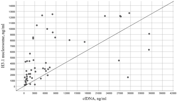
The most significant correlations of Nu.H3.1 were recorded with lactate level (direct moderate strength relationship: rxy = 0.4; p = 0.004) and SOFA scale (direct moderate strength relationship: rxy = 0.41; p = 0.002). Direct moderate strength relationship was also noted between Nu.H3.1 and IL-6 level (rxy = 0.39; p = 0.04). Additionally, it should be noted that correlation between two NETs markers—cfDNA and Nu.H3.1—showed strong direct relationship (rxy = 0.67; p = 0.0001) (Figure 2).
Figure 2. The regression function showing the dependence of the cfDNA level on the Nu.H3.1.
Subsequently, using paired and multiple linear regression, assessment of dependence of statistically significant parameters identified in correlation analysis on Nu.H3.1 concentration in patients with septic shock included in the study was performed (Table 3).
Table 3. Paired and multiple regression of the nucleosome concentration dependence.

Simple Regression

R\R2

ꞵ

р

Multiple Regression

р

р

R\R2

Adjusted R

SOFA

0,41/0,17

414,2

0,02

Apache II

0,003

0,0001

0,74/0,51

0,47

Apache II

0,29/0,08

175,3

0,04

Lactate

0,003

Oxygenation index

0,37/0,13

-10,5

0,008

Bilirubin

0,02

Lactate

0,41/0,17

656

0,002

ALT

0,009

IL-6

0,39/0,15

4,2

0,04

von Willebrand factor

0,35/0,12

6,0

0,02

Bilirubin

0,35/0,12

26,2

0,02

ALT

0,38/0,15

2,6

0,005

AST

0,46/0,21

1,0

0,001

С3

0,39/0,15

-4965

0,005

Note: SOFA — Sequential Organ Failure Assessment, APACHE II — Acute Physiology and Chronic Health Evaluation II, IL-6 — Interleukin-6, ALT— alanine aminotransferase, АСТ — aspartate aminotransferase, C3 — Complement components C3.
The most significant relationships of Nu.H3.1 in paired regression were identified with SOFA scale total score, mixed venous blood lactate levels, and AST. However, according to multiple regression results, only APACHE II, lactate, bilirubin, and ALT retained their dependence on NETs in the final regression model, which showed close relationship by Chaddock scale (correlation coefficient rxy = 0.74). Significance level of this model was p < 0.0001, and combination of these factors in the identified model determined 47% of Nu.H3.1 variance.
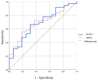
According to ROC analysis results, AUC of Nu.H3.1 showed statistical significance regarding 28-day hospital mortality and AKI in patients with septic shock (Table 4, Figure 3, Figure 4).
Figure 3. The area under the characteristic curve of the concentration of Nu.H3.1 in relation to 28-day mortality in patients with septic shock.
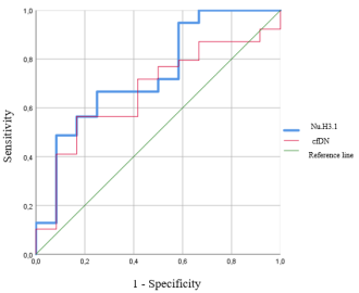
Figure 4. The area under the characteristic curve of the concentration of Nu.H3.1 in relation to AKI in patients with septic shock.
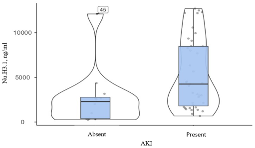
Table 4. AUC concentrations of Nu.H3.1 in relation to 28-day mortality and development of AKI in patients with septic shock.

Parameter

AKI

DeLong Test

28-day Mortality

DeLong Test

AUC (95% CI)

р

AUC (95% CI)

р

Nu.H3.1

0,74 (0,57-0,90)

0,014

Р1-2 0,37

0,68 (0,53-0,84)

0,037

Р1-2 0,92

cfDNA

0,67(0,51-0,83)

0,076

0,69 (0,54-0,84)

0,031

Note: AKI — Acute Kidney Injury, AUC — Area Under the Curve, CI — Confidence Interval, cfDNA — Cell-free DNA, Nu.H3.1 — Nucleosomes containing histones of the H3.1 type.
When comparing AUC of Nu.H3.1 and cfDNA, no statistically significant differences were identified, which generally allows considering these indicators as equivalent in predicting clinical events—AKI and 28-day mortality.
When dividing the sample of patients with septic shock into presence/absence of AKI, median Nu.H3.1 concentration in the AKI group differed statistically significantly compared to the group without AKI (4252 [1700–8488] vs 2283 [369–3034] ng/mL; p = 0.014) (Figure 5, Figure 6). Similar difference was noted when comparing Nu.H3.1 concentration in patients with septic shock who died within 28 days of hospitalization versus those who survived during this period (1925 [933–4084] vs 4153 [1939–8949] ng/mL; p = 0.037) (Figure 7, Figure 8).
Figure 5. Comparative analysis of median Nu.H3.1 in patients with septic shock with or without AKI.
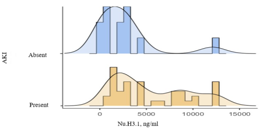
Figure 6. Comparative analysis of median Nu.H3.1 in patients with septic shock with or without AKI.
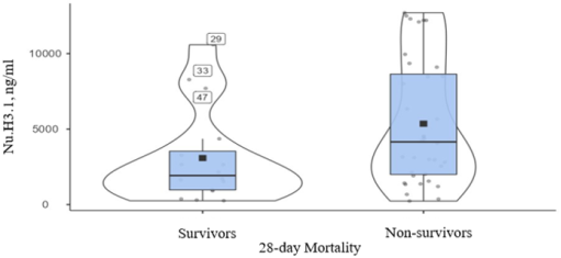
Figure 7. Comparative analysis of median Nu.H3.1 in patients with septic shock who died and survived for a period of 28 days.
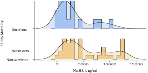
Figure 8. Comparative analysis of median Nu.H3.1 in patients with septic shock who died and survived for a period of 28 days.

Odds ratio for AKI development at Nu.H3.1 concentration >2731 ng/mL was 6.0 (95% CI 1.6–26.0; p < 0.018), sensitivity and specificity — 67% and 75% respectively. For 28-day mortality at Nu.H3.1 concentration >2731 ng/mL, OR was 4.4 (95% CI 1.2–15.6; p = 0.03), sensitivity and specificity — 67% and 79% respectively.
4. Discussion
Consistently high morbidity and mortality in sepsis determine the relevance of searching for new diagnostic strategies and therapeutic targets . Use of diagnostic and prognostic markers in sepsis is associated with several difficulties, the dominant one being complex pathogenesis including hyperinflammation, coagulation disorders, endothelial dysfunction, vasodilation, and organ damage . In this context, DNA-containing structures—cfDNA and NETs—appear as promising biomarkers, as their pathophysiological role in sepsis is associated with activation of both immune and coagulation systems, as well as endothelial and microcirculatory damage and, consequently, multiple organ injury. DNA-containing structures possess properties of so-called ideal septic process biomarker, and modern diagnostic capabilities for their determination are represented by numerous laboratory techniques that can be used not only for research purposes but also in real clinical practice .
In septic shock, cellular, tissue damage and hyperinflammation occur synchronously with mutual potentiation. Presumably, parallel monitoring of both cfDNA as a marker of cellular damage and inflammatory response intensity, and NETs level characterizing cellular immunity and phagocytosis activity, would undoubtedly provide diagnostic advantages for patients. Component composition and etiology of DNA-containing structures in septic patients remain insufficiently studied .
Our work demonstrated an interesting strong direct correlational relationship between cfDNA and Nu.H3.1 concentrations (rxy = 0.67; p = 0.0001). We were able to find only one study where both cfDNA concentration and NETs marker—MPO-DNA—were measured in sepsis patients, but correlation between them was not studied.
The correlations we observed between NETs and classical prognostic indicators (including APACHE II and SOFA scores, oxygenation index, and markers of endothelial dysfunction, hyperinflammation, and organ dysfunction) are well-supported by current pathophysiological models. Specifically, recent literature indicates that NET-mediated organ injury arises through a dual mechanism: the direct cytotoxic effect of neutrophil extracellular traps on host tissues, compounded by secondary ischemic damage and hypoperfusion resulting from NET-driven microvascular thrombosis and uncontrolled systemic inflammation .
In paired regression, the strongest association with elevated NETs levels was shown by SOFA scale total score (p = 0.02), mixed venous blood lactate level (p = 0.002), and AST (p = 0.001), but in multiple regression, only APACHE II, lactate, bilirubin, and ALT retained their dependence on NETs (correlation coefficient rxy = 0.74). We did not find clinical studies describing dependence of nucleosome concentration and liver dysfunction parameters in available literature sources, but it should be emphasized the known role of hepatic macrophage system in clearance of DNA-containing structures; it should also be mentioned the significant contribution of hepatocyte DNA to the total cfDNA pool in sepsis . Thus, liver damage in septic shock plays a dual role regarding DNA-containing structures, increasing their formation due to cytolysis and reducing elimination rate .
The significant association of lactate and NETs level shown in our work corresponds to the accepted concept of hyperinflammation influence on reduced tissue perfusion in sepsis. However, similar results were shown only for cfDNA .
Direct relationships of NETs level with severity of multiple organ dysfunction determined by APACHE II and SOFA scales were shown in retrospective study by Su Y. et al., which included 120 patients with sepsis. Nucleosome concentration in the group with APACHE II score >20 was higher than in groups 11–20 and 0–10 points (all p < 0.001). Nucleosome level in the group with SOFA score >10 points was higher than in groups 6–10 and 0–5 points (all p < 0.001) .
Similarly to our results, in the largest study to date by Filippini D. F. L. et al., which included 1700 ICU patients, direct correlations of NETs formation markers and proinflammatory cytokines, as well as weak positive associations with CRP, were described . This can be explained by proinflammatory effects of neutrophil traps capable of inducing cytokine release into bloodstream by other cells, as well as entry into bloodstream through disrupted neutrophil membrane of vesicles with inflammatory mediators, which occurs with chromatin release from nucleus during NETs formation . Thus, content of DNA-containing structures in bloodstream may be considered as a new diagnostic factor of hyperinflammatory sepsis phenotype .
We showed inverse correlation of NETs level and complement system protein C3. This may be explained by the ability of neutrophil traps to directly activate proteins of this system to enhance anti-infective protection in bacterial infections, as shown in studies by Leffler J. et al. and Wang H. et al. . Complement also participates in natural elimination of NETs by opsonizing them, which facilitates recognition and phagocytosis of NETs by macrophages . Thus, excessive NETs formation may lead to depletion of complement protein pool in bloodstream .
Interaction of NETs with von Willebrand factor normally represents one of key integrations of hemostasis and is necessary for localization of infectious agent; in sepsis, it acquires uncontrolled character, leading to endothelial damage, deposition of NETs components in its glycocalyx, development of immunothrombosis. NETs components cause reduced activity of metalloproteinase ADAMTS13, which maintains high von Willebrand factor level . This explains the direct association of NETs and von Willebrand factor found in our study. We did not find clinical studies demonstrating relationships between these two indicators; however, in work by Mao J. Y. et al., correlation of NETs levels and marker of endothelial glycocalyx damage syndecan-1 was shown (r = 0.6383, p < 0.0001). Among significant results of this study, association of DIC syndrome presence with higher nucleosome level (140 [47–233] vs 307 [199–415] ng/mL; [p < 0.0001]) and identification of NETs as independent risk factor for its development (OR 1.0104; 95% CI 1.0032–1.0176) should be highlighted . In works by Su Y. et al. and Filippini D. F. L. et al., correlations of excessive NETs levels with high frequency of sepsis-induced coagulopathy and DIC syndrome development were also described .
Correlational relationship of pulmonary oxygenation function and NETs level shown in our study is described in numerous works—in patient populations with both sepsis and acute respiratory distress syndrome and COVID-19 . For example, in study by Middleton E. A. et al., MPO-DNA complexes in plasma increased with COVID-19 progression and were associated with decreased oxygenation index . NETs components (histones, granular proteins) and DNA itself are capable of causing both direct and secondary (due to inflammation) damage to both pulmonary microcirculation vessel endothelium and alveolar epithelial cells, causing pulmonary tissue edema, which may explain adverse effect of neutrophil traps on respiratory function in sepsis .
Our study demonstrated the significant prognostic value of both cfDNA and NETs levels in predicting lethal outcomes and the development of acute kidney injury (AKI). This aligns with established pathophysiological concepts, as recent literature confirms the critical role of NETs in the progression of renal diseases and their emerging utility as prognostic biomarkers, particularly in conditions such as sepsis-induced AKI . However, while the general involvement of neutrophil extracellular traps in kidney damage is well-documented, the specific predictive ability of the Nu.H3.1 marker for AKI development remains largely unexplored in current studies, highlighting the novelty of our findings.
Regarding risk of lethal outcome, in study by Filippini D. F. L. et al., it was shown that H3.1 nucleosome level was significantly associated with 28-day mortality when adjusted for age and sex, but this association disappeared after adjustment for SOFA. In patients with initial concentration of nucleosomes containing histones type H3.1 >1000 ng/mL, higher probability of 28-day mortality was observed (RR 1.68; 95% CI 1.35–2.10; p < 0.001) . DNA-containing structures represent considerable interest as potential targets for extracorporeal therapy, especially in connection with emergence of selective hemosorbents capable of eliminating them from bloodstream . Despite all assumed clinical advantages of such intervention, substantial questions remain unclear regarding the level of DNA-containing structures constituting indication for initiation of extracorporeal therapy and timing of its commencement. Our study showed association of NETs with two most important clinical events—mortality and AKI development. At Nu.H3.1 level >2731 ng/mL, odds of first event increased 4-fold, of second—6-fold. Similar discrete level was demonstrated in the largest study to date on nucleosomes in sepsis by Haem Rahimi M. et al. with participation of 151 patients: optimal threshold value predicting lethal outcome was 4639 ng/mL. In study by Morimont L. et al., which included 20 patients with septic shock, mean level of H3.1 nucleosomes associated with lethal outcome was 1776 (402–16,032) ng/mL. In study by Cavalier E. et al. on cohort of COVID-19 patients, threshold level determining disease severity and need for intensive care unit treatment was 1250 ng/mL .
Obvious hypothesis is that indicated levels constitute indications for initiation of extracorporeal elimination of DNA-containing structures. However, further research in this direction is necessary.
5. Conclusion
The study showed that the marker of NETs formation—the level of Nu.H3.1—is an independent predictor of mortality and development of AKI in septic shock. Its threshold concentration associated with significant increase in probability of these significant clinical events was identified, which may be considered as a laboratory benchmark in determining indications for extracorporeal elimination of DNA-containing structures from bloodstream.
6. Recommendations
The indicated unresolved issues in septic shock treatment and obvious motivation to optimize its intensive care through searching for significant mediators (potential targets for extracorporeal elimination) could be a source of subjective biases in this study. These doubts may be reinforced by the fact that similar studies are extremely scarce, and obtained results are largely unique. However, transparent study design, including measurement of studied mediator levels outside clinical setting (in separate laboratory), excludes such possibility. Based on this, discovered associations of studied biomarkers with other clinical-laboratory parameters and sepsis treatment outcomes are considered as objective evidence of prognostic value of cfDNA and nucleosome levels. On the other hand, feasibility of eliminating these substances in septic shock remains only a working hypothesis, since their significance as marker or as active mediator of septic process cannot be identified.
Another more obvious limitation is the single-center nature of our work. Influence on results of both explicit (in-hospital orders) and "latent" (so-called style, manner, and subjective preferences in conducting therapeutic measures) hospital protocols of patient management in intensive care unit cannot be excluded. At the same time, study design is easily reproducible in other clinics and within multicenter study, necessity of which is beyond doubt.
Abbreviations

AKI

Acute Kidney Injury

ALT

Alanine Aminotransferase

APACHE II

Acute Physiology and Chronic Health Evaluation II

AST

Aspartate Aminotransferase

BMI

Body Mass Index

cfDNA

Cell Free DNA

CRP

C-reactive Protein

ICU

Intensive Care Unit

IL-6

Interleukin-6

LVEDV

Left Ventricular End-Diastolic Volume

LVEF

Left Ventricular Ejection Fraction

NETs

Neutrophil Extracellular Traps

Nu.H3.1

Nucleosomes Containing Histones of the H3.1 Type

PCT

Procalcitonin

SOFA

Sequential Organ Failure Assessment

sPAP

Systolic Pulmonary Artery Pressure

Author Contributions
Marina Grinenko: Conceptualization, Data curation, Formal Analysis, Investigation, Methodology, Project administration, Visualization, Writing – original draft, Writing – review & editing
Anastasia Doronenkova: Data curation, Investigation, Methodology
Anastasia Marysheva: Investigation, Visualization, Writing – original draft
Ivan Zaigraev: Data curation, Formal Analysis, Software, Validation
Nikolay Krotenko: Conceptualization, Formal Analysis, Investigation, Methodology, Validation, Writing – review & editing
Nikolay Pokrovskiy: Investigation, Resources, Validation
Olga Ignatenko: Data curation, Visualization, Writing – review & editing
Lev Krichevskiy: Conceptualization, Supervision, Validation, Writing – review & editing
Conflict of Interest
The authors declare no conflict of interest.
References
[1] Bottari G., Ranieri V. M., Ince C., et al. Use of extracorporeal blood purification therapies in sepsis: the current paradigm, available evidence, and future perspectives. Crit Care. 2024; 28(1): 432.
[2] Kellum J. A., Ronco C. The role of endotoxin in septic shock. Crit Care. 2023; 27(1): 400.
[3] Payen D., Dupuis C., Deckert V., et al. Endotoxin Mass Concentration in Plasma Is Associated With Mortality in a Multicentric Cohort of PeritonitisInduced Shock. Front Med (Lausanne). 2021; 8: 749405.
[4] Xie Y., Zhuang D., Chen H., et al. 28?day sepsis mortality prediction model from combined serial interleukin-6, lactate, and procalcitonin measurements: a retrospective cohort study. Eur J Clin Microbiol Infect Dis. 2023; 42(1): 77–85.
[5] Song J., Park D. W., Moon S., et al. Diagnostic and prognostic value of interleukin-6, pentraxin 3, and procalcitonin levels among sepsis and septic shock patients: a prospective controlled study according to the Sepsis-3 definitions. BMC Infect Dis. 2019; 19(1): 968.
[6] Li X. Y., Liu M., Fu Y. J., et al. Alterations in levels of cytokine following treatment to predict outcome of sepsis: A meta-analysis. Cytokine. 2023; 161: 156056.
[7] Brinkmann V. Neutrophil Extracellular Traps in the Second Decade J Innate Immun. 2018; 10(5–6): 414–21.
[8] Charoensappakit A., Sae-Khow K., Rattanaliam P., et al. Cell-free DNA as diagnostic and prognostic biomarkers for adult sepsis: a systematic review and meta-analysis. Sci Rep. 2023; 13(1): 19624.
[9] Sorbini M., Carradori T., Togliatto G. M., et al. Technical Advances in Circulating Cell-Free DNA Detection and Analysis for Personalized Medicine in Patients’ Care. Biomolecules. 2024; 14(4): 498.
[10] Pisarev V. M., Chumachenko A. G., Filev A. D., et al. Combination of DNA Molecular Biomarkers in the Prediction of Critical Illness Outcome. General Reanimatology. 2019; 15(3): 31–47.
[11] Filippini D. F. L., Jiang M., Kramer L., et al. Plasma H3.1 nucleosomes as biomarkers of infection, inflammation and organ failure. Crit Care. 2025; 29(1): 198.
[12] Czaikoski P. G., Mota J. M., Nascimento D. C., et al. Neutrophil Extracellular Traps Induce Organ Damage during Experimental and Clinical Sepsis. PLoS One. 2016; 11(2): e0148142.
[13] Weng W., Hu Z., Pan Y. Macrophage Extracellular Traps: Current Opinions and the State of Research regarding Various Diseases. J Immunol Res. 2022; 2022: 7050807.
[14] Chen Y., Yang T., Wang S., et al. Hemocompatible nucleosome-inspired heparin-mimicking hydrogel microspheres for safe and efficient extracorporeal removal of circulating histones in critically ill patients. J Mater Chem B. 2025; 13(7): 2366–81.
[15] Aseeva E. A., Pokrovsky N. S., Soloviev S. K., et al. The first clinical experience with selective DNA plasmasorption using the NucleoCapture Device in the treatment of systemic lupus erythematosus. Sovremennaya Revmatologiya = Modern Rheumatology Journal. 2024; 18(2): 75–80.
[16] Kirov M. Yu., Kuzkov V. V., Protsenko D. N., et al. Septic shock in adults: guidelines of the All-Russian public organization “Federation of Anesthesiologists and Reanimatologists”. Annals of Critical Care. 2023; 4: 7–42.
[17] Evans L., Rhodes A., Alhazzani W., et al. Surviving Sepsis Campaign: International Guidelines for Management of Sepsis and Septic Shock 2021. Intensive Care Med. 2021; 47: 1181–247.
[18] Mostafa M., Hamed L., Mokhtar S., et al. Analysis of mortality factors in ICU patients with sepsis and septic shock: a retrospective study. Egypt. J. Crit. Care Med. 2025; 12(1): 1.
[19] Rudd K. E., Johnson S. C., Agesa K. M., et al. Global, regional, and national sepsis incidence and mortality, 1990–2017: analysis for the Global Burden of Disease Study. The Lancet. 2020; 395(10219): 200–11.
[20] He R-R., Yue G-L., Dong M-L., et al. Sepsis Biomarkers: Advancements and Clinical Applications — A Narrative Review. 2024; 25(16): 9010.
[21] de Miranda F. S., Claudio L. M. A. M., de Almeida D. S. M., et al. Cell-Free Nuclear and Mitochondrial DNA as Potential Biomarkers for Assessing Sepsis Severity. Biomedicines. 2024; 12(5): 933.
[22] Zhang D., Guo J., Shi C., et al. MPO-DNA Complexes and cf-DNA in Patients with Sepsis and Their Clinical Value. Biomedicines. 2024; 2(10): 2190.
[23] Su Y., Li D., Deng S., et al. Prognostic value of coagulation and fibrinolysis function indexes and NETs for sepsis patients. Am J Transl Res. 2023; 15(6): 4164–71.
[24] Cahilog Z., Zhao H., Wu L., et al. The Role of Neutrophil NETosis in Organ Injury: Novel Inflammatory Cell Death Mechanisms. Inflammation. 2020; 43(6): 2021–32.
[25] Von Meijenfeldt F. A., Lisman T. Unravelling the Role of Neutrophil Extracellular Traps in Acute Liver Failure. Cell Mol Gastroenterol Hepatol. 2022; 14(3): 720–1.
[26] Cavalier E., Guiot J., Lechner K., et al. Circulating Nucleosomes as Potential Markers to Monitor COVID-19 Disease Progression. Front Mol Biosci. 2021; 8: 600881.
[27] Khier S., Gahan P. B. Hepatic Clearance of Cell-Free DNA: Possible Impact on Early Metastasis Diagnosis. Mol Diagn Ther. 2021; 25(6): 677–82.
[28] Du Clos T. W., Volzer M. A., Hahn F. F., et al. Chromatin clearance in C57Bl/10 mice: interaction with heparan sulphate proteoglycans and receptors on Kupffer cells. Clin Exp Immunol. 1999; 117(2): 403–11.
[29] Cano-Gamez K., Maclean P., Inoue M., et al. The circulating cell-free DNA landscape in sepsis is dominated by impaired liver clearance. Cell Genom. 2025.
[30] Kung C. T., Hsiao S. Y., Tsai T. C., et al. Plasma nuclear and mitochondrial DNA levels as predictors of outcome in severe sepsis patients in the emergency room. J Transl Med. 2012; 10: 130.
[31] Margraf S., Lögters T., Reipen J., et al. Neutrophil-derived circulating free DNA (cf-DNA/NETs): a potential prognostic marker for posttraumatic development of inflammatory second hit and sepsis. Shock. 2008; 30(4): 352–8.
[32] Haem Rahimi M., Bidar F., Lukaszewicz A. C., et al. Association of pronounced elevation of NET formation and nucleosome biomarkers with mortality in patients with septic shock. Ann Intensive Care. 2023; 13(1): 102.
[33] Wang H., Kim S. J., Lei Y., et al. Neutrophil extracellular traps in homeostasis and disease. Signal Transduct Target Ther. 2024; 9(1): 235.
[34] Yang Q., Langston J. C., Prosniak R., et al. Distinct functional neutrophil phenotypes in sepsis patients correlate with disease severity. Front Immunol. 2024; 15: 1341752.
[35] Leffler J., Martin M., Gullstrand B., et al. Neutrophil Extracellular Traps That Are Not Degraded in Systemic Lupus Erythematosus Activate Complement Exacerbating the Disease. J Immunol. 2012; 188(7): 3522–31.
[36] Wang H., Wang C., Zhao M. H., et al. Neutrophil extracellular traps can activate alternative complement pathways. Clin Exp Immunol. 2015; 181(3): 518–27.
[37] Yuen J., Pluthero F. G., Douda D. N., et al. NETosing Neutrophils Activate Complement Both on Their Own NETs and Bacteria via Alternative and Nonalternative Pathways. Front Immunol. 2016; 7: 137.
[38] Santocki M., Kolaczkowska E. On Neutrophil Extracellular Trap (NET) Removal: What We Know Thus Far and Why So Little. Cells. 2020; 9(9): 2079.
[39] Yang J., Wu Z., Long Q., et al. Insights Into Immunothrombosis: The Interplay Among Neutrophil Extracellular Trap, von Willebrand Factor, and ADAMTS13. Front Immunol. 2020; 11: 610696.
[40] Mao J. Y., Zhang J. H., Cheng W., et al. Effects of Neutrophil Extracellular Traps in Patients With Septic Coagulopathy and Their Interaction With Autophagy. Front Immunol. 2021; 12: 757041.
[41] Middleton E. A., He X. Y., Denorme F., et al. Neutrophil extracellular traps contribute to immunothrombosis in COVID-19 acute respiratory distress syndrome. Blood. 2020; 136(10): 1169–79.
[42] Zhou X., Jin J., Lv T., et al. A Narrative Review: The Role of NETs in Acute Respiratory Distress Syndrome/Acute Lung Injury. Int J Mol Sci. 2024; 25(3): 1464.
[43] Aitbaev K. A., Murkamilov I. T., Fomin V. V., et al. Neutrophil Extracellular Traps (NETs) in Kidney Disease: Role in Pathogenesis and Possibilities of NET Regulatory Therapy. Nephrology (Saint-Petersburg). 2023; 27(4): 22–33.
[44] He J., Zheng F., Qiu L., et al. Plasma neutrophil extracellular traps in patients with sepsis-induced acute kidney injury serve as a new biomarker to predict 28?day survival outcomes of disease. Front Med (Lausanne). 2024; 11: 1496966.
[45] Salazar-Gonzalez H., Zepeda-Hernandez A., Melo Z., et al. Neutrophil Extracellular Traps in the Establishment and Progression of Renal Diseases. Medicina (Kaunas). 2019; 55(8): 431.
[46] Morimont L., Dechamps M., David C., et al. NETosis and Nucleosome Biomarkers in Septic Shock and Critical COVID-19 Patients: An Observational Study. Biomolecules. 2022; 12(8): 1038.
Cite This Article
  • APA Style

    Grinenko, M., Doronenkova, A., Marysheva, A., Zaigraev, I., Krotenko, N., et al. (2026). Prognostic Role of Nucleosomes Containing Histones of the H3.1 Type in Septic Shock: Results of a Prospective Study. International Journal of Clinical and Experimental Medical Sciences, 12(5), 82-95. https://doi.org/10.11648/j.ijcems.20261205.11

    Copy | Download

    ACS Style

    Grinenko, M.; Doronenkova, A.; Marysheva, A.; Zaigraev, I.; Krotenko, N., et al. Prognostic Role of Nucleosomes Containing Histones of the H3.1 Type in Septic Shock: Results of a Prospective Study. Int. J. Clin. Exp. Med. Sci. 2026, 12(5), 82-95. doi: 10.11648/j.ijcems.20261205.11

    Copy | Download

    AMA Style

    Grinenko M, Doronenkova A, Marysheva A, Zaigraev I, Krotenko N, et al. Prognostic Role of Nucleosomes Containing Histones of the H3.1 Type in Septic Shock: Results of a Prospective Study. Int J Clin Exp Med Sci. 2026;12(5):82-95. doi: 10.11648/j.ijcems.20261205.11

    Copy | Download

  • @article{10.11648/j.ijcems.20261205.11,
      author = {Marina Grinenko and Anastasia Doronenkova and Anastasia Marysheva and Ivan Zaigraev and Nikolay Krotenko and Nikolay Pokrovskiy and Olga Ignatenko and Lev Krichevskiy},
      title = {Prognostic Role of Nucleosomes Containing Histones of the H3.1 Type in Septic Shock: Results of a Prospective Study},
      journal = {International Journal of Clinical and Experimental Medical Sciences},
      volume = {12},
      number = {5},
      pages = {82-95},
      doi = {10.11648/j.ijcems.20261205.11},
      url = {https://doi.org/10.11648/j.ijcems.20261205.11},
      eprint = {https://article.sciencepublishinggroup.com/pdf/10.11648.j.ijcems.20261205.11},
      abstract = {Cell-free DNA (cfDNA) and neutrophil extracellular traps (NETs), specifically marked by histone type 3.1, play a dual pathogenic role in sepsis by reflecting cellular damage and directly exacerbating tissue injury, highlighting their clinical relevance for extracorporeal elimination therapies. To evaluate their prognostic value, we conducted a prospective, single-center observational study quantifying nucleosomes containing histones of the H3.1 type (Nu.H3.1) in 52 adult patients with septic shock admitted to an intensive care unit between August 2023 and May 2024. The cohort (64% male) had a mean SOFA score of 10±4 and an APACHE II score of 22±7. The H3.1-nuc concentration was 4658 ± 3962 ng/mL, and the median cfDNA level was 3041 (876–7815) ng/mL. Statistical analysis revealed that Nu.H3.1 levels significantly and positively correlated with cfDNA (r=0.67; p=0.0001), serum lactate (r=0.4; p=0.004), and SOFA scores (r=0.41; p=0.002). Furthermore, Nu.H3.1 demonstrated significant predictive performance for both 28-day mortality (AUC=0.68; 95% CI: 0.53–0.84; p=0.037) and the development of acute kidney injury (AKI) (AUC=0.74; 95% CI: 0.57–0.90; p=0.014). In conclusion, Nu.H3.1 serves as an independent predictor of adverse outcomes in septic shock, with established threshold values identifying patients at high risk for mortality and AKI. These findings suggest that Nu.H3.1 is a promising biomarker for clinical risk stratification and for substantiating indications for the extracorporeal elimination of toxic DNA-associated structures from the systemic circulation.},
     year = {2026}
    }
    

    Copy | Download

  • TY  - JOUR
    T1  - Prognostic Role of Nucleosomes Containing Histones of the H3.1 Type in Septic Shock: Results of a Prospective Study
    AU  - Marina Grinenko
    AU  - Anastasia Doronenkova
    AU  - Anastasia Marysheva
    AU  - Ivan Zaigraev
    AU  - Nikolay Krotenko
    AU  - Nikolay Pokrovskiy
    AU  - Olga Ignatenko
    AU  - Lev Krichevskiy
    Y1  - 2026/10/23
    PY  - 2026
    N1  - https://doi.org/10.11648/j.ijcems.20261205.11
    DO  - 10.11648/j.ijcems.20261205.11
    T2  - International Journal of Clinical and Experimental Medical Sciences
    JF  - International Journal of Clinical and Experimental Medical Sciences
    JO  - International Journal of Clinical and Experimental Medical Sciences
    SP  - 82
    EP  - 95
    PB  - Science Publishing Group
    SN  - 2469-8032
    UR  - https://doi.org/10.11648/j.ijcems.20261205.11
    AB  - Cell-free DNA (cfDNA) and neutrophil extracellular traps (NETs), specifically marked by histone type 3.1, play a dual pathogenic role in sepsis by reflecting cellular damage and directly exacerbating tissue injury, highlighting their clinical relevance for extracorporeal elimination therapies. To evaluate their prognostic value, we conducted a prospective, single-center observational study quantifying nucleosomes containing histones of the H3.1 type (Nu.H3.1) in 52 adult patients with septic shock admitted to an intensive care unit between August 2023 and May 2024. The cohort (64% male) had a mean SOFA score of 10±4 and an APACHE II score of 22±7. The H3.1-nuc concentration was 4658 ± 3962 ng/mL, and the median cfDNA level was 3041 (876–7815) ng/mL. Statistical analysis revealed that Nu.H3.1 levels significantly and positively correlated with cfDNA (r=0.67; p=0.0001), serum lactate (r=0.4; p=0.004), and SOFA scores (r=0.41; p=0.002). Furthermore, Nu.H3.1 demonstrated significant predictive performance for both 28-day mortality (AUC=0.68; 95% CI: 0.53–0.84; p=0.037) and the development of acute kidney injury (AKI) (AUC=0.74; 95% CI: 0.57–0.90; p=0.014). In conclusion, Nu.H3.1 serves as an independent predictor of adverse outcomes in septic shock, with established threshold values identifying patients at high risk for mortality and AKI. These findings suggest that Nu.H3.1 is a promising biomarker for clinical risk stratification and for substantiating indications for the extracorporeal elimination of toxic DNA-associated structures from the systemic circulation.
    VL  - 12
    IS  - 5
    ER  - 

    Copy | Download

Author Information