Background: Ameloblastic fibrosarcoma (AFS) is a rare malignant odontogenic tumor characterized by benign epithelial components and a malignant mesenchymal stroma. Accurate diagnosis is challenging, especially when initial biopsy samples are limited. Case Presentation: A 35-year-old male presented with a progressively enlarging swelling on the left side of his face. Clinical examination revealed a firm, non-tender mass in the left mandibular region, resulting in mild facial asymmetry. Intraoral evaluation showed buccal cortical expansion in the posterior mandible, accompanied by slight mobility of the adjacent teeth. Panoramic radiograph and cone beam computed tomography (CBCT) revealed an ill-defined, multilocular radiolucent lesion involving the left mandibular body and ramus, with cortical bone perforation and mild soft tissue extension. An incisional biopsy initially diagnosed ameloblastic fibroma (AF). The patient underwent segmental mandibulectomy, and final histopathological evaluation revealed low-grade ameloblastic fibrosarcoma. Reconstruction was performed using a reconstruction plate and autogenous iliac bone graft. The postoperative course was uneventful, with no recurrence observed during a 6-month follow-up. Conclusions: This case underscores the diagnostic challenges of AFS and highlights the importance of comprehensive histopathological evaluation. Early detection, accurate diagnosis, and complete surgical excision with clear margins are critical for best outcomes.
This is an Open Access article, distributed under the terms of the Creative Commons Attribution 4.0 International License (http://creativecommons.org/licenses/by/4.0/), which permits unrestricted use, distribution and reproduction in any medium or format, provided the original work is properly cited.
. Histologically, it comprises benign ameloblastic epithelial islands within a malignant mesenchymal stroma. AFS predominantly affects the mandible of young adults and exhibits locally aggressive behavior with a high recurrence rate. Diagnosing AFS is challenging due to its rarity and histological resemblance to benign lesions such as AF. Limited biopsy samples may fail to capture the malignant components, leading to potential misdiagnosis. A comprehensive histopathological evaluation of the entire lesion is essential for an accurate diagnosis
[3]
Munisekhar M, Shylaja S, Kumar RV, Rao KA, Patil SR, Alam MK. Ameloblastic fibrosarcoma - A rarity? JPRAS Open. 2019 Jun 21; 21: 56-62.
. This case report presents a rare instance of low-grade AFS in a 35-year-old male, initially misdiagnosed as AF based on incisional biopsy. The final diagnosis was established post-surgical resection, emphasizing the importance of thorough histopathological assessment.
2. Case Presentation
A 35-year-old male presented to the Department of Oral and Maxillofacial Surgery with a complaint of progressive swelling on the left side of his face over several months. There was no associated pain or paresthesia. The patient’s medical history was unremarkable.
Clinical examination revealed a firm, non-tender swelling in the left mandibular region, causing mild facial asymmetry (Figure 1). Intraorally, buccal cortical expansion was observed in the posterior mandibular area. The overlying mucosa appeared intact, but mild tooth mobility was noted in the region adjacent to the lesion. No cervical lymphadenopathy was present.
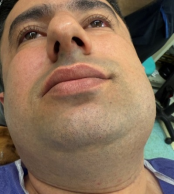
Figure 1. The image shows a firm, non-tender swelling on the left side of the mandible.
Panoramic radiograph and CBCT revealed an ill-defined, multilocular radiolucent lesion involving the left mandibular body and ramus. CBCT images showed cortical bone thinning and areas of perforation, along with mild soft tissue extension (Figure 2).
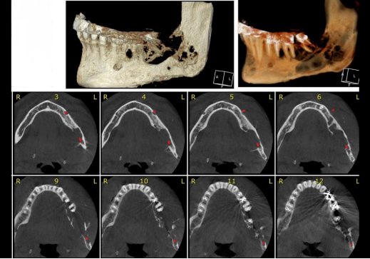
Figure 2. CBCT images showed cortical bone thinning and areas of perforation.
An incisional biopsy was performed under local anesthesia. Histopathological examination suggested AF (Figure 3). Based on this diagnosis, a segmental mandibulectomy was planned.
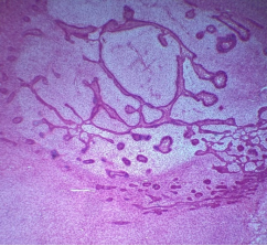
Figure 3. H&E sections (10×10) show epithelial islands and strands within a loose, cellular fibroblastic stroma resembling primitive dental papilla. Peripheral epithelial cells are low columnar. Hyaline-like material is present adjacent to the epithelial structures.
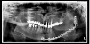
The patient underwent segmental mandibulectomy under general anesthesia. Reconstruction was performed using a reconstruction plate and an autogenous iliac crest bone graft (Figure 4). The postoperative course was uneventful.
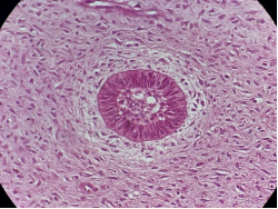
Figure 5. H&E stain (40×10 magnification) shows a mesenchymal area composed of spindle and stellate cells exhibiting mild to moderate nuclear pleomorphism, cytologic atypia, vesicular hyperchromatic nuclei, and numerous mitotic figures.
Final histopathological evaluation of the resected specimen revealed a biphasic tumor composed of benign ameloblastic epithelial islands and a malignant mesenchymal component exhibiting cellular atypia and increased mitotic activity, consistent with low-grade ameloblastic fibrosarcoma (Figure 5). Immunohistochemical analysis using the Ki-67 proliferation marker demonstrated a labeling index of [e.g., 15%], supporting the diagnosis of a low-grade neoplasm with limited proliferative activity (Figure 6).
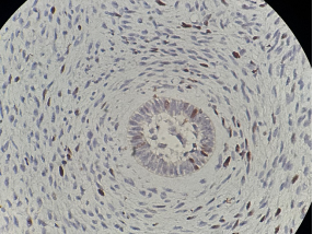
Figure 6. Immunohistochemical staining with Ki-67 (40×10 magnification) shows that approximately 15% of mesenchymal cells express the marker. Most epithelial cells exhibited only a small number of Ki-67-positive cells.
The patient was followed up for 12 months postoperatively, with no evidence of recurrence.
3. Discussion
Ameloblastic fibrosarcoma is a rare malignant odontogenic tumor characterized by benign epithelial components and a malignant mesenchymal stroma. It often arises de novo or from the malignant transformation of a pre-existing ameloblastic fibroma. The mandible is the most commonly affected site, and the tumor typically presents in the third to fourth decades of life. Diagnosing AFS is challenging due to its rarity and histological similarities to benign lesions. Limited biopsy samples (as our case) may not capture the malignant mesenchymal component, leading to misdiagnosis. For instance, a study by Kobayashi et al. reported a case where a patient initially diagnosed with AF underwent curettage but later developed AFS, highlighting the risk of misdiagnosis and the importance of thorough histopathological evaluation. Similarly, Chrcanovic et al. emphasized that incisional biopsies might not represent the entire lesion, potentially missing areas of malignant transformation
[2]
Chrcanovic BR, Brennan PA, Rahimi S, Gomez RS. Ameloblastic fibroma and ameloblastic fibrosarcoma: a systematic review. Journal of Oral Pathology & Medicine. 2018 Apr; 47(4): 315-25.
. In this case, the initial incisional biopsy suggested AF, highlighting the importance of comprehensive histopathological evaluation. “Ameloblastic fibrosarcoma is histologically graded based on the cellular atypia and mitotic activity of the mesenchymal component, with low-grade tumors showing mild atypia and limited mitotic figures, while high-grade tumors demonstrate marked pleomorphism and high proliferative indices
[4]
WHO Classification of Head and Neck Tumours. 5th ed. Lyon: International Agency for Research on Cancer (IARC); 2022. p. 317-319.
[4]
. Immunohistochemical analysis using the Ki-67 proliferation marker plays a critical role in determining the malignant potential and aggressiveness of tumors
[5]
Li LT, Jiang G, Chen Q and Zheng JN: Ki67 is a promising molecular target in the diagnosis of cancer (Review). Mol Med Rep 11: 1566-1572, 2015.
[5]
. In AFS a Ki-67 labeling index below 20% is generally indicative of a low-grade neoplasm with less aggressive clinical behavior, whereas an index exceeding 30% reflects high proliferative activity and correlates with a more aggressive phenotype and higher risk of recurrence. A Ki-67 index below 10% in AFS mirrors the low proliferative activity characteristic of AF and can lead to diagnostic confusion between these entities
[6]
Mohsenifar Z, Behrad S, Abbas FM. Epithelial Dysplasia in Ameloblastic Fibrosarcoma Arising from Recurrent Ameloblastic Fibroma in a 26-Year-Old Iranian Man. Am J Case Rep. 2015 Aug 18; 16: 548-53.
. Diagnosing AFS can be challenging, especially when relying on incisional biopsies that sample only a limited portion of the lesion. Such limited sampling may not capture the malignant features of the mesenchymal component, leading to potential misdiagnosis as AF
[7]
Gilani SM, Raza A, Al-Khafaji BM. Ameloblastic fibrosarcoma: a rare malignant odontogenic tumor. Eur Ann Otorhinolaryngol Head Neck Dis. 2014 Feb; 131(1): 53-6.
Radiographically, AFS often presents as an ill-defined, multilocular radiolucent lesion with cortical bone destruction and soft tissue extension. CBCT is valuable in assessing the extent of bone involvement and planning surgical intervention. The primary treatment for AFS is wide surgical excision with clear margins. Given the tumor’s aggressive nature and high recurrence rate, complete resection is essential. In this case, a segmental mandibulectomy was performed, followed by immediate reconstruction using a reconstruction plate and autogenous iliac bone graft. Although immediate mandibular reconstruction using an iliac crest bone graft was performed based on the initial pathological diagnosis, it is important to emphasize that such an approach is generally not recommended in malignant cases.
Immediate bone grafting in mandibular malignancies poses several concerns, including the risk of tumor recurrence, and interference with postoperative radiotherapy or radiological follow-up. Therefore, delayed reconstruction is often the preferred approach until histopathological confirmation of tumor-free margins and disease control is achieved
[8]
Coletti DP, Ord RA, Liu X. Nonvascularized bone graft reconstruction of the mandible: outcomes and factors influencing success. J Oral Maxillofac Surg. 2009 Feb; 67(2): 400-4.
[9]
Carroll C, Gill M, Bowden E, O'Connell JE, Shukla R, Sweet C. Ameloblastic Fibroma of the Mandible Reconstructed with Autogenous Parietal Bone: Report of a Case and Literature Review. Case Rep Dent. 2019 Jun 18; 2019: 5149219.
. Adjuvant radiotherapy and chemotherapy have limited roles in AFS management, with surgical excision remaining the mainstay of treatment. Regular follow-up is crucial to monitor for recurrence.
4. Conclusion
Low-grade ameloblastic fibrosarcoma is a rare malignant odontogenic tumor that may initially mimic a benign lesion both clinically and histologically. Diagnosing AFS can be challenging, especially when relying on incisional biopsies that sample only a limited portion of the lesion. Such limited sampling may not capture the malignant features of the mesenchymal component, leading to potential misdiagnosis as AF. Given these challenges, it’s crucial to correlate clinical, radiographic, and histopathological findings to ensure accurate diagnosis. When AFS is suspected, complete surgical excision with clear margins is recommended to prevent recurrence and ensure optimal treatment outcomes. Accordingly, bone grafting in cases of AF as our case, should be undertaken with caution and only after definitive histopathological confirmation of the diagnosis.
This case highlights the diagnostic challenges associated with limited biopsy samples and underscores the necessity of comprehensive histopathological evaluation. Early detection, accurate diagnosis, and complete surgical excision with clear margins are critical for optimal patient outcomes. Long-term follow-up is essential due to the risk of recurrence.
Abbreviations
AF
Ameloblastic Fibroma
AFS
Ameloblastic Fibrosarcoma
CBCT
Cone Beam Computed Tomography
Acknowledgments
The authors would like to thank the Department of Oral and Maxillofacial Surgery at Azarbayjan Hospital for their support.
Ethics Approval and Consent to Participate
Not applicable.
Consent for Publication
Written informed consent was obtained from the patient for publication of this case report and accompanying images.
Availability of Data and Materials
Not applicable.
Funding
No funding was received for this study.
Conflicts of Interest
The authors declare no conflicts of interest.
References
[1]
Heath C. Lectures on certain diseases of the jaws. Br Med J. 1887; 2(1383): 5-13.
[2]
Chrcanovic BR, Brennan PA, Rahimi S, Gomez RS. Ameloblastic fibroma and ameloblastic fibrosarcoma: a systematic review. Journal of Oral Pathology & Medicine. 2018 Apr; 47(4): 315-25.
WHO Classification of Head and Neck Tumours. 5th ed. Lyon: International Agency for Research on Cancer (IARC); 2022. p. 317-319.
[5]
Li LT, Jiang G, Chen Q and Zheng JN: Ki67 is a promising molecular target in the diagnosis of cancer (Review). Mol Med Rep 11: 1566-1572, 2015.
[6]
Mohsenifar Z, Behrad S, Abbas FM. Epithelial Dysplasia in Ameloblastic Fibrosarcoma Arising from Recurrent Ameloblastic Fibroma in a 26-Year-Old Iranian Man. Am J Case Rep. 2015 Aug 18; 16: 548-53.
Coletti DP, Ord RA, Liu X. Nonvascularized bone graft reconstruction of the mandible: outcomes and factors influencing success. J Oral Maxillofac Surg. 2009 Feb; 67(2): 400-4.
[9]
Carroll C, Gill M, Bowden E, O'Connell JE, Shukla R, Sweet C. Ameloblastic Fibroma of the Mandible Reconstructed with Autogenous Parietal Bone: Report of a Case and Literature Review. Case Rep Dent. 2019 Jun 18; 2019: 5149219.
Motlagh, M. F., Mostafazadhe, S., Motlagh, F. F. (2025). Low-Grade Ameloblastic Fibrosarcoma of the Mandible: A Case Report with Initial Misdiagnosis. International Journal of Clinical Oral and Maxillofacial Surgery, 11(2), 58-62. https://doi.org/10.11648/j.ijcoms.20251102.11
Motlagh, M. F.; Mostafazadhe, S.; Motlagh, F. F. Low-Grade Ameloblastic Fibrosarcoma of the Mandible: A Case Report with Initial Misdiagnosis. Int. J. Clin. Oral Maxillofac. Surg.2025, 11(2), 58-62. doi: 10.11648/j.ijcoms.20251102.11
Motlagh MF, Mostafazadhe S, Motlagh FF. Low-Grade Ameloblastic Fibrosarcoma of the Mandible: A Case Report with Initial Misdiagnosis. Int J Clin Oral Maxillofac Surg. 2025;11(2):58-62. doi: 10.11648/j.ijcoms.20251102.11
@article{10.11648/j.ijcoms.20251102.11,
author = {Masoud Fallahi Motlagh and Samira Mostafazadhe and Farbod Fallahi Motlagh},
title = {Low-Grade Ameloblastic Fibrosarcoma of the Mandible: A Case Report with Initial Misdiagnosis
},
journal = {International Journal of Clinical Oral and Maxillofacial Surgery},
volume = {11},
number = {2},
pages = {58-62},
doi = {10.11648/j.ijcoms.20251102.11},
url = {https://doi.org/10.11648/j.ijcoms.20251102.11},
eprint = {https://article.sciencepublishinggroup.com/pdf/10.11648.j.ijcoms.20251102.11},
abstract = {Background: Ameloblastic fibrosarcoma (AFS) is a rare malignant odontogenic tumor characterized by benign epithelial components and a malignant mesenchymal stroma. Accurate diagnosis is challenging, especially when initial biopsy samples are limited. Case Presentation: A 35-year-old male presented with a progressively enlarging swelling on the left side of his face. Clinical examination revealed a firm, non-tender mass in the left mandibular region, resulting in mild facial asymmetry. Intraoral evaluation showed buccal cortical expansion in the posterior mandible, accompanied by slight mobility of the adjacent teeth. Panoramic radiograph and cone beam computed tomography (CBCT) revealed an ill-defined, multilocular radiolucent lesion involving the left mandibular body and ramus, with cortical bone perforation and mild soft tissue extension. An incisional biopsy initially diagnosed ameloblastic fibroma (AF). The patient underwent segmental mandibulectomy, and final histopathological evaluation revealed low-grade ameloblastic fibrosarcoma. Reconstruction was performed using a reconstruction plate and autogenous iliac bone graft. The postoperative course was uneventful, with no recurrence observed during a 6-month follow-up. Conclusions: This case underscores the diagnostic challenges of AFS and highlights the importance of comprehensive histopathological evaluation. Early detection, accurate diagnosis, and complete surgical excision with clear margins are critical for best outcomes.},
year = {2025}
}
TY - JOUR
T1 - Low-Grade Ameloblastic Fibrosarcoma of the Mandible: A Case Report with Initial Misdiagnosis
AU - Masoud Fallahi Motlagh
AU - Samira Mostafazadhe
AU - Farbod Fallahi Motlagh
Y1 - 2025/07/24
PY - 2025
N1 - https://doi.org/10.11648/j.ijcoms.20251102.11
DO - 10.11648/j.ijcoms.20251102.11
T2 - International Journal of Clinical Oral and Maxillofacial Surgery
JF - International Journal of Clinical Oral and Maxillofacial Surgery
JO - International Journal of Clinical Oral and Maxillofacial Surgery
SP - 58
EP - 62
PB - Science Publishing Group
SN - 2472-1344
UR - https://doi.org/10.11648/j.ijcoms.20251102.11
AB - Background: Ameloblastic fibrosarcoma (AFS) is a rare malignant odontogenic tumor characterized by benign epithelial components and a malignant mesenchymal stroma. Accurate diagnosis is challenging, especially when initial biopsy samples are limited. Case Presentation: A 35-year-old male presented with a progressively enlarging swelling on the left side of his face. Clinical examination revealed a firm, non-tender mass in the left mandibular region, resulting in mild facial asymmetry. Intraoral evaluation showed buccal cortical expansion in the posterior mandible, accompanied by slight mobility of the adjacent teeth. Panoramic radiograph and cone beam computed tomography (CBCT) revealed an ill-defined, multilocular radiolucent lesion involving the left mandibular body and ramus, with cortical bone perforation and mild soft tissue extension. An incisional biopsy initially diagnosed ameloblastic fibroma (AF). The patient underwent segmental mandibulectomy, and final histopathological evaluation revealed low-grade ameloblastic fibrosarcoma. Reconstruction was performed using a reconstruction plate and autogenous iliac bone graft. The postoperative course was uneventful, with no recurrence observed during a 6-month follow-up. Conclusions: This case underscores the diagnostic challenges of AFS and highlights the importance of comprehensive histopathological evaluation. Early detection, accurate diagnosis, and complete surgical excision with clear margins are critical for best outcomes.
VL - 11
IS - 2
ER -