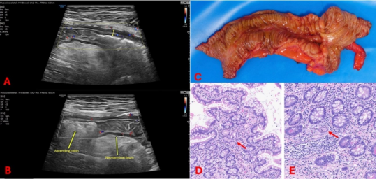
Crohn’s disease is a chronic and relapsing inflammatory condition that often requires surgical intervention. Post-operative recurrence of Crohn’s disease remains common after surgery occurring in up to 75% of patients. The role of disease-free surgical margins in post-operative recurrence has been debated, however, it has been identified as an independent risk factor for recurrence. We present a case of a woman with obstructive Crohn’s disease who underwent ileocecectomy and was maintained on uninterrupted infliximab therapy. Despite operative and pathology reports describing grossly normal surgical margins, three months post-operation the patient was experiencing new intermittent lower abdominal pain and intestinal ultrasound revealed active ileitis involving a 10cm segment immediately proximal to the ileocolonic anastomosis. Due to concern of incomplete resection, microscopic re-evaluation of the margins was conducted and confirmed histologically positive margins of active Crohn’s disease. Current guidelines have called for additional studies to examine the role of disease-free margins. Our case demonstrates the limitations of gross evaluation of disease-free margins and suggests a potential application of intestinal ultrasound intraoperatively for such assessment. While intestinal ultrasound has been utilized intraoperatively for colonic malignant margin identification, it has not yet been studied in inflammatory bowel disease and requires further evaluation.
| Published in | International Journal of Gastroenterology (Volume 9, Issue 2) |
| DOI | 10.11648/j.ijg.20250902.11 |
| Page(s) | 90-93 |
| Creative Commons |
This is an Open Access article, distributed under the terms of the Creative Commons Attribution 4.0 International License (http://creativecommons.org/licenses/by/4.0/), which permits unrestricted use, distribution and reproduction in any medium or format, provided the original work is properly cited. |
| Copyright |
Copyright © The Author(s), 2025. Published by Science Publishing Group |
Intestinal Ultrasound, Crohn’s Disease, Post-operative Recurrence, Surgical Margins, Inflammatory Bowel Disease
POR | Post-Operative Recurrence |
CD | Crohn’s Disease |
IUS | Ntestinal Ultrasound |
TI | Terminal Ileum |
| [1] | Orlando A, Mocciaro F, Renna S, et al. Early post-operative endoscopic recurrence in Crohn's disease patients: data from an Italian Group for the study of inflammatory bowel disease (IG-IBD) study on a large prospective multicenter cohort. J Crohns Colitis. 2014 Oct; 8(10): 1217-21. |
| [2] | Gisbert JP, Chaparro M. Anti-TNF agents and new biological agents (vedolizumab and ustekinumab) in the prevention and treatment of postoperative recurrence after surgery in Crohn's disease. Drugs. 2023 Sep; 83(13): 1179-1205. |
| [3] | Wolff BG. Resection margins in Crohn's disease. Br J Surg. 2001; 88(6): 771-2. |
| [4] | de Buck van Overstraeten A, Eshuis EJ, Vermeire S, et al. Short- and medium-term outcomes following primary ileocaecal resection for Crohn's disease in two specialist centres. Br J Surg. 2017 Nov; 104(12): 1713-22. |
| [5] | Maaser C, Sturm A, Vavricka SR, Kucharzik T, Fiorino G, Annese V, Calabrese E, Baumgart DC, Bettenworth D, Borralho Nunes P, Burisch J, Castiglione F, Eliakim R, Ellul P, González-Lama Y, Gordon H, Halligan S, Katsanos K, Kopylov U, Kotze PG, Krustinš E, Laghi A, Limdi JK, Rieder F, Rimola J, Taylor SA, Tolan D, van Rheenen P, Verstockt B, Stoker J; European Crohn’s and Colitis Organisation [ECCO] and the European Society of Gastrointestinal and Abdominal Radiology [ESGAR]. ECCO-ESGAR Guideline for Diagnostic Assessment in IBD Part 1: Initial diagnosis, monitoring of known IBD, detection of complications. J Crohns Colitis. 2019 Feb 1; 13(2): 144-164. |
| [6] | Gionchetti P, Dignass A, Danese S, et al. 3rd European evidence-based consensus on the diagnosis and management of Crohn’s disease 2016: Part 2: Surgical management and special situations. J Crohns Colitis. 2017 Feb; 11(2): 135-49. |
| [7] | Fazio VW, Marchetti F, Church M, et al. Effect of resection margins on the recurrence of Crohn's disease in the small bowel: a randomized controlled trial. Ann Surg. 1996; 224(4): 563-73. |
| [8] | Nguyen GC, Loftus EV Jr, Hirano I, et al. American Gastroenterological Association Institute guideline on the management of Crohn's disease after surgical resection. Gastroenterology. 2017; 152(1): 271-5. |
| [9] | Kelm M, Benatzky C, Buck V, et al. Positive resection margins in Crohn's disease are a relevant risk factor for postoperative disease recurrence. Sci Rep. 2024 May 11; 14(1): 10823. |
| [10] | Wasmann KATGM, van Amesfoort J, van Montfoort ML, et al. The predictive value of inflammation at ileocecal resection margins for postoperative Crohn’s recurrence: a cohort study. Inflamm Bowel Dis. 2020 Nov; 26(11): 1691-9. |
| [11] | Riault C, Diouf M, Chatelain D, et al. Positive histologic margins as a risk factor of recurrence after ileocaecal resection in Crohn's disease. Clin Res Hepatol Gastroenterol. 2021 Sep; 45(5): 101569. |
| [12] | Poredska K, Kunovsky L, Marek F, et al. The influence of microscopic inflammation at resection margins on early postoperative endoscopic recurrence after ileocaecal resection for Crohn’s disease. J Crohns Colitis. 2020 Mar; 14(3): 361-8. |
| [13] | Yzet C, Riault C, Brazier F, et al. Positive margins and plexitis increase the risk of recurrence after ileocecal resection: a systematic review and meta-analysis. Dig Liver Dis. 2023; 55(12): 1611-20. |
| [14] | Ryan JM, Rogers AC, O'Toole A, Burke JP. Meta-analysis of histological margin positivity in the prediction of recurrence after Crohn's resection. Dis Colon Rectum. 2019; 62(7): 882-92. |
| [15] | Tandon P, Malhi G, Abdali D, et al. Active margins, plexitis, and granulomas increase postoperative Crohn's recurrence: systematic review and meta-analysis. Clin Gastroenterol Hepatol. 2021; 19(3): 451-62. |
| [16] | Ripollés T, Poza J, Suarez Ferrer C, Martínez-Pérez MJ, Martín-Algíbez A, de Las Heras Paez B. Evaluation of Crohn’s disease activity: development of an ultrasound score in a multicenter study. Inflamm Bowel Dis. 2021; 27(1): 145–154. |
| [17] | Greif F, Aranovich D, Hananel N, et al. Intraoperative ultrasound in colorectal surgery. J Clin Ultrasound. 2009 Jul; 37(7): 375-9. |
| [18] | Panaro F, Casaccia M, Cavaliere D, Torelli P. Laparoscopic colon resection with intraoperative polyp localisation with high resolution ultrasonography coupled with colour power Doppler. Postgrad Med J. 2003; 79(935): 533-4. |
APA Style
Cleveland, N. K., Duty, C. M., Bhondwe, K. S., Alpert, L. (2025). Rethinking Intra-operative Management of Crohn’s Disease: Intestinal Ultrasound Detects Microscopic Disease Unidentified at Ileal Margin Resection. International Journal of Gastroenterology, 9(2), 90-93. https://doi.org/10.11648/j.ijg.20250902.11
ACS Style
Cleveland, N. K.; Duty, C. M.; Bhondwe, K. S.; Alpert, L. Rethinking Intra-operative Management of Crohn’s Disease: Intestinal Ultrasound Detects Microscopic Disease Unidentified at Ileal Margin Resection. Int. J. Gastroenterol. 2025, 9(2), 90-93. doi: 10.11648/j.ijg.20250902.11
@article{10.11648/j.ijg.20250902.11,
  author = {Noa Krugliak Cleveland and Charlotte Mary Duty and Khushi Snehdhan Bhondwe and Lindsay Alpert},
  title = {Rethinking Intra-operative Management of Crohn’s Disease: Intestinal Ultrasound Detects Microscopic Disease Unidentified at Ileal Margin Resection
},
  journal = {International Journal of Gastroenterology},
  volume = {9},
  number = {2},
  pages = {90-93},
  doi = {10.11648/j.ijg.20250902.11},
  url = {https://doi.org/10.11648/j.ijg.20250902.11},
  eprint = {https://article.sciencepublishinggroup.com/pdf/10.11648.j.ijg.20250902.11},
  abstract = {Crohn’s disease is a chronic and relapsing inflammatory condition that often requires surgical intervention. Post-operative recurrence of Crohn’s disease remains common after surgery occurring in up to 75% of patients. The role of disease-free surgical margins in post-operative recurrence has been debated, however, it has been identified as an independent risk factor for recurrence. We present a case of a woman with obstructive Crohn’s disease who underwent ileocecectomy and was maintained on uninterrupted infliximab therapy. Despite operative and pathology reports describing grossly normal surgical margins, three months post-operation the patient was experiencing new intermittent lower abdominal pain and intestinal ultrasound revealed active ileitis involving a 10cm segment immediately proximal to the ileocolonic anastomosis. Due to concern of incomplete resection, microscopic re-evaluation of the margins was conducted and confirmed histologically positive margins of active Crohn’s disease. Current guidelines have called for additional studies to examine the role of disease-free margins. Our case demonstrates the limitations of gross evaluation of disease-free margins and suggests a potential application of intestinal ultrasound intraoperatively for such assessment. While intestinal ultrasound has been utilized intraoperatively for colonic malignant margin identification, it has not yet been studied in inflammatory bowel disease and requires further evaluation.},
 year = {2025}
}
TY  - JOUR
T1  - Rethinking Intra-operative Management of Crohn’s Disease: Intestinal Ultrasound Detects Microscopic Disease Unidentified at Ileal Margin Resection

AU  - Noa Krugliak Cleveland
AU  - Charlotte Mary Duty
AU  - Khushi Snehdhan Bhondwe
AU  - Lindsay Alpert
Y1  - 2025/07/23
PY  - 2025
N1  - https://doi.org/10.11648/j.ijg.20250902.11
DO  - 10.11648/j.ijg.20250902.11
T2  - International Journal of Gastroenterology
JF  - International Journal of Gastroenterology
JO  - International Journal of Gastroenterology
SP  - 90
EP  - 93
PB  - Science Publishing Group
SN  - 2640-169X
UR  - https://doi.org/10.11648/j.ijg.20250902.11
AB  - Crohn’s disease is a chronic and relapsing inflammatory condition that often requires surgical intervention. Post-operative recurrence of Crohn’s disease remains common after surgery occurring in up to 75% of patients. The role of disease-free surgical margins in post-operative recurrence has been debated, however, it has been identified as an independent risk factor for recurrence. We present a case of a woman with obstructive Crohn’s disease who underwent ileocecectomy and was maintained on uninterrupted infliximab therapy. Despite operative and pathology reports describing grossly normal surgical margins, three months post-operation the patient was experiencing new intermittent lower abdominal pain and intestinal ultrasound revealed active ileitis involving a 10cm segment immediately proximal to the ileocolonic anastomosis. Due to concern of incomplete resection, microscopic re-evaluation of the margins was conducted and confirmed histologically positive margins of active Crohn’s disease. Current guidelines have called for additional studies to examine the role of disease-free margins. Our case demonstrates the limitations of gross evaluation of disease-free margins and suggests a potential application of intestinal ultrasound intraoperatively for such assessment. While intestinal ultrasound has been utilized intraoperatively for colonic malignant margin identification, it has not yet been studied in inflammatory bowel disease and requires further evaluation.
VL  - 9
IS  - 2
ER  -
Inflammatory Bowel Disease Center, University of Chicago Medicine, Chicago, United States
Inflammatory Bowel Disease Center, University of Chicago Medicine, Chicago, United States
Inflammatory Bowel Disease Center, University of Chicago Medicine, Chicago, United States
Department of Pathology, University of Chicago Medical Center and Biological Sciences, Chicago, United States
Information