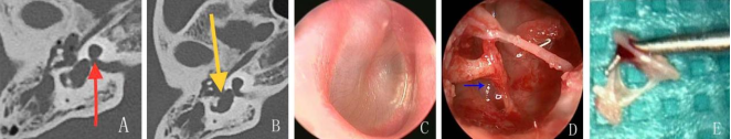
Objective: To summarize the experience in diagnosis and treatment of patients with cerebrospinal fluid otorrhea secondary to congenital inner ear malformations and surgical operation treatment methods. Methods: The clinical data of a patient with cerebrospinal fluid otorrhea secondary to congenital inner ear malformations, Mondini dysplasia, was retrospectively analyzed, and the cerebrospinal otorrhea packing repair was performed transcanal endoscopic. Results: The patient was misdiagnosed and improperly treated before the surgery. The patient was successfully treated with transcanal endoscopic surgical exploration revealed a missing leak in the patella floor plate, patellar muscle and myofascial membrane were used to pack the inner ear, seal the vestibular window. There was no cerebrospinal fluid otorrhea and recurrence of meningitis after postoperative follow-up of 1 year. Conclusions: It reminds that patient who has hearing impairment associated with recurrent meningitis, otorrhea, or rhinorrhea should be evaluated for the possibility of this congenital dysplasia, and early diagnosis and prompt surgical intervention may prevent further episodes. It a condition that requires elevated clinical vigilance.
| Published in | International Journal of Otorhinolaryngology (Volume 11, Issue 2) |
| DOI | 10.11648/j.ijo.20251102.15 |
| Page(s) | 28-31 |
| Creative Commons |
This is an Open Access article, distributed under the terms of the Creative Commons Attribution 4.0 International License (http://creativecommons.org/licenses/by/4.0/), which permits unrestricted use, distribution and reproduction in any medium or format, provided the original work is properly cited. |
| Copyright |
Copyright © The Author(s), 2025. Published by Science Publishing Group |
Cerebrospinal Fluid, Inner Ear Malformations, Recurrent Meningitis, Sensorineural Hearing Loss
CFO | Cerebrospinal Fluid Otorrhea |
IEM | Inner Ear Malformations |
CT | Computed Tomography |
SFF | Stapes Floor Plate Fistula |
ABR | Auditory Brainstem Response |
| [1] | Masuda S., Usui S., Matsunaga T. High prevalence of inner-ear and/or internal auditory canal malformation in children with unilateral sensorineural hearing loss. Int J Pediatr Otorhinolaryngol, 2013, 77: 228-232. |
| [2] | Sennaroğlu L., Bajin MD. Classification and Current Management of Inner Ear Malformations. Balkan Med J, 2017, 34: 397-411. |
| [3] | Wu H, Cai XZ. Progress in diagnosis and treatment of cerebrospinal fluid otorrhea caused by inner ear malformation. Lin Chuang Er Bi Yan Hou Tou Jing Wai Ke Za Zhi. 2021; 35(11): 1048-1052. |
| [4] | Sennaroğlu L., Bajin MD. Management of stapes footplate fistula in inner ear malformations. Int J Pediatr Otorhinolaryngol, 2021, 140: 110525. |
| [5] | Ohlms LA., Edwards MS., Mason EO., Igarashi M., Alford BR., Smith RJ. Recurrent meningitis and Mondini dysplasia. Arch Otolaryngol Head Neck Surg, 1990, 116: 608-612. |
| [6] | Muzzi E., Battelino S., Gregori M., Pellegrin A., Orzan E. Life-threatening unilateral hearing impairments. Review of the literature on the association between inner ear malformation and meningitis. Int J Pediatr Otorhinolaryngol, 2015, 79: 1969-1974. |
| [7] | Deng W., Liu J., Pang F., Zhang XM. Diagnosis and management of pediatric cerebrospinal fluid leakage secondary to inner ear malformations: A report of 13 case.Int J Pediatr Otorhinolaryngol, 2020, 135: 110049. |
| [8] | Thomassin JM., Korchia D., Doris JM. Endoscopic-guided otosurgery in the prevention of residual cholesteatomas. Laryngoscope, 1993, 103: 939-943. |
| [9] | Kou YF, Zhu VF, Kutz JW, et al. Transcanal Endoscopic Management of Cerebrospinal Fluid Otorrhea Secondary to Congenital Inner Ear Malformations. Otology & neurotology. 2016; 37(1): 62-5. |
| [10] | Bois E, Demondion S, Elmaleh M, et al. Surgical Treatment for Cerebrospinal Fluid Leaks in Patients With Inner Ear Malformations. Otology & neurotology. 2020; 41(8): 1102-1107. |
| [11] | Tyagi I., Syal R., Goyal A. Cerebrospinal fluid otorhinorrhoea due to inner-ear Malformations: clinical presentation and new perspectives in management. J Laryngol Otol, 2005, 119: 714-718. |
| [12] | Lien TH., Fu CM., Hsu CJ., Lu L., Peng SS., Chang LY. Recurrent bacterial meningitis associated with Mondini dysplasia. Pediatr Neonatol, 2011, 52: 294-296. |
| [13] | Isaacson B., Booth T., Kutz Jr JW., Lee KH., Roland PS. Labyrinthitis ossificans: how accurate is MRI in predicting cochlear obstruction? Otolaryngol Head Neck Surg, 2009, 140: 692-696. |
| [14] | Adams GL, McCoid G, Weisbeski D. Cerebrospinal fluid otorrhea presenting as serous otitis media. Minn Med. 1982; 65(7): 410-5. PMID: 7202112. |
| [15] | Stone JA, Castillo M, Neelon B, et al. Evaluation of CSF leaks: high-resolution CT compared with contrast-enhanced CT and radionuclide cisternography. AJNR Am J Neuroradiol. 1999; 20(4): 706-12. PMID: 10319986. |
APA Style
Yang, M., Guan, Y., Zhang, B., Liang, X., Chen, H. (2025). Cerebrospinal Fluid Otorrhea from Congenital Inner Ear Malformations: Clinical Experience and Lessons of a Case Report. International Journal of Otorhinolaryngology, 11(2), 28-31. https://doi.org/10.11648/j.ijo.20251102.15
ACS Style
Yang, M.; Guan, Y.; Zhang, B.; Liang, X.; Chen, H. Cerebrospinal Fluid Otorrhea from Congenital Inner Ear Malformations: Clinical Experience and Lessons of a Case Report. Int. J. Otorhinolaryngol. 2025, 11(2), 28-31. doi: 10.11648/j.ijo.20251102.15
@article{10.11648/j.ijo.20251102.15,
  author = {Mingbao Yang and Yafeng Guan and Bei Zhang and Xiuni Liang and Heng Chen},
  title = {Cerebrospinal Fluid Otorrhea from Congenital Inner Ear Malformations: Clinical Experience and Lessons of a Case Report
},
  journal = {International Journal of Otorhinolaryngology},
  volume = {11},
  number = {2},
  pages = {28-31},
  doi = {10.11648/j.ijo.20251102.15},
  url = {https://doi.org/10.11648/j.ijo.20251102.15},
  eprint = {https://article.sciencepublishinggroup.com/pdf/10.11648.j.ijo.20251102.15},
  abstract = {Objective: To summarize the experience in diagnosis and treatment of patients with cerebrospinal fluid otorrhea secondary to congenital inner ear malformations and surgical operation treatment methods. Methods: The clinical data of a patient with cerebrospinal fluid otorrhea secondary to congenital inner ear malformations, Mondini dysplasia, was retrospectively analyzed, and the cerebrospinal otorrhea packing repair was performed transcanal endoscopic. Results: The patient was misdiagnosed and improperly treated before the surgery. The patient was successfully treated with transcanal endoscopic surgical exploration revealed a missing leak in the patella floor plate, patellar muscle and myofascial membrane were used to pack the inner ear, seal the vestibular window. There was no cerebrospinal fluid otorrhea and recurrence of meningitis after postoperative follow-up of 1 year. Conclusions: It reminds that patient who has hearing impairment associated with recurrent meningitis, otorrhea, or rhinorrhea should be evaluated for the possibility of this congenital dysplasia, and early diagnosis and prompt surgical intervention may prevent further episodes. It a condition that requires elevated clinical vigilance.
},
 year = {2025}
}
TY  - JOUR
T1  - Cerebrospinal Fluid Otorrhea from Congenital Inner Ear Malformations: Clinical Experience and Lessons of a Case Report

AU  - Mingbao Yang
AU  - Yafeng Guan
AU  - Bei Zhang
AU  - Xiuni Liang
AU  - Heng Chen
Y1  - 2025/09/03
PY  - 2025
N1  - https://doi.org/10.11648/j.ijo.20251102.15
DO  - 10.11648/j.ijo.20251102.15
T2  - International Journal of Otorhinolaryngology
JF  - International Journal of Otorhinolaryngology
JO  - International Journal of Otorhinolaryngology
SP  - 28
EP  - 31
PB  - Science Publishing Group
SN  - 2472-2413
UR  - https://doi.org/10.11648/j.ijo.20251102.15
AB  - Objective: To summarize the experience in diagnosis and treatment of patients with cerebrospinal fluid otorrhea secondary to congenital inner ear malformations and surgical operation treatment methods. Methods: The clinical data of a patient with cerebrospinal fluid otorrhea secondary to congenital inner ear malformations, Mondini dysplasia, was retrospectively analyzed, and the cerebrospinal otorrhea packing repair was performed transcanal endoscopic. Results: The patient was misdiagnosed and improperly treated before the surgery. The patient was successfully treated with transcanal endoscopic surgical exploration revealed a missing leak in the patella floor plate, patellar muscle and myofascial membrane were used to pack the inner ear, seal the vestibular window. There was no cerebrospinal fluid otorrhea and recurrence of meningitis after postoperative follow-up of 1 year. Conclusions: It reminds that patient who has hearing impairment associated with recurrent meningitis, otorrhea, or rhinorrhea should be evaluated for the possibility of this congenital dysplasia, and early diagnosis and prompt surgical intervention may prevent further episodes. It a condition that requires elevated clinical vigilance.

VL  - 11
IS  - 2
ER  -
Department of Otorhinolaryngology, The University of Hong Kong-Shenzhen Hospital, Shenzhen, China
Information