Spindle cell carcinoma (SpCC) is a rare and aggressive variant of squamous cell carcinoma characterized by a distinctive spindle-cell morphology and variable epithelial differentiation. It most frequently develops in the mucosal surfaces of the upper aerodigestive tract, particularly the larynx, hypopharynx, and oral cavity. Although maxillofacial involvement can occur, recurrent SpCC involving the cheek and mandibular region represents an uncommon clinical presentation and may pose significant diagnostic and therapeutic challenges. We report a case of recurrent spindle cell carcinoma involving the cheek and mandibular region following previous treatment. The patient presented with clinical features suggestive of a recurrent malignant lesion, and radiological evaluation demonstrated tumour involvement of the affected maxillofacial region. Histopathological examination revealed a highly atypical spindle-cell proliferation with malignant cytological features, raising consideration of several differential diagnoses, including spindle cell sarcoma, malignant fibrous histiocytoma, and other mesenchymal neoplasms. Given the unusual morphology and anatomical location, immunohistochemical evaluation was performed using a broad panel of epithelial and mesenchymal markers. The immunoprofile demonstrated epithelial differentiation and supported the diagnosis of spindle cell carcinoma, allowing distinction from primary mesenchymal spindle-cell malignancies. The recurrent nature of the lesion further emphasized the aggressive biological behaviour and potential for local recurrence associated with SpCC. Management of these tumours requires careful correlation of clinical, radiological, histopathological, and immunohistochemical findings, particularly when conventional squamous differentiation is inconspicuous. This case highlights the diagnostic complexity of recurrent SpCC in an uncommon cheek and mandibular location and emphasizes the importance of maintaining a high index of suspicion when evaluating recurrent spindle-cell lesions of the maxillofacial region. Early recognition, accurate pathological diagnosis, appropriate assessment of local and regional disease, and multidisciplinary management are essential for optimizing treatment and follow-up. Awareness of this rare presentation may facilitate timely diagnosis and improve clinical decision-making in patients with recurrent maxillofacial spindle cell carcinoma.
This is an Open Access article, distributed under the terms of the Creative Commons Attribution 4.0 International License (http://creativecommons.org/licenses/by/4.0/), which permits unrestricted use, distribution and reproduction in any medium or format, provided the original work is properly cited.
Spindle Cell Carcinoma, Spindle Cell Lesion, Sarcomatous Lesions, Case Report
1. Introduction
Spindle cell carcinoma (SpCC) is an uncommon, poorly differentiated variant of squamous cell carcinoma characterized by a mixture of malignant epithelial and spindle-cell features
[1]
Wernheden E, Trøstrup H, Pedersen Pilt A. Unusual Presentation of Cutaneous Spindle Cell Squamous Cell Carcinoma: A Case Report. Case Rep Dermatol. 2020; 12(1): 70-75.
Leonardi E, Dalri P, Pusiol T, Valdagni R, Piscioli F. Spindle-Cell Squamous Carcinoma of Head and Neck Region: a Clinicopathologic and Immunohistochemical Study of Eight Cases. ORL. 1986; 48(5): 275-281.
Histologically, SpCC may be dominated by spindle-shaped malignant cells that resemble soft-tissue sarcomas, making the diagnosis challenging. Immunohistochemical markers, particularly epithelial markers such as cytokeratins together with mesenchymal markers such as vimentin, can help distinguish SpCC from other spindle-cell neoplasms
[2]
Leonardi E, Dalri P, Pusiol T, Valdagni R, Piscioli F. Spindle-Cell Squamous Carcinoma of Head and Neck Region: a Clinicopathologic and Immunohistochemical Study of Eight Cases. ORL. 1986; 48(5): 275-281.
Alem HB, AlNoury MK. Management of spindle cell carcinoma of the maxillary sinus: a case report and literature review. Am J Case Rep. 2014; 15: 454-458.
. Although SpCC most often involves the larynx and hypopharynx, simultaneous involvement of the cheek and mandibular region is uncommon
[1]
Wernheden E, Trøstrup H, Pedersen Pilt A. Unusual Presentation of Cutaneous Spindle Cell Squamous Cell Carcinoma: A Case Report. Case Rep Dermatol. 2020; 12(1): 70-75.
Leonardi E, Dalri P, Pusiol T, Valdagni R, Piscioli F. Spindle-Cell Squamous Carcinoma of Head and Neck Region: a Clinicopathologic and Immunohistochemical Study of Eight Cases. ORL. 1986; 48(5): 275-281.
Ohba S, Yoshimura H, Matsuda S, Imamura Y, Sano K. Spindle cell carcinoma arising at the buccal mucosa: a case report and review of the literature. CRANIO®. 2015; 33(1): 42-45.
The pathogenesis of SpCC remains incompletely understood. Reported associations include established risk factors for squamous cell carcinoma, particularly tobacco and alcohol exposure, as well as previous radiation. Because maxillofacial presentations are uncommon, published evidence concerning their clinical presentation, diagnosis, and management remains limited. This report describes a recurrent spindle-cell carcinoma in the cheek region and emphasizes the diagnostic challenges associated with this unusual presentation.
The case also emphasizes the value of integrating clinical examination, imaging, histopathology, and immunohistochemistry when evaluating rare spindle-cell malignancies of the head and neck.
2. Case Presentation
A 26-year-old Asian, Muslim, male from Nowshera, Punjab,, Pakistan, presented to the OPD tertiary care hospital in Gujranwala on May 5, 2025, with the complaint of recurrent swelling over the right cheek. The patient also had swelling over the right cheek three years ago, for which he underwent surgical excision 2.5 years prior. The patient is a known smoker and currently on no medication. He now reports a recurrence of the lump at the same site. There is a history of previous surgery for a similar lump in the right cheek area.
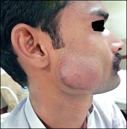
An ovoid lump was noted over the right cheek on physical examination and shown in Figure 1, suggesting possible deep tissue involvement. The swelling is non-tender and is warm compared to the rest of the body. The skin over the swelling is non-mobile, and the swelling is hard in consistency. Moreover, the buccal mucosa was intact with no visible lesions.
Ultrasonography (USG) of the right parotid gland shows an ill-defined fixed echogenic lesion of size 4.4 x 1.8cm seen in the lump. The right parotid region with internal cystic compartment is seen.
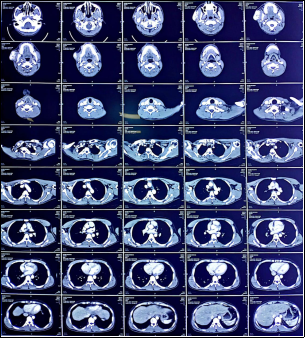
Magnetic resonance imaging (MRI) of the face and neck with contrast revealed T1 intermediate, T2 bright signal intensity noted in the right cheek of the subcutaneous plane bulge as shown in Figure 2. The overlying skin causes pushing and compressing of the underlying muscles and infiltrating the masseter muscle. The lesion caused a visible bulge and compressed the underlying structures. It infiltrated the right masseter muscle and measured approximately 5.1 × 2.5 cm. On post-contrast, images show intense enhancement.
Figure 2. Magnetic resonance imaging of the face and neck demonstrating the right cheek lesion with extension toward the masseter muscle.
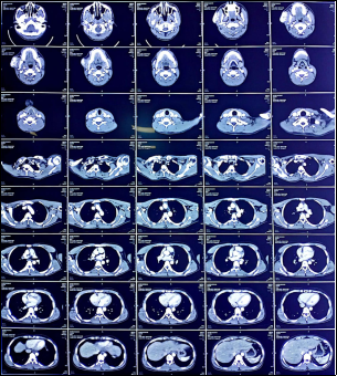
A CT scan of the chest without contrast showed a well-defined, avidly enhancing soft tissue density lesion as shown in Figure 3, measuring 4.8 × 2.8 × 4.2 cm (AP × TR × CC), located deep to the subcutaneous tissue, caudal to the superior lobe of the right parotid gland, with evidence of infiltration into the right masseter muscle. There were bilateral round subcentimetric cervical lymph nodes at level ׀ and ׀׀, more on the right side.
Figure 3. Computed tomography demonstrating the soft-tissue lesion in the right cheek region.
Additionally, on CT scan, a subpleural patch of ground-glass haze was noted in the basal segments of the bilateral lower lobes, likely due to the gravitational effect as shown in Figure 3.
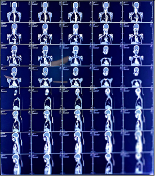
The CT scan findings are suggestive of a well-defined avidly enhancing soft tissue density lesion seen just deep to the subcutaneous region in the right cheek as shown in Figure 4.
Fine-needle aspiration cytology (FNAC) confirmed the diagnosis of a low-grade spindle cell neoplasm. Given the recurrence, the patient underwent re-excision of the swelling.
After surgical excision of the lump, it was sent for histopathological examination with the following gross features: formalin dipped with an oriented skin-covered excision measuring 80mm from superior to inferior, 48mm from medial to lateral, and 30mm from surface to deep. The overlying skin ellipse measures 80 x 45mm. It did not show any ulceration or scar mark. The specimen was sliced from medial to lateral into 6 slices. A well-defined loculated lesion was noted just below the skin in the subcutaneous plane, measuring 46 x 28 x 21 mm.
The lesion is present at a distance of 20mm from inferior margin (inked green), 10mm from superior margin (inked red), 2mm from skin, 6mm from deep margin (inked black), 8mm from medial margin (inked yellow) and 5mm from lateral margin (inked blue). The portion of the salivary gland is also noted at the deep margin, which appears grossly unremarkable.
On micro, sections of the lesion are composed of varying-sized blood vessels lined by endothelial cells. Background stroma is sclerosed/fibrosed. No atypical features were seen.
On immunohistochemical staining, CD31 is positive, but CAMTA is negative.
Figure 4. Computed tomography demonstrating the right cheek soft-tissue lesion and associated findings.
3. Discussion
Spindle cell carcinoma is a malignant epithelial neoplasm with prominent spindle-cell morphology and the capacity for local invasion and metastasis. Sarcomatoid (spindle-cell) carcinoma is an uncommon variant of squamous cell carcinoma and has been reported to account for approximately 3% of squamous carcinomas of the head and neck
[6]
Thompson LDR, Wieneke JA, Miettinen M, Heffner DK. Spindle Cell (Sarcomatoid) Carcinomas of the Larynx: A Clinicopathologic Study of 187 Cases. Am J Surg Pathol. 2002; 26(2).
. Various historical terms—including carcinosarcoma, pseudosarcoma, and squamous cell carcinoma with pseudosarcoma—have been used for this lesion. Current terminology recognizes it as a morphologically biphasic tumour in which surface epithelial dysplasia or carcinoma is associated with an underlying malignant spindle-cell proliferation
[7]
Thompson LDR. Squamous cell carcinoma variants of the head and neck. Curr Diagnostic Pathol. 2003; 9(6): 384-396.
Tobacco exposure, including both smoked and smokeless forms, and alcohol consumption are recognized risk factors for squamous cell carcinoma and have also been associated with sarcomatoid carcinoma
[6]
Thompson LDR, Wieneke JA, Miettinen M, Heffner DK. Spindle Cell (Sarcomatoid) Carcinomas of the Larynx: A Clinicopathologic Study of 187 Cases. Am J Surg Pathol. 2002; 26(2).
Evidence from clinicopathologic and molecular studies supports a monoclonal relationship between the sarcomatoid and conventional squamous components, with the spindle-cell component considered to represent dedifferentiation and progression from the conventional carcinoma
[10]
Lambert PR, Ward PH, Berci G. Pseudosarcoma of the Larynx: A Comprehensive Analysis. Arch Otolaryngol. 1980; 106(11): 700-708.
. The differential diagnosis can include fibrosarcoma and other spindle-cell tumours; the distinction relies on morphology and, when necessary, immunohistochemical evidence of epithelial differentiation
[11]
Rath R, Das BK, Das S, Baisakh M. Spindle cell carcinoma of maxilla: Histomorphological and immunohistochemical analysis of a case. J Oral Maxillofac Pathol. 2014; 18(2): 256-261.
Spindle cell carcinoma may have a different prognosis and treatment response from conventional squamous cell carcinoma
[12]
Gupta R, Singh S, Hedau S, et al. Spindle cell carcinoma of head and neck: an immunohistochemical and molecular approach to its pathogenesis. J Clin Pathol. 2007; 60(5): 472-475.
. Its substantial morphologic and immunohistochemical overlap with other benign and malignant spindle-cell lesions can create a significant diagnostic challenge
[13]
Anderson CE, Al-Nafussi A. Spindle cell lesions of the head and neck: an overview and diagnostic approach. Diagnostic Histopathol. 2009; 15(5): 264-272.
Despite immunohistochemical, ultrastructural, and molecular investigations, the histogenesis of SpCC remains incompletely resolved
[11]
Rath R, Das BK, Das S, Baisakh M. Spindle cell carcinoma of maxilla: Histomorphological and immunohistochemical analysis of a case. J Oral Maxillofac Pathol. 2014; 18(2): 256-261.
. Cytokeratin and vimentin staining can be useful in identifying epithelial differentiation, particularly when an obvious epithelial component is absent
[14]
Lewis JSJ. Spindle cell lesions--neoplastic or non-neoplastic?: spindle cell carcinoma and other atypical spindle cell lesions of the head and neck. Head Neck Pathol. 2008; 2(2): 103-110.
. Because cytokeratin expression in spindle cells is not universal, a negative result does not by itself exclude SpCC, and diagnosis should integrate morphology with the complete immunohistochemical profile
[14]
Lewis JSJ. Spindle cell lesions--neoplastic or non-neoplastic?: spindle cell carcinoma and other atypical spindle cell lesions of the head and neck. Head Neck Pathol. 2008; 2(2): 103-110.
No single standardized treatment strategy has been established for SpCC, and management has varied among published series
[15]
Feng L, Cai D, Muhetaer A, et al. Spindle cell carcinoma: the general demographics, basic clinico-pathologic characteristics, treatment, outcome and prognostic factors. Oncotarget. 2017; 8(26): 43228-43236.
. Surgery remains the principal treatment, ranging from local excision to more extensive resection according to tumour extent
[16]
Chang NJ, Kao DS, Lee LY, et al. Sarcomatoid carcinoma in head and neck: a review of 30 years of experience--clinical outcomes and reconstructive results. Ann Plast Surg. 2013; 71 Suppl 1: S1-7.
. Radiotherapy and chemotherapy may be added according to adverse prognostic features such as involved or uncertain margins, poor differentiation, and advanced stage
[17]
Olsen KD, Lewis JE, Suman VJ. Spindle cell carcinoma of the larynx and hypopharynx. Otolaryngol neck Surg Off J Am Acad Otolaryngol Neck Surg. 1997; 116(1): 47-52.
SpCC is an uncommon and potentially aggressive malignancy with risks of local recurrence and metastasis. Management should be guided by tumour extent and stage, generally following principles used for conventional squamous cell carcinoma while recognizing the distinctive behaviour of sarcomatoid lesions. Accurate diagnosis requires careful assessment of histomorphology, clinical findings, imaging, and an appropriate immunohistochemical panel to distinguish SpCC from other spindle-cell neoplasms.
5. Recommendations
In recurrent spindle-cell lesions of the head and neck, early tissue diagnosis should be pursued, with careful histopathological assessment and an appropriate immunohistochemical panel. Multidisciplinary evaluation integrating clinical findings, imaging, pathology, and surgical planning is recommended to support accurate diagnosis and individualized management.
Abbreviations
SpCC
Spindle Cell Carcinoma
Author Contributions
Tooba Noor: Conceptualization, Data curation, Writing – original draft
Kashif Shahzad: Investigation, Methodology
Muhammad Mudassar: Supervision, Validation, Writing – review & editing
No new data were created or analyzed in this study. Data sharing is not applicable to this article.
Conflicts of Interest
The authors declare that they have no competing interests.
References
[1]
Wernheden E, Trøstrup H, Pedersen Pilt A. Unusual Presentation of Cutaneous Spindle Cell Squamous Cell Carcinoma: A Case Report. Case Rep Dermatol. 2020; 12(1): 70-75.
Leonardi E, Dalri P, Pusiol T, Valdagni R, Piscioli F. Spindle-Cell Squamous Carcinoma of Head and Neck Region: a Clinicopathologic and Immunohistochemical Study of Eight Cases. ORL. 1986; 48(5): 275-281.
Alem HB, AlNoury MK. Management of spindle cell carcinoma of the maxillary sinus: a case report and literature review. Am J Case Rep. 2014; 15: 454-458.
Ohba S, Yoshimura H, Matsuda S, Imamura Y, Sano K. Spindle cell carcinoma arising at the buccal mucosa: a case report and review of the literature. CRANIO®. 2015; 33(1): 42-45.
Thompson LDR, Wieneke JA, Miettinen M, Heffner DK. Spindle Cell (Sarcomatoid) Carcinomas of the Larynx: A Clinicopathologic Study of 187 Cases. Am J Surg Pathol. 2002; 26(2).
Rath R, Das BK, Das S, Baisakh M. Spindle cell carcinoma of maxilla: Histomorphological and immunohistochemical analysis of a case. J Oral Maxillofac Pathol. 2014; 18(2): 256-261.
Gupta R, Singh S, Hedau S, et al. Spindle cell carcinoma of head and neck: an immunohistochemical and molecular approach to its pathogenesis. J Clin Pathol. 2007; 60(5): 472-475.
Anderson CE, Al-Nafussi A. Spindle cell lesions of the head and neck: an overview and diagnostic approach. Diagnostic Histopathol. 2009; 15(5): 264-272.
Lewis JSJ. Spindle cell lesions--neoplastic or non-neoplastic?: spindle cell carcinoma and other atypical spindle cell lesions of the head and neck. Head Neck Pathol. 2008; 2(2): 103-110.
Feng L, Cai D, Muhetaer A, et al. Spindle cell carcinoma: the general demographics, basic clinico-pathologic characteristics, treatment, outcome and prognostic factors. Oncotarget. 2017; 8(26): 43228-43236.
Chang NJ, Kao DS, Lee LY, et al. Sarcomatoid carcinoma in head and neck: a review of 30 years of experience--clinical outcomes and reconstructive results. Ann Plast Surg. 2013; 71 Suppl 1: S1-7.
Olsen KD, Lewis JE, Suman VJ. Spindle cell carcinoma of the larynx and hypopharynx. Otolaryngol neck Surg Off J Am Acad Otolaryngol Neck Surg. 1997; 116(1): 47-52.
Noor, T., Shahzad, K., Mudassar, M., Atif, S., Mudassar, A., et al. (2026). Recurrent Spindle Cell Carcinoma in the Region of the Cheek: A Case Report. International Journal of Surgical Research and Practice, 1(1), 7-11. https://doi.org/10.11648/j.ijsrp.20260101.12
Noor, T.; Shahzad, K.; Mudassar, M.; Atif, S.; Mudassar, A., et al. Recurrent Spindle Cell Carcinoma in the Region of the Cheek: A Case Report. Int. J. Surg. Res. Pract.2026, 1(1), 7-11. doi: 10.11648/j.ijsrp.20260101.12
Noor T, Shahzad K, Mudassar M, Atif S, Mudassar A, et al. Recurrent Spindle Cell Carcinoma in the Region of the Cheek: A Case Report. Int J Surg Res Pract. 2026;1(1):7-11. doi: 10.11648/j.ijsrp.20260101.12
@article{10.11648/j.ijsrp.20260101.12,
  author = {Tooba Noor and Kashif Shahzad and Muhammad Mudassar and Samaviya Atif and Affan Mudassar and Naveera Waseem},
  title = {Recurrent Spindle Cell Carcinoma in the Region of the Cheek: A Case Report},
  journal = {International Journal of Surgical Research and Practice},
  volume = {1},
  number = {1},
  pages = {7-11},
  doi = {10.11648/j.ijsrp.20260101.12},
  url = {https://doi.org/10.11648/j.ijsrp.20260101.12},
  eprint = {https://article.sciencepublishinggroup.com/pdf/10.11648.j.ijsrp.20260101.12},
  abstract = {Spindle cell carcinoma (SpCC) is a rare and aggressive variant of squamous cell carcinoma characterized by a distinctive spindle-cell morphology and variable epithelial differentiation. It most frequently develops in the mucosal surfaces of the upper aerodigestive tract, particularly the larynx, hypopharynx, and oral cavity. Although maxillofacial involvement can occur, recurrent SpCC involving the cheek and mandibular region represents an uncommon clinical presentation and may pose significant diagnostic and therapeutic challenges. We report a case of recurrent spindle cell carcinoma involving the cheek and mandibular region following previous treatment. The patient presented with clinical features suggestive of a recurrent malignant lesion, and radiological evaluation demonstrated tumour involvement of the affected maxillofacial region. Histopathological examination revealed a highly atypical spindle-cell proliferation with malignant cytological features, raising consideration of several differential diagnoses, including spindle cell sarcoma, malignant fibrous histiocytoma, and other mesenchymal neoplasms. Given the unusual morphology and anatomical location, immunohistochemical evaluation was performed using a broad panel of epithelial and mesenchymal markers. The immunoprofile demonstrated epithelial differentiation and supported the diagnosis of spindle cell carcinoma, allowing distinction from primary mesenchymal spindle-cell malignancies. The recurrent nature of the lesion further emphasized the aggressive biological behaviour and potential for local recurrence associated with SpCC. Management of these tumours requires careful correlation of clinical, radiological, histopathological, and immunohistochemical findings, particularly when conventional squamous differentiation is inconspicuous. This case highlights the diagnostic complexity of recurrent SpCC in an uncommon cheek and mandibular location and emphasizes the importance of maintaining a high index of suspicion when evaluating recurrent spindle-cell lesions of the maxillofacial region. Early recognition, accurate pathological diagnosis, appropriate assessment of local and regional disease, and multidisciplinary management are essential for optimizing treatment and follow-up. Awareness of this rare presentation may facilitate timely diagnosis and improve clinical decision-making in patients with recurrent maxillofacial spindle cell carcinoma.},
 year = {2026}
}
TY  - JOUR
T1  - Recurrent Spindle Cell Carcinoma in the Region of the Cheek: A Case Report
AU  - Tooba Noor
AU  - Kashif Shahzad
AU  - Muhammad Mudassar
AU  - Samaviya Atif
AU  - Affan Mudassar
AU  - Naveera Waseem
Y1  - 2026/09/22
PY  - 2026
N1  - https://doi.org/10.11648/j.ijsrp.20260101.12
DO  - 10.11648/j.ijsrp.20260101.12
T2  - International Journal of Surgical Research and Practice
JF  - International Journal of Surgical Research and Practice
JO  - International Journal of Surgical Research and Practice
SP  - 7
EP  - 11
PB  - Science Publishing Group
UR  - https://doi.org/10.11648/j.ijsrp.20260101.12
AB  - Spindle cell carcinoma (SpCC) is a rare and aggressive variant of squamous cell carcinoma characterized by a distinctive spindle-cell morphology and variable epithelial differentiation. It most frequently develops in the mucosal surfaces of the upper aerodigestive tract, particularly the larynx, hypopharynx, and oral cavity. Although maxillofacial involvement can occur, recurrent SpCC involving the cheek and mandibular region represents an uncommon clinical presentation and may pose significant diagnostic and therapeutic challenges. We report a case of recurrent spindle cell carcinoma involving the cheek and mandibular region following previous treatment. The patient presented with clinical features suggestive of a recurrent malignant lesion, and radiological evaluation demonstrated tumour involvement of the affected maxillofacial region. Histopathological examination revealed a highly atypical spindle-cell proliferation with malignant cytological features, raising consideration of several differential diagnoses, including spindle cell sarcoma, malignant fibrous histiocytoma, and other mesenchymal neoplasms. Given the unusual morphology and anatomical location, immunohistochemical evaluation was performed using a broad panel of epithelial and mesenchymal markers. The immunoprofile demonstrated epithelial differentiation and supported the diagnosis of spindle cell carcinoma, allowing distinction from primary mesenchymal spindle-cell malignancies. The recurrent nature of the lesion further emphasized the aggressive biological behaviour and potential for local recurrence associated with SpCC. Management of these tumours requires careful correlation of clinical, radiological, histopathological, and immunohistochemical findings, particularly when conventional squamous differentiation is inconspicuous. This case highlights the diagnostic complexity of recurrent SpCC in an uncommon cheek and mandibular location and emphasizes the importance of maintaining a high index of suspicion when evaluating recurrent spindle-cell lesions of the maxillofacial region. Early recognition, accurate pathological diagnosis, appropriate assessment of local and regional disease, and multidisciplinary management are essential for optimizing treatment and follow-up. Awareness of this rare presentation may facilitate timely diagnosis and improve clinical decision-making in patients with recurrent maxillofacial spindle cell carcinoma.
VL  - 1
IS  - 1
ER  -
Noor, T., Shahzad, K., Mudassar, M., Atif, S., Mudassar, A., et al. (2026). Recurrent Spindle Cell Carcinoma in the Region of the Cheek: A Case Report. International Journal of Surgical Research and Practice, 1(1), 7-11. https://doi.org/10.11648/j.ijsrp.20260101.12
Noor, T.; Shahzad, K.; Mudassar, M.; Atif, S.; Mudassar, A., et al. Recurrent Spindle Cell Carcinoma in the Region of the Cheek: A Case Report. Int. J. Surg. Res. Pract.2026, 1(1), 7-11. doi: 10.11648/j.ijsrp.20260101.12
Noor T, Shahzad K, Mudassar M, Atif S, Mudassar A, et al. Recurrent Spindle Cell Carcinoma in the Region of the Cheek: A Case Report. Int J Surg Res Pract. 2026;1(1):7-11. doi: 10.11648/j.ijsrp.20260101.12
@article{10.11648/j.ijsrp.20260101.12,
  author = {Tooba Noor and Kashif Shahzad and Muhammad Mudassar and Samaviya Atif and Affan Mudassar and Naveera Waseem},
  title = {Recurrent Spindle Cell Carcinoma in the Region of the Cheek: A Case Report},
  journal = {International Journal of Surgical Research and Practice},
  volume = {1},
  number = {1},
  pages = {7-11},
  doi = {10.11648/j.ijsrp.20260101.12},
  url = {https://doi.org/10.11648/j.ijsrp.20260101.12},
  eprint = {https://article.sciencepublishinggroup.com/pdf/10.11648.j.ijsrp.20260101.12},
  abstract = {Spindle cell carcinoma (SpCC) is a rare and aggressive variant of squamous cell carcinoma characterized by a distinctive spindle-cell morphology and variable epithelial differentiation. It most frequently develops in the mucosal surfaces of the upper aerodigestive tract, particularly the larynx, hypopharynx, and oral cavity. Although maxillofacial involvement can occur, recurrent SpCC involving the cheek and mandibular region represents an uncommon clinical presentation and may pose significant diagnostic and therapeutic challenges. We report a case of recurrent spindle cell carcinoma involving the cheek and mandibular region following previous treatment. The patient presented with clinical features suggestive of a recurrent malignant lesion, and radiological evaluation demonstrated tumour involvement of the affected maxillofacial region. Histopathological examination revealed a highly atypical spindle-cell proliferation with malignant cytological features, raising consideration of several differential diagnoses, including spindle cell sarcoma, malignant fibrous histiocytoma, and other mesenchymal neoplasms. Given the unusual morphology and anatomical location, immunohistochemical evaluation was performed using a broad panel of epithelial and mesenchymal markers. The immunoprofile demonstrated epithelial differentiation and supported the diagnosis of spindle cell carcinoma, allowing distinction from primary mesenchymal spindle-cell malignancies. The recurrent nature of the lesion further emphasized the aggressive biological behaviour and potential for local recurrence associated with SpCC. Management of these tumours requires careful correlation of clinical, radiological, histopathological, and immunohistochemical findings, particularly when conventional squamous differentiation is inconspicuous. This case highlights the diagnostic complexity of recurrent SpCC in an uncommon cheek and mandibular location and emphasizes the importance of maintaining a high index of suspicion when evaluating recurrent spindle-cell lesions of the maxillofacial region. Early recognition, accurate pathological diagnosis, appropriate assessment of local and regional disease, and multidisciplinary management are essential for optimizing treatment and follow-up. Awareness of this rare presentation may facilitate timely diagnosis and improve clinical decision-making in patients with recurrent maxillofacial spindle cell carcinoma.},
 year = {2026}
}
TY  - JOUR
T1  - Recurrent Spindle Cell Carcinoma in the Region of the Cheek: A Case Report
AU  - Tooba Noor
AU  - Kashif Shahzad
AU  - Muhammad Mudassar
AU  - Samaviya Atif
AU  - Affan Mudassar
AU  - Naveera Waseem
Y1  - 2026/09/22
PY  - 2026
N1  - https://doi.org/10.11648/j.ijsrp.20260101.12
DO  - 10.11648/j.ijsrp.20260101.12
T2  - International Journal of Surgical Research and Practice
JF  - International Journal of Surgical Research and Practice
JO  - International Journal of Surgical Research and Practice
SP  - 7
EP  - 11
PB  - Science Publishing Group
UR  - https://doi.org/10.11648/j.ijsrp.20260101.12
AB  - Spindle cell carcinoma (SpCC) is a rare and aggressive variant of squamous cell carcinoma characterized by a distinctive spindle-cell morphology and variable epithelial differentiation. It most frequently develops in the mucosal surfaces of the upper aerodigestive tract, particularly the larynx, hypopharynx, and oral cavity. Although maxillofacial involvement can occur, recurrent SpCC involving the cheek and mandibular region represents an uncommon clinical presentation and may pose significant diagnostic and therapeutic challenges. We report a case of recurrent spindle cell carcinoma involving the cheek and mandibular region following previous treatment. The patient presented with clinical features suggestive of a recurrent malignant lesion, and radiological evaluation demonstrated tumour involvement of the affected maxillofacial region. Histopathological examination revealed a highly atypical spindle-cell proliferation with malignant cytological features, raising consideration of several differential diagnoses, including spindle cell sarcoma, malignant fibrous histiocytoma, and other mesenchymal neoplasms. Given the unusual morphology and anatomical location, immunohistochemical evaluation was performed using a broad panel of epithelial and mesenchymal markers. The immunoprofile demonstrated epithelial differentiation and supported the diagnosis of spindle cell carcinoma, allowing distinction from primary mesenchymal spindle-cell malignancies. The recurrent nature of the lesion further emphasized the aggressive biological behaviour and potential for local recurrence associated with SpCC. Management of these tumours requires careful correlation of clinical, radiological, histopathological, and immunohistochemical findings, particularly when conventional squamous differentiation is inconspicuous. This case highlights the diagnostic complexity of recurrent SpCC in an uncommon cheek and mandibular location and emphasizes the importance of maintaining a high index of suspicion when evaluating recurrent spindle-cell lesions of the maxillofacial region. Early recognition, accurate pathological diagnosis, appropriate assessment of local and regional disease, and multidisciplinary management are essential for optimizing treatment and follow-up. Awareness of this rare presentation may facilitate timely diagnosis and improve clinical decision-making in patients with recurrent maxillofacial spindle cell carcinoma.
VL  - 1
IS  - 1
ER  -





 Share This Article
 Twitter
 Linked In
 Facebook
 Copy
Copy Download