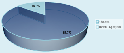
Background: Commonly, primary hyperparathyroidism is caused by a single parathyroid adenoma; a smaller fraction are hyperplasia, or multigland disease. Prelocalization of the diseased parathyroid gland is key to effective parathyroidectomy. Focused parathyroidectomy has become the standard treatment for selected patients with primary hyperparathyroidism. The aim is to assess the role of preoperative localization before doing focused parathyroidectomy. Methods: From July 2022 to June 2023, this cross-sectional study was conducted at Bangabandhu Sheikh Mujib Medical University's general surgery department in Dhaka. 14 patients with primary hyperparathyroidism had focused parathyroidectomy. Statistical analysis of the results was obtained by using window-based computer software with IBM SPSS statistics (Version 22). Results: The male-to-female ratio was 2.5:1, and the mean age was 32.36 years (SD = 9.76). The sensitivity, specificity, and accuracy for the ultrasound’s validity test were 75.0%, 50.0%, and 71.4%, respectively. The validity test for the Sestamibi scan has an accuracy of 85.7%, a specificity of 0.0%, and a sensitivity of 100.0%. The biochemical frozen section validity test's sensitivity, specificity, accuracy, and positive and negative predictive values were all 100%. The majority of individuals (85.7%) in the histopathology study investigation had parathyroid adenoma. The range of the postoperative hospital stay was 2 to 19 days, with an average of 8.2±6.26 days. Not all patients who underwent surgery had ugly scars. Conclusion: Preoperative localization enables patients to have focused parathyroidectomy, which is potentially cost effective, reduce postoperative complication, hospital stay with good cosmesis and patient satisfaction; ultimately improve postoperative outcome.
| Published in | Journal of Surgery (Volume 13, Issue 5) |
| DOI | 10.11648/j.js.20251305.12 |
| Page(s) | 126-133 |
| Creative Commons |
This is an Open Access article, distributed under the terms of the Creative Commons Attribution 4.0 International License (http://creativecommons.org/licenses/by/4.0/), which permits unrestricted use, distribution and reproduction in any medium or format, provided the original work is properly cited. |
| Copyright |
Copyright © The Author(s), 2025. Published by Science Publishing Group |
Primary Hyperparathyroidism, Parathyroid Adenoma, Focused Parathyroidectomy
Demographic Profile | Number | Percentage |
|---|---|---|
Age (in years) | ||
11-20 | 3 | 21.4 |
21-30 | 3 | 21.4 |
31-40 | 5 | 35.6 |
41-50 | 3 | 21.4 |
Mean±SD | 32.36 ± 9.76 | |
Range (min, max) | 18, 45 | |
Gender | ||
Female | 4 | 28.6 |
Male | 10 | 71.4 |
Presenting Complaints | ||
Renal symptoms | 9 | 64.3 |
Bone symptoms | 5 | 35.7 |
Abdominal symptoms | 5 | 35.7 |
Muscle weakness | 4 | 28.6 |
Fatigue | 4 | 28.6 |
Memory impairment | 3 | 21.4 |
Past history | ||
Renal disease | 5 | 35.7 |
HTN | 3 | 21.4 |
Bone disease | 3 | 21.4 |
DM | 1 | 7.1 |
Chronic calculus cholecystitis and pancreatitis | 1 | 7.1 |
No past history | 3 | 21.4 |
USG of neck (n=14) | Sestamibi (n=14) | Peroperative (n=14) | ||||
|---|---|---|---|---|---|---|
n | % | n | % | N | % | |
Left | 4 | 28.6 | - | - | - | - |
Left lower | - | - | 6 | 42.9 | 6 | 42.9 |
Left upper | - | - | 1 | 7.1 | 1 | 7.1 |
Right | 4 | 28.6 | - | - | - | - |
Right lower | - | - | 4 | 28.6 | 3 | 21.4 |
Right upper | - | - | - | - | 1 | 7.1 |
Right & left | 1 | 7.1 | - | - | - | - |
Superior medistinal | - | - | 3 | 21.4 | 3 | 21.4 |
Normal findings | 4 | 28.6 | - | - | - | - |
Right (medial & lateral) | 1 | 7.1 | - | - | - | - |
USG of neck | Peroperative | |||||||||||||||||||||
|---|---|---|---|---|---|---|---|---|---|---|---|---|---|---|---|---|---|---|---|---|---|---|
Left | Left lower | Left upper | Right | Right lower | Right upper | Superior mediastinal | Right & left | Right (medial & lateral) | Normal | Total | ||||||||||||
- | (n=6) | (n=1) | - | (n=3) | (n=1) | (n=3) | - | - | - | (n=14) | ||||||||||||
n | % | n | % | n | % | n | % | n | % | n | % | n | % | n | % | n | % | n | % | n | % | |
Left | - | - | 4 | 66.6 | - | - | - | - | - | - | - | - | - | - | - | - | - | - | - | - | 4 | 28.6 |
Left lower | - | - | - | - | - | - | - | - | - | - | - | - | - | - | - | - | - | - | - | - | - | - |
Left upper | - | - | - | - | - | - | - | - | - | - | - | - | - | - | - | - | - | - | - | - | - | - |
Right | - | - | - | - | - | - | - | - | 2 | 66.7 | 1 | 100.0 | 1 | 33.3 | - | - | - | - | - | - | 4 | 28.6 |
Right lower | - | - | - | - | - | - | - | - | - | - | - | - | - | - | - | - | - | - | - | - | - | - |
Right upper | - | - | - | - | - | - | - | - | - | - | - | - | - | - | - | - | - | - | - | - | - | - |
Superior mediastinal | - | - | - | - | - | - | - | - | - | - | - | - | - | - | - | - | - | - | - | - | - | - |
Right & left | - | - | 1 | 16.7 | - | - | - | - | - | - | - | - | - | - | - | - | - | - | - | - | 1 | 7.1 |
Right (medial & lateral) | - | - | - | - | - | - | - | - | 1 | 33.3 | - | - | - | - | - | - | - | - | - | - | 1 | 7.1 |
Normal | - | - | 1 | 16.7 | 1 | 100.0 | - | - | - | - | - | - | 2 | 66.7 | - | - | - | - | - | - | 4 | 28.6 |
Sestamibi | Per-operative | |||||||||||
|---|---|---|---|---|---|---|---|---|---|---|---|---|
Left lower | Left upper | Right lower | Right upper | Superior mediastinal | Total | |||||||
(n=6) | (n=1) | (n=3) | (n=1) | (n=3) | ||||||||
n | % | n | % | n | % | n | % | n | % | n | % | |
Left lower | 6 | 100.0 | - | - | - | - | - | - | - | - | 6 | 42.9 |
Left upper | - | - | 1 | 100.0 | - | - | - | - | - | - | 1 | 7.1 |
Right lower | - | - | - | - | 3 | 100.0 | 1 | 100.0 | - | - | 4 | 28.6 |
Right upper | - | - | - | - | - | - | - | - | - | - | - | - |
Superior Mediastinal | - | - | - | - | - | - | - | - | 3 | 100.0 | 3 | 21.4 |
Test of validity | USG | Sestamibi | Biochemical Frozen section |
|---|---|---|---|
Sensitivity | 75.0 | 100.0 | 100.0 |
Specificity | 50.0 | 0.0 | 100.0 |
Accuracy | 71.4 | 85.7 | 100.0 |
PPV | 90.0 | 85.7 | 100.0 |
NPV | 25.0 | 0.0 | 100.0 |
Postoperative outcome | Number | Percentage |
|---|---|---|
Complication (n=7) | ||
Bone pain | 4 | 57.1 |
Circumoral paresthesia | 4 | 57.1 |
Carpopedal spasm | 1 | 14.3 |
Digital paresthesia | 3 | 42.9 |
Cosmesis (n=14) | ||
Poor | 0 | 0.0 |
Good | 14 | 100.0 |
Postoperative hospital stay in days (n=14) | 8.2±6.26 | |
Range (min, max) | 2, 19 | |
PHPT | Primary Hyperparathyroidism |
SGD | Single Gland Disease |
MGD | Multi Gland Disease |
BNE | Bilateral Neck Exploration |
USG | Ultrasonography |
PTH | Parathyroid Hormone |
SPSS | Statistical Package for the Social Sciences |
SD | Standard Deviation |
PPV | Positive Predictive Value |
NPV | Negative Predictive Value |
HTN | Hypertension |
DM | Diabetes Mellitus |
| [1] | Tay D, Das JP, Yeh R. Preoperative Localization for Primary Hyperparathyroidism: A Clinical Review. Biomedicines. 2021 Apr 6; 9(4): 390. |
| [2] | Williams N, O’Connell PR and McCaskie A. Bailey & Love’ s Short practice of Surgery, 27th edition. CRC PressTaylor & Francis Group, Boca Raton, FL. 2018; page number 823, 825, 829, 830. Text Book of Surgery. |
| [3] | Taterra D, Wong LM, Vikse J, Sanna B, Pękala P, Walocha J, Cirocchi R, Tomaszewski K, Henry BM. The prevalence and anatomy of parathyroid glands: a meta-analysis with implications for parathyroid surgery. Langenbecks Arch Surg. 2019 Feb; 404(1): 63-70. |
| [4] | John P. Bilezikian, Maria Luisa Brandi, Richard Eastell, Shonni J. Silverberg, Robert Udelsman, Claudio Marcocci, John T. Potts, Guidelines for the Management of Asymptomatic Primary Hyperparathyroidism: Summary Statement from the Fourth International Workshop, The Journal of Clinical Endocrinology & Metabolism, Volume 99, Issue 10, 1 October 2014, Pages 3561-3569, |
| [5] | Udelsman R, Åkerström G, Biagini C, Duh QY, Miccoli P, Niederle B, Tonelli F. The surgical management of asymptomatic primary hyperparathyroidism: proceedings of the Fourth International Workshop. J Clin Endocrinol Metab. 2014 Oct; 99(10): 3595-606. |
| [6] | Egan RJ, Scott-Coombes DM. The surgical management of sporadic primary hyperparathyroidism. Best Practice & research. Clinical Endocrinology & Metabolism. 2018 Dec; 32(6): 847-859. |
| [7] | Topal, Ugur, Kubilay Dalci, Ayse Unal, Ahmet Saritas, Isa Gunay, Aysun Uguz, and Gurhan Sakman. "Clinical and surgical approach to parathyroid adenomas: A single-center experience." Annals of Medical Research 26, no. 10 (2019): 2259. |
| [8] | Wilhelm SM, Wang TS, Ruan DT, Lee JA, Asa SL, Duh QY, Doherty GM, Herrera MF, Pasieka JL, Perrier ND, Silverberg SJ, Solórzano CC, Sturgeon C, Tublin ME, Udelsman R, Carty SE. The American Association of Endocrine Surgeons Guidelines for Definitive Management of Primary Hyperparathyroidism. JAMA Surg. 2016 Oct 1; 151(10): 959-968. |
| [9] | Varadharajan K, Choudhury N. Current practice in the surgical management of parathyroid disorders: a United Kingdom survey. Eur Arch Otorhinolaryngol. 2018 Oct; 275(10): 2549-2553. |
| [10] | Tunca F, Akici M, Işcan Y, Cem Sormaz I, Giles Senyurek Y, Terzioğlu T. The impact of combined interpretation of localization studies on image-guided surgical approaches for primary hyperparathyroidism. Minerva Endocrinol. 2017 Sep; 42(3): 213-222. |
| [11] | Horányi J, Duffek L, Szlávik R, Takács I, Tóth M, Romics L Jr. Intraoperative determination of PTH concentrations in fine needle tissue aspirates to identify parathyroid tissue during parathyroidectomy. World J Surg. 2010 Mar; 34(3): 538-43. |
| [12] | Mahbub, S., Biswas, S. S., Dey, B. K., Alam, M. S., & Hoq, J. (2021). Parathyroid Adenoma: an experience in BIRDEM General Hospital. Bangladesh Journal of Otorhinolaryngology, 27(1), 44-51. |
| [13] | Khan A, Bilezikian J, Bone H, Gurevich A, Lakatos P, Misiorowski W, Rozhinskaya L, Trotman ML, Tóth M. Cinacalcet normalizes serum calcium in a double-blind randomized, placebo-controlled study in patients with primary hyperparathyroidism with contraindications to surgery. Eur J Endocrinol. 2015 May; 172(5): 527-35. |
| [14] | Noureldine SI, Gooi Z, Tufano RP. Minimally invasive parathyroid surgery. Gland Surg. 2015 Oct; 4(5): 410-9. |
| [15] | Thomas PR, Beggs AD, Han TS. Utility of surgeon-performed pre-operative ultrasound in the localisation of parathyroid adenomas. JRSM Cardiovasc Dis. 2019 Jun 19; 8: 2048004019856950. |
| [16] | Budoor Alemadi, Maryam Ahmad Alsaeed, Fatima Alsayyah, Salma Rahma, Fatheya Al Awadi, Fauzia Rashid; Correlation between Surgical Outcomes of Primary Hyperparathyroidism with Neck Ultrasound and Parathyroid Scan-Tc99m/MIBI Localization Studies in Dubai Hospital. Dubai Diabetes Endocrinol J 28 December 2022; 28(4): 136-142. |
| [17] | Khorasani N, Mohammadi A. Effective factors on the sensitivity of preoperative sestamibi scanning for primary hyperparathyroidism. Int J Clin Exp Med. 2014 Sep 15; 7(9): 2639-44. |
| [18] | Scattergood S, Marsden M, Kyrimi E, Ishii H, Doddi S, Sinha P. Combined ultrasound and Sestamibi scintigraphy provides accurate preoperative localisation for patients with primary hyperparathyroidism. Ann R Coll Surg Engl. 2019 Feb; 101(2): 97-102. |
| [19] | Cheung K, Wang TS, Farrokhyar F, Roman SA, Sosa JA. A meta-analysis of preoperative localization techniques for patients with primary hyperparathyroidism. Ann Surg Oncol. 2012 Feb; 19(2): 577-83. |
| [20] | Solorzano, C. C., Carneiro-Pla, D. M. and Irvin, G. L. (2006) Surgeon-Performed Ultrasonography as the Initial and Only Localizing Study in Sporadic Primary Hyperparathyroidism. Journal of the American College of Surgeons, 202, 18-24. |
APA Style
Uddin, M. N., Joarder, M. A. I., Alam, F., Mohal, N., Aktar-Uj-Jaman, et al. (2025). Role of Preoperative Localization Before Focused Parathyroidectomy in Case of Parathyroid Adenoma. Journal of Surgery, 13(5), 126-133. https://doi.org/10.11648/j.js.20251305.12
ACS Style
Uddin, M. N.; Joarder, M. A. I.; Alam, F.; Mohal, N.; Aktar-Uj-Jaman, et al. Role of Preoperative Localization Before Focused Parathyroidectomy in Case of Parathyroid Adenoma. J. Surg. 2025, 13(5), 126-133. doi: 10.11648/j.js.20251305.12
@article{10.11648/j.js.20251305.12,
  author = {Md. Nadim Uddin and Md. Aminul Islam Joarder and Ferdous Alam and Noor Mohal and Aktar-Uj-Jaman and Soniya Akter},
  title = {Role of Preoperative Localization Before Focused Parathyroidectomy in Case of Parathyroid Adenoma
},
  journal = {Journal of Surgery},
  volume = {13},
  number = {5},
  pages = {126-133},
  doi = {10.11648/j.js.20251305.12},
  url = {https://doi.org/10.11648/j.js.20251305.12},
  eprint = {https://article.sciencepublishinggroup.com/pdf/10.11648.j.js.20251305.12},
  abstract = {Background: Commonly, primary hyperparathyroidism is caused by a single parathyroid adenoma; a smaller fraction are hyperplasia, or multigland disease. Prelocalization of the diseased parathyroid gland is key to effective parathyroidectomy. Focused parathyroidectomy has become the standard treatment for selected patients with primary hyperparathyroidism. The aim is to assess the role of preoperative localization before doing focused parathyroidectomy. Methods: From July 2022 to June 2023, this cross-sectional study was conducted at Bangabandhu Sheikh Mujib Medical University's general surgery department in Dhaka. 14 patients with primary hyperparathyroidism had focused parathyroidectomy. Statistical analysis of the results was obtained by using window-based computer software with IBM SPSS statistics (Version 22). Results: The male-to-female ratio was 2.5:1, and the mean age was 32.36 years (SD = 9.76). The sensitivity, specificity, and accuracy for the ultrasound’s validity test were 75.0%, 50.0%, and 71.4%, respectively. The validity test for the Sestamibi scan has an accuracy of 85.7%, a specificity of 0.0%, and a sensitivity of 100.0%. The biochemical frozen section validity test's sensitivity, specificity, accuracy, and positive and negative predictive values were all 100%. The majority of individuals (85.7%) in the histopathology study investigation had parathyroid adenoma. The range of the postoperative hospital stay was 2 to 19 days, with an average of 8.2±6.26 days. Not all patients who underwent surgery had ugly scars. Conclusion: Preoperative localization enables patients to have focused parathyroidectomy, which is potentially cost effective, reduce postoperative complication, hospital stay with good cosmesis and patient satisfaction; ultimately improve postoperative outcome.
},
 year = {2025}
}
TY  - JOUR
T1  - Role of Preoperative Localization Before Focused Parathyroidectomy in Case of Parathyroid Adenoma

AU  - Md. Nadim Uddin
AU  - Md. Aminul Islam Joarder
AU  - Ferdous Alam
AU  - Noor Mohal
AU  - Aktar-Uj-Jaman
AU  - Soniya Akter
Y1  - 2025/09/25
PY  - 2025
N1  - https://doi.org/10.11648/j.js.20251305.12
DO  - 10.11648/j.js.20251305.12
T2  - Journal of Surgery
JF  - Journal of Surgery
JO  - Journal of Surgery
SP  - 126
EP  - 133
PB  - Science Publishing Group
SN  - 2330-0930
UR  - https://doi.org/10.11648/j.js.20251305.12
AB  - Background: Commonly, primary hyperparathyroidism is caused by a single parathyroid adenoma; a smaller fraction are hyperplasia, or multigland disease. Prelocalization of the diseased parathyroid gland is key to effective parathyroidectomy. Focused parathyroidectomy has become the standard treatment for selected patients with primary hyperparathyroidism. The aim is to assess the role of preoperative localization before doing focused parathyroidectomy. Methods: From July 2022 to June 2023, this cross-sectional study was conducted at Bangabandhu Sheikh Mujib Medical University's general surgery department in Dhaka. 14 patients with primary hyperparathyroidism had focused parathyroidectomy. Statistical analysis of the results was obtained by using window-based computer software with IBM SPSS statistics (Version 22). Results: The male-to-female ratio was 2.5:1, and the mean age was 32.36 years (SD = 9.76). The sensitivity, specificity, and accuracy for the ultrasound’s validity test were 75.0%, 50.0%, and 71.4%, respectively. The validity test for the Sestamibi scan has an accuracy of 85.7%, a specificity of 0.0%, and a sensitivity of 100.0%. The biochemical frozen section validity test's sensitivity, specificity, accuracy, and positive and negative predictive values were all 100%. The majority of individuals (85.7%) in the histopathology study investigation had parathyroid adenoma. The range of the postoperative hospital stay was 2 to 19 days, with an average of 8.2±6.26 days. Not all patients who underwent surgery had ugly scars. Conclusion: Preoperative localization enables patients to have focused parathyroidectomy, which is potentially cost effective, reduce postoperative complication, hospital stay with good cosmesis and patient satisfaction; ultimately improve postoperative outcome.

VL  - 13
IS  - 5
ER  -
Department of General Surgery, Bangabandhu Sheikh Mujib Medical University, Dhaka, Bangladesh
Department of General Surgery, Bangabandhu Sheikh Mujib Medical University, Dhaka, Bangladesh
Department of General Surgery, Bangabandhu Sheikh Mujib Medical University, Dhaka, Bangladesh
Department of Anesthesiology, Islami Bank Medical College, Rajshahi, Bangladesh
Department of General Surgery, Bangabandhu Sheikh Mujib Medical University, Dhaka, Bangladesh
Department of Casualty, Sir Salimullah Medical College & Mitford Hospital, Dhaka, Bangladesh
Information