Research Article | | Peer-Reviewed

Diseases of Citrus (Citrus spp. L.) and Their Integrated Management

Received: 3 November 2025     Accepted: 13 November 2025     Published: 24 July 2026
Views:       Downloads:
Abstract

Citrus (Citrus spp. L.) is one of the most popular fruits in the world as a good source of vitamin C, and belongs to the Family: Rutaceae. Citrus most likely originated from species native to southeastern Asia and India and spread to the other parts of the world. The worldwide spread of the numerous citrus varieties in commercial planting has entered pests, diseases and disorders causing yield losses. Brazil, Mediterranean countries, China and the United States about two-thirds of the total citrus production. China is the first largest citrus producer and second to Brazil. Citrus fruits are growing in over 140 countries. North America has the highest consumption per capita of citrus fruits in the world followed by South America and Europe. Oranges occupy the major portion of the world followed by mandarins. Citrus production is always threatened by pathogens that cause economic losses. In India area under citrus are about one lakh hectares and production of approximately 14 lakh tones. India ranks sixth among the top citrus-producing countries in the world. Important species of citrus varieties grown in India as loose-jacket mandarin oranges/santras (Citrus reticulata), tight jacket sweet oranges (Citrus sinensis); musambi oranges and acid limes/ kaghzi limes (Citrus aurantifolia). Other species of citrus varieties cultivated in limited areas viz., grapefruit, pummelo (Citrus grandis), sweet lime (Citrus limettioides) and lemon. Citrus diseases harm fruit production as well as the fruit quality of the citrus tree. Citrus is susceptible to a large number of diseases caused by fungi, bacteria, viruses, viroids, phytoplasma, nutrient deficiencies, phanerogamic parasites and environmental effects leading to different physiological disorders. The major citrus diseases are citrus gummosis, citrus canker, huanglongbing, citrus variegated chlorosis, citrus decline, citrus greening, slow decline, and spreading decline are cause significant reductions in production.

Published in Science Discovery Food (Volume 1, Issue 2)
DOI 10.11648/j.sdf.20260102.12
Page(s) 89-100
Creative Commons

This is an Open Access article, distributed under the terms of the Creative Commons Attribution 4.0 International License (http://creativecommons.org/licenses/by/4.0/), which permits unrestricted use, distribution and reproduction in any medium or format, provided the original work is properly cited.

Copyright

Copyright © The Author(s), 2026. Published by Science Publishing Group

Keywords

Citrus spp. Economic Importance, Diseases, Cause, Symptoms, Management

1. Introduction
Citrus (Citrus spp.) belongs to the family: Rutaceae and is one of the economically important fruit crops in India. Citrus spp. occupy 953.4 thousand ha with annual productivity of 11655 thousand tonnes of fruits ranking 4th at a global level . Citrus is the third most important fruit crop in the world after apple and banana, the production of about 135.76 million tonnes spreading over 9.68 million hectares worldwide . Brazil is the second largest citrus producer after China. It is believed that most species under the genus Citrus originated from tropical and subtropical regions of Southeast Asia particularly India, South China, Indonesia and Peninsular Malaysia. The North-eastern states of India are recognized as one of the primary gene centres or natural homes and reservoirs of 17 Citrus species of which 8 species are indigenous to the region . The citrus crop is commercially cultivated in all four geographical zones viz. northwest, central, south and northeast, producing different kinds of mandarins (Citrus reticulata), sweet orange (C. sinensis), acid lime (C. aurantifolia), sweet lemon (C. limettoides) and lemon (C. lemon) . They are universally propagated by budding onto seedling rootstocks, seed and propagating materials influencing the chances of pathogen infection, thereby, affecting citrus production nationally and internationally. Citrus production is subjected to a variety of diseases caused by various microorganisms’ viz. fungi, bacteria, viruses, viroids and nematodes. Bacterial diseases constant threat to citrus cultivation and cause substantial economic impacts in all growing areas around the world. Citrus canker, citrus variegated chlorosis and Huanglongbing cause significant reductions in production . The present review gives a brief highlight of citrus diseases viz. canker, greening, quick decline, gummosis, exocortis, slow decline etc along with their characteristic symptoms, causal organisms, disease cycle, epidemiology as well as integrated management approaches.
2. Gummosis and Root Rot of Citrus
(Phytophthora nicotianae var. parasitica, P. palmivora, P. citropthora)
2.1. Economic Importance
Citrus foot rot, often known as gummosis of citrus or brown rot of citrus trees, is a major disease that wreaks havoc on citrus trees around the world. Gummosis is one of the soil-borne diseases of the citrus worldwide and causes mortality of newly planted trees as well as the slow decline of mature trees. The disease is widely known as gummosis, brown rot, collar rot, leaf fall, root rot, brown rot gummosis, trunk rot, foot rot, fruit rot and fibrous root rot. The first appearance of Phytophthora epidemics of citrus was first reported from Azore Island (1832-1836). It can cause serious economic losses by causing damping-off of seedlings in seedbeds, root and crown rot in nurseries, gummosis in trees, brown rot and fruit drop in orchards as well as post-harvest decay in storage . Rootstock with graft union at or below soil surface exposing scion tissues to a pathogen can cause the susceptibility to citrus gummosis or root rot. In India, the disease is widespread in Punjab and Assam, where lemons are more susceptible than grapefruit and rough lemon, while in South India, it is common in sweet oranges. The disease incidence of more than 20% seedling mortality and 20-50% decline of Nagpur mandarin have been reported from central India (Maharashtra), 20-100% decline of acid lime in South India (Andhra Pradesh) and 10-80% of Kinnow mandarin (C. sinensis) from North-Western India (Punjab) . The Citrus species affected included sweet orange (C. sinensis), mandarin (C. reticulata), lime (C. aurantifolia), lemon (C. limon) and grapefruit (C. paradisi) .
2.2. Symptoms
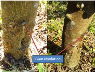
The Phytophthora species causing gummosis develops rapidly under moist, cool conditions. Phytophthora fungi are present in almost all citrus orchards, early symptoms of Phytophthora gummosis is sap oozing from small cracks in the infected bark, and the tree a bleeding appearance. The gumming may be washed off during heavy rain. Lesions spread around the circumference of the trunk, slowly girdling the tree. The typical symptom associated with the disease was the exudation of gum in the affected area which later dried on the bark surfaces. Symptoms caused by Gummosis (Phytophthora spp.) disease are as follows.
Damping-off- Damping-off of citrus seedlings in a nursery bed, the fungus penetrates the stem just above the soil line causing the seedlings to topple leading to seed rot/pre-emergence rot. With the abundance of moisture and favourable temperature, infected seedlings are killed rapidly . Footrot and gummosis- The dead bark of the lesions appears brown and active lesions ooze out gum from the trunk, the phenomenon known as gummosis. The infection starts from the basal portion of the stem infection near the ground level. Water-soaked patches turn into dark brown and produce lesions on the trunk as well as crown roots. The infection progresses into wood and bark dries up, and shrinks, with a small longitudinal crack through which abundant amber to reddish brown coloured gum exudation occurs . (Figure 1)
Figure 1. Exudation of gum on the citrus trunk.
Fibrosis root rot- The root cortex starts decaying fibrous roots soft. The cortex of fibrous roots is white thread-like the roots stringy appearance. The infection extends downward to the crown roots causing fibrous root rot ultimately drying and tree decline. The affected collar region of the trunk is girdled and finally, the infected tree dies. The tree usually blossoms heavily into profuse flowering, producing small fruits dropping before maturity. Under advanced stages of decline, the tree is unable to maintain water, mineral uptake and nutrient reservoirs, resulting in a reduction in fruit size, loss of leaves and twig-dieback . Die-back- This usually results in premature leaf fall and heavy defoliation. The chlorotic foliage has yellow mid-rib, lateral veins and leaf tissues and branches of symptoms dieback and slow decline of citrus . The leaves of the affected trees show symptoms similar to that of nutritional deficiency. Brown rot of fruit- The fungus infects unripe, ripening and ripe fruits in storage conditions and causes brown rot. The infected fruits show light brown, leathery and affected areas adjacent to rind growing up of white mycelium under humid conditions. The characteristic symptoms are water-soaked spots on fruit skin initially, where fruits get softened, rotten, and brown, releasing a pungent and rancid odour, and white fungus grows on rotten fruits .
2.3. Causal Organism
Anton de Bary discovered the fungus as “Phytophthora” meaning “plant destroyer”. Gummosis, brown-rot gummosis, collar-rot, brown-rot, trunk-rot and foot-rot are the same disease. Citrus gummosis and root rot disease are caused by three species of oomycetes fungi Phytophthora viz. P. palmivora, P. nicotianae var. parasitica and P. citropthora. In southern parts of India, P. palmivora has been the major cause of citrus gummosis and root rot while, in Karnataka and other part of India, P. parasitica is the major disease causing agent. In USA, P. parasitica and P. citropthora are major diseases causing pathogen of root rot, In some cases, P. hibernalis and P. syringae affect citrus fruits. In tropical areas, P. citricola has also been reported to cause fruit rot in citrus. The fungus has slender, sympodially branched sporangiophores bearing papillate, broadly ovoid, ellipsoid, It bears amphigynous, spherical or oval antheridia measuring and oogonia with rough, thick-walled, yellowish brown diameter. The fungus has intercellular mycelia with haustoria and sporangiospores having inverted in pear shaped.
2.4. Disease Cycle
The primary infection to initiated by oospores and chlamydospores surviving on fallen fruits, twigs, leaves and cracks on standing trees. P. nicotinae and P. citropthora begin the production of sporangia, which releases a large number of zoospores in soil by repeated infection of fibrous roots. The presence of host root exudates, nutrients, optimal soil moisture, temperature and aeration stimulates the germination of oospores and chlamydospores. The germination of sporangia to produce zoospores requires free water and is an important factor in root disease and the severity of Phytophthora root rot. Zoospores are attracted to the root surface by root exudates, germinate and infect by penetrating cortical cells area of the root elongation zone resulting in the rot of the entire rootlet . The rotted cortex fungus produces chlamydospore under unfavourable conditions and is persistent in soil for longer periods. Under favourable conditions viz. temperature (27-32°C), citrus root extracts stimulate the indirect germination of chlamydospores into sporangia and zoospores or direct germination into mycelia. P. palmivora shows more colonization efficiency than P. parasitica in the susceptible host viz. sour orange (C. aurantium) than trifoliate orange (P. trifoliata). The primary mode of dispersal by citrus orchards is the use of infected nurseries, rootstock, equipment and vehicles from infected to non-infected groves. Rainwater, irrigation water, wind, and raindrop splashes mediate the dispersal of sporangia and zoospores onto the trunk above the bud union. The dispersal from root-to-root in soil, surface water, splash from soil to aerial plant parts, as well as by human activities . The optimum temperature required by P. citropthora and P. parasitica are 24-28°C and 28-32°C respectively while the abundant sporangia production of this fungus. Irrigation plays an important role in increasing the propagules number of the fungus from the initial population .
3. Citrus Canker- Xanthomonas Citri Subsp. Citri
3.1. Economic Importance
Citrus canker is one the most devastating disease of citrus worldwide. The disease has been reported to be originated in tropical areas of Asia such as South China, India, Japan and Indonesia , it was also reported in Florida in the USA. The pathogen is widely distributed through planting materials in the citrus growing countries viz. USA, Europe, South Africa, New Zealand, Australia, Japan, India, Jawa and China. Citrus canker is considered one of the important diseases under strict international quarantine and prohibits the import of citrus plants and fruits from canker-affected countries.
3.2. Symptoms
Symptoms appear on all the above-ground plant parts such as leaves, twigs, thorns, older branches and fruits. Symptoms on leaves start appearing from 15-20 days after the bud burst. Initially, lesions are small, round, water-soaked, dark green to yellowish brown with raised convex surface first appearing on the lower surface and then on the upper surface of the leaves . The spots are raised initially corky with an oily appearance but later turn into brown with glossy dark brown margins. In a later stage, spots turn grayish and finally rupture exposing a light brown spongy mass forming rough, corky and canker. The centre of the spots falls off giving a shot-hole effect and eventually such spots coalesce giving a scabby appearance. Spots appearing on midrib or petioles of leaves cause premature defoliation. Symptoms on twigs, branches and thorns appear as rough, irregular, corky lesions surrounded by oily or water-soaked margins varying in size . Usually observed on susceptible crops like grapefruit, lime, sweet orange and trifoliate orange, leading to severe defoliation. The infected trees extensively lead to premature leave senesces and shoot dieback. The fruits about 2-4 cm in diameter are highly susceptible but become resistant once they attain maturity due to the thickening of the cuticle and hardening of tissues. On fruits, symptoms appear as rough, corky lesions having prominent crater-like depressions confined to the rind. Severely infected fruits become highly blemished but without any visible effect on pulp or juice . This is a latent disease as the length of time between infection and appearance of the first symptoms in the field, the latency period increases with the onset of cool weather . Different types of citrus canker are known to affect citrus trees worldwide as Asiatic/ True citrus canker (Cancrosis A)- Originally found in Asia and one of the most severe and widespread diseases worldwide affecting a large number of citrus species. False canker (Cancrosis B) - Originated from South America affecting lemons and lesser extent on lime, sour orange, sweet orange, citron, grapefruit etc. and also reported from Argentina, Paraguay and Uruguay. Mexican Lime Canker (Cancrosis C) - Originally found in the Sao Paulo state of Brazil and affecting Mexican lime, Galego lemon and lesser extent on Tahiti lemon. Canker D - First time reported in 1981 their pustules on leaves and young shoots of Mexican lime but not on fruits on the Pacific coast of Mexico. Canker E - First time reported in 1984 in canker in nurseries of Florida affecting grapefruit, sweet orange, trifoliate orange, tangor, tangerine, tangelo, mandarin, citrumelo and citron. The disease affects on leaves and shoots of nursery plants and produces flat, sunken, necrotic, water-soaked and chlorotic lesions known as Xanthomonas leaf spots.
3.3. Causal Organism
The causal organisms for different forms of citrus canker viz; Xanthomonas citri pv. citri, X. fuscans pv. aurantifolii, X. alfalfa pv. Citrumelonis. Asiatic/ True citrus canker/ Cancrosis A- Xanthomonas citri subsp. citri , subsp. nov. (syn. Xa. Citri, Xa. campestris pv. citri, Xa. axonopodis pv. citri). Cancrosis B, C and D- Xanthomonas fuscans subsp. aurantifolii , subsp. nov. (syn. Xa. campestris aurantifolii). Cancrosis E- Xanthomonas alfalfa subsp. citrumelonis , subsp. nov. (syn. Xa. campestris pv. citrumelo). Bacteria are gram-negative, obligatory aerobic, single-celled, non-spore formers, monotrichous, non-encapsulated, chemoautotrophic and straight rods measuring 0.5-0.75 x 1.5-2.0 µm. The optimum temperature required by the bacterium is 28°C with a minimum temperature of 6-7°C and a maximum of 36-38°C having a doubling time of 79 minutes . The colonies formed in nutrient agar media are circular, slightly raised, glistening, creamy straw to amber yellow with copious slime due to the production of yellow-coloured Xanthomonadin pigment .
3.4. Disease Cycle
The canker lesions are formed by rapid enlargement of hypertrophic cells occupying intercellular spaces leading to expansion of the epidermis and exposure of internal callus tissue. Citrus canker is characterized by overgrowth of spongy parenchymatous cells and an increase in amount near the site of infection at the early stage of invasion. The bacteria manifests itself and multiplies in the lesions formed on leaves, stems, twigs, branches and fruits . The bacteria penetrate the host through natural opening viz; wounds caused by pruning, rainstorms, stomata, movement of thorns and insects. The susceptibility to canker bacteria depends on younger age, longer wound healing time, and more number and size of stomata per unit leaf area. The invasion is more in younger organs such as leaves, stems and fruits due to wider stomatal openings than in older organs with a thicker cuticular layer having a narrow opening. The bacteria invasion into host intercellular spaces adheres to cell walls through interaction between exopolysaccharides (EPS) and citrus agglutinins multipy rapidly while dissolving middle lamella . EPS stimulates the bacteria for ethylene biosynthesis followed and cell membrane damage within the periphery of canker lesion zones. The high level of ethylene produced induces the formation yellow halo zone surrounding lesions and an abscission layer at the base of the leaf petiole resulting in defoliation. In addition, the bacterium secretes a transcriptional activator that interacts with the host machinery to induce transcription for genes that regulate plant hormones such as gibberellin and auxin . The survival of the bacterium from one season to the next in lesions formed on leaves, stems and branches. In stem cankers, bacteria may viable for 5-7 years whereas in leaf lesions for 2-3 months under dry conditions. In the presence of active host tissue of fallen leaf debris, the bacterium survives at most 3 weeks under wet conditions at a soil depth of 3-6 cm. The bacteria ooze from the lesions and disperse to new citrus growth by rain, wind, insects, humans and planting material . The rainstorms lead to dispersal and infection of bacteria by water congestion between plant surface and mesophyll cells of citrus leaf tissues, leading to forced entry of bacteria through stomata openings. The disease severity depends on strong winds, rains and rainstorms that aid disease outbreaks by causing host-tissue injuries, increasing humidity and dispersal of inoculums . The dissemination of bacteria by rain, wind and also insects such as citrus leaf miner Phyllocnistis citrella Stainton (Lepidoptera: Gracillariidae). Leaf miners cause maximum injury and the resulting increased infection takes place by the third instar and pupal stages of leaf miners . Citrus leaf miners cause wounds and provide an avenue for entry of bacterium into the host. Resistant cultivars have natural barriers to palisade parenchyma and spongy mesophyll cells .
Citrus canker favoured mild temperature (20°-30°C) and wet weather (4-24 hrs) of rains moistening the host surface for at least 20 minutes is essential for causing successful infection. The maximum infection of canker bacteria was reported to occur at maximum and minimum temperatures of 40°C and 12°C respectively with leaf wetness duration of 24 hours . In tropical and sub-tropical climatic conditions in India, Kagzi lime is the most susceptible but few mandarin oranges are resistant.
4. Citrus Greening (Huanglongbing) (Candidatus Liberobacter Asiaticus)
4.1. Economic Importance
Citrus huanglongbing (HLB) is the most destructive disease of citrus throughout the world and originated in Africa named “citrus greening”, and spreads to Europe, India, China and other Southeast Asian countries . The disease was first described in china by Reinking [19] as “Huanglungbin” meaning “yellow shoot and yellow dragon” due to the appearance of yellow shoots , and accepted the name of disease by the International Organization of Citrus Virologists in 1995. Other names are also known as “mottled leaf disease” in the Philippines, “Likubin” in Taiwan and “phloem necrosis and vein phloem degeneration” in Indonesia and “die-back and decline” in India . Greening disease occurs widely in Andhra Pradesh, Karnataka, Uttar Pradesh, Himachal Pradesh, Rajasthan, Jammu, Bihar, West Bengal and Sikkim causing catastrophic losses in yield as high as 30-100% . Huanglongbing is associated with the bacteria genus Candidatus Liberibacter, detected in Brazil in 2004. Another bacterium was characterized as a phytoplasma belonging to the 16SrIX group. Phytoplasma is 99% similar to Pigeon pea witches broom phytoplasma of the 16Sr IX group . Candidatus Liberibacter asiaticus and Candidatus Liberibacter americanus, both transmitted by the Asian citrus psyllid (Diaphorina citri) Kuwayama, and Candidatus Liberibacter africanus, transmitted by the psyllid (Trioza erytreae) (Del Guercio) .
4.2. Symptoms
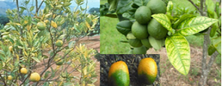
The characteristic symptoms of the citrus greening disease vary with cultivar, host age, infection time as well as biotic and abiotic factors affecting the growth of trees. Foliage symptoms include blotchy mottling and yellowing of leaves developing as a pattern of asymmetrical yellow-dark green areas on two halves lacking clear limits between the colours. Yellow leaves appear on single shoots at any section of the tree canopy, and yellowing spreads gradually to the entire tree giving it a “yellow shoot disease” appearance. Include yellowing of leaf veins, mottling and eventual fall of leaves, loss of fibrous rootlets and ultimately death of the plant. The leaf chlorosis symptoms in younger leaves resemble zinc deficiency with yellow venation at an early stage. Older leaves have green mottle, an appearance of “scattered green islands” in yellow tissue of leaf lamina. The yellow areas are surrounded by midrib on one side and lateral veins on another, yellowing gradually expand towards the margin of the leaf . (Figure 2)
Figure 2. Citrus greening symptoms on leaves and fruits.
The diseased trees showed reduced foliage, thickened leaves, severe twig dieback, shorter internodes, stunted growth, bushy appearance, and flowers earlier than healthy trees producing small fruits. The infected fruits are small and drop prematurely having a sour bitter taste .
4.3. Causal Organism
The citrus greening disease is caused by Candidatus liberobacter asiaticus, Candidatus liberobacter africanus, and Candidatus liberobacter americanus. It is phloem-limited fastidious vascular bacteria, having gram-negative rigid rod-shaped cells measuring 1.0-2.0 x 0.2-0.5 µm in size with two layered envelopes (20-25 nm thick) consisting of the cell wall and inner cytoplasmic membrane . Pathogens are obligate and heat tolerant. The bacterium was taxonomically identified as Candidatus (Ca) Liberobacter asiaticus, Ca. L. africanus and Ca. L. americanus for Asian, African and American citrus greening disease respectively . The Asian strain disease is found in Asia, Europe (Netherlands), the Middle East, North America, South America and Central America, whereas the African strain is found in the Middle East as well as the American strain is found in Brazil. Liberobacter differs in their reaction to a temperature such L. asiaticus is heat-tolerant surviving at a temperature more than 30°C, whereas, L. africanus is preferring a temperature range of 22-25°C . The Asian form is more destructive and lowers fruit yield and quality in tropical and subtropical Asian regions, with a tree life span of fewer than 10 years. Diseases detection through biological indexing on budwood collected from 1-year-old greening-suspected plants viz. sweet orange (mosambi) as indicator plants.
4.4. Disease Cycle
The primary infection takes place by the spread of bacterium via infected nursery plants, diseased bud wood grafts and dodder . The secondary infection by an insect vector, citrus psylla viz Diphorina citri Kuwayama as Asian and American psyllid vector as Trioza erytreae Del Guercio as well as African psyllid vector. A single female can lay 800-1000 eggs during her life hatching into nymphs at the temperature range of 30°-50°C, its entire life span of 50-80 days. The acquisition period of bacterium ranges from 15-30 minutes to a minimum of 5 hrs for D. citri and 24 hrs for T. erytreae of feeding with a latent period of 8-12 days, prior become infective. Nymphs acquire the bacteria and are persistently transmitted by adults; a single individual transmits the disease from infected to healthy plants within one hour. The adult and nymphs of the insect vector produce an extra amount of honeydew that causes sooty mould . Citrus huanglongbing is a multiyear disease, and disease rate progress is influenced by the inoculums reservoir, local vector populations, host age and environmental factors. Ca. L. asiaticus are heat tolerant as it can able to cause disease symptoms even at a temperature greater than 35°C, therefore, considered the most destructive pathogen. Ca. L. africanius shows severe symptoms of African greening under cool conditions ranging from 22-24°C . It’s more severe on mandarins, and sweet oranges while less susceptible on lime, lemons, pummelos and grapefruit. Mosambi sweet orange and Darjeeling orange (C. reticulata) are good indicator plants for citrus greening disease.
5. Quick Decline (Citrus Tristeza Virus)
5.1. Economic Importance
Citrus Tristeza is one of the most widespread disease of citrus. Its first reported from Italy and Florida in C. aurantifolia and C. sinensis. The name “Tristeza” means sadness in Spanish, describing the sad appearance of diseased citrus trees. The first epidemic decline of sweet orange plantings and sour orange rootstock was recorded in Argentina (1930) as “root disease”, in Brazil (1937) as “Tristeza”, in Africa (1938) as “dieback of sour limes”, in California (1939) as “quick decline” . In India, citrus tristeza disease is more prevalent in Andhra Pradesh, Tamil Nadu, Karnataka, Madhya Pradesh, Maharashtra, Punjab, Bihar, West Bengal and Sikkim.
5.2. Symptoms
The characteristic symptoms of citrus tristeza (quick decline) are seedling yellows, severe chlorosis, sick appearance and dwarfing primarily in sour orange, lemon and grapefruit . The leaves of such trees turn yellow by gradual wilting and dropping off leaving behind defoliated twigs showing severe dieback symptoms while the fruits continue to hang on dead trees . The specific symptom of the disease i.e. honeycomb pitting in the form of fine pitting of the inner face of bark in the trunk portion just below the bud union has been observed in sweet orange while in acid lime, it shows linear pits in the wood cylinder. The younger leaves of acid lime (C. aurantifolia) show the characteristic symptoms of vein clearing or vein flecking . Affected citrus species also show stem-pitting symptoms; trees exhibit deep longitudinal pits in the wood under bark, trunks, branches and twigs regardless of the rootstock resulting from cambial tissue disorders . Stem-pitting symptoms show reduced plant growth, stunting, and decreased branch development, brittle twigs, set fewer fruits as well as small and deformed fruits of poor quality. Diseased trees blossom heavily and gradually wilt either partially or completely as the fruits develop .
5.3. Causal Organism
Citrus Tristeza virus belongs to the Genus: Closterovirus, Species: Citrus tristeza virus cause quick decline, phloem-limiting, filamentous thread-like particles i.e. Citrus Tristeza Virus (CTV) of 2000 x 10-12 nm in size . Each particle consists of one single-stranded positive RNA (+ssRNA) and a single polypeptide coat protein.
5.4. Disease Cycle
The characteristic symptoms depend on host species, scion-rootstock combinations, vector activity as well as virus strains . The Citrus tristeza virus survives in the infected fruit trees as a perennial source of infection for new growths. It is transmitted neither by seeds nor mechanically with the sap of infected plants. Primary infection in citrus orchards takes place by Citrus tristeza virus infected propagating materials both scion or rootstocks such as bud wood, plants, stem-cut inoculations and grafts are used for planting as transmit the virus. Graft-transmitted from citrus to citrus taking a minimum of 8 days required for it to pass by grafting to receptor tissue . Citrus tristeza virus is also transmitted by two dodder species viz. Cuscuta subinclusa and C. americana. Secondary infection of Citrus tristeza virus is carried by tropical brown citrus aphids viz. Toxoptera citricida (Hemiptera: Aphididae), A. gossypii, and A. citricola facilitating horizontal spread of virus . Aphids transmit Citrus tristeza virus in a semi-persistent manner with an acquisition period of 30-60 minutes for feeding on infected trees to acquire the virus in the foregut and an inoculation period of 4-6 hours while probing phloem tissue. Once an aphid becomes viruliferous, it can remain infective for about 24 hours while transmitting to secondary hosts, thereby losing its infectivity within 48 hours of acquisition . Brown citrus aphid (T. citricida) is the most efficient aphid vector for the citrus tristeza virus.
6. Citrus Exocortis Viroid
6.1. Economic Importance
Citrus exocortis viroid
6.2. Symptoms
The symptoms of citrus exocortis viroid disease depend on the virulence strain and susceptibility of rootstocks. Susceptible plants show vertical splits in the bark, outer bark partially loosened into narrow, vertical, thin strips cracked and scaly appearance . The rootstocks of most plants are exocortis-susceptible viz. trifoliate oranges show enlarged scaly appearance as compared to the scions, therefore giving the name “scale butt”. The young infected stems of citrus plants show yellow blotches, leaf epinasty, stem epinasty, cracking, and darkening of leaf veins and petioles. Symptoms of gummy pitting of pockets have also been observed in the trifoliate rootstock . Infected plants become stunted with shorter internodes, and chlorotic leaves resulting in necrosis and ultimately death of rootstock tissues. Trifoliate and rangpur lime rootstocks show peculiar symptoms ranging from mild bark cracking to very severe bark scaling with various degrees of stunting and chlorotic stem blotching symptoms, bark cracking, leaf epinasty and vein necrosis is observed in highly susceptible citrons .
6.3. Causal Organism
Citrus exocortis disease is caused by Citrus Exocortis Viroid consisting of only ribonucleic acid (RNA) without any protein coat. The RNA is single-stranded (ssRNA) with a known sequence of 371 nucleotides arranged in a linear, closed and circular fashion . These RNA species replicate through a rolling circle mechanism involving only RNA intermediates without coding for proteins, thus, infectious agents interact directly with host factors to exert pathogenic effects . The strains of Citrus Exocortis Viroid can be differentiated based on symptom severity, incubation period and stunting degree in infected plants, where mild strains cause only stunting of plants but virulent strains lead to bark scaling .
6.4. Disease Cycle
Citrus exocortis viroid once come in contact with a susceptible ahost, viroid enters the phloem elements and gets associated with a host. Viroid infects the plants by perennating into the living plant parts surviving permanently in infected and herbaceous host plants. The viroid spreads throughout the plant and brings several metabolic changes to increased sugar content, enzyme production, and respiration rate and oxygen uptake. The viroid can be mechanically transmitted from diseased to healthy plants by the propagation of infected bud wood and grafting as well as mechanically by budding knives and pruning shears. First demonstrated the mechanical transmission of Citrus exocortis viroid and showed that contaminated tools can be disinfected by a mixture of 2% sodium hydroxide and 2% formaldehyde . Citrus exocortis viroid retains infectivity on knife blades for up to 8 days . It is also transmitted by dodder and sap to Petunia and other herbaceous plants. The viroid can also survive on the surface of contaminated blades treated with almost all commonly available chemical sterilants except sodium hypochlorite solution. The temperature for symptom development ranges from warm to hot viz. maximum temperature of 32-40°C during daytime and minimum temperature of 27-30°C during the night. Poncirus trifoliata rootstock is highly susceptible to citrus exocortis while C. limonia rootstock has the ability to milder symptoms but some citrus species viz. sweet orange, sour orange, rough lemon and grapefruit are highly tolerant.
7. Slow Decline of Citrus (Tylenchulus Semipenetrans)
7.1. Economic Importance
Citrus root nematode is a plant-parasitic nematode that is widely occurring in all the citrus growing regions of the world causing around 10-30% loss in citrus production . Field infestations of about 50-90% have been reported due to citrus nematodes from various citrus orchards worldwide viz. Arizona, California, Florida and Texas United States . It was discovered for the first time in 1913 by J. R. Hodges from California, later Nathan Cobb identified it as the causal agent of the slow decline of citrus .
7.2. Symptoms
The characteristic symptoms of citrus slow decline include nutrient deficiency, poor growth, yellowing of leaves, leaf curling, early dropping of leaves, sparse canopy, twig die-back, smaller dull green trees and reduced fruit production . Infected roots are thicker, darker, and decayed, around the feeding sites and development of dark necrotic lesions after invasion by secondary bacteria and fungi. Juveniles along with clay and sand particles cling to the gelatinous substance of egg mass resulting in dark, rough and young roots. Gradually nematodes reach a higher population in case of severe infections leading to a slow decline of citrus orchards within 3-5 years after initial infection . In addition to that, nematode injury facilitates secondary infection of bacteria and fungi viz. Phytophthora and Fusarium through nematode feeding sites result in cortical sloughing and death of feeder roots .
7.3. Causal Organism
Tylenchulu semipenetrans Cobb is a sedentary plant parasitic nematodes, about 0.25-0.4 mm long and 18-80 µm in diameter swollen females, vermiform-like juveniles and males with semi-endoparasitic feeding behaviour . The males are smaller with significantly reduced oesophagus and stylet, having a hyaline band corresponding to the copulatory system at the posterior end of the body. Whereas, female nematodes have a single ovary with a sub-terminal vulva and posterior excretory pore . The life cycle of T. semipenetrans is a dimorphic species comprised of egg, four juvenile stages and an adult sexual dimorphism having distinct male and female individuals at both the juvenile and adult stages. Eggs hatche under suitable environmental conditions, the first-stage juvenile (J1) moults inside the egg without having stylet while the second-stage juvenile hatch from eggs with distinguished sex. J2 males are short and further undergo molting into J3, and J4 with weaker stylet to become sedentary males without feeding and only for reproduction . While, J2 females are comparatively longer and further moults into J3, J4 and sedentary adults with swollen posterior ends upon feeding on epidermal layers of root cells. The life span of male nematodes is about 7-10 days, while female nematode lives for about 6-8 weeks at 24°C reproducing by amphimixis (parthenogenesis). The second-instar (J2) females are the infective stage of nematodes and adult females lay up to 75-100 eggs and are deposited in a gelatinous matrix secreted from the excretory pore.
7.4. Disease Cycle
The female second stage (J2) juvenile nematodes attack younger feeder roots by penetrating through the outer surface of roots into deep cortical layers and pericycle cells creating a tiny cavity on the root surface. While feeding ectoparasitically on epidermal root cells, it becomes sedentary and establishes a permanent feeding site consisting of specialized parenchyma nurse cells (3-6 nurse cells) as the main source of nutrients. The feeding by adult females causes swelling of the posterior end of the body that will protrude out from the root surface while its elongated neck and head remain embedded into the cortex . The eggs of citrus nematodes can survive for up to nine years in the soil until the depth of 4 meters. The mode of dispersal of citrus nematode from infected to healthy orchard takes place through nematode-infested propagative plant materials, nursery stocks and the use of infested irrigation water. The occurrence of nematodes varies between summer, winter and rainfall as their movement is relatively slower in the soil .
8. Integrated Diseases Managing of Citrus
8.1. Exclusion
Very strict quarantine measures should be practised to prevent the entry of pathogens in the form of infected planting materials into disease-free areas. Selection of a proper site for a citrus orchard with adequate drainage and preventive phytosanitary measures such as cleanliness, sound hygiene, soil fumigation and treated irrigation water should be practised. Use of tested and certified disease-free nursery trees and resistant rootstocks such as sour orange or khatta orange, Rangpur lime, rough lemon and trifoliate orange for propagating popular/ commercial varieties with high budded grafts having bud union at about 30-45 cm above the base at the time of planting . Citrus budwood grafting and shoot-tip grafting can be used for the production of virus-free planting materials. Tristeza-tolerant rootstocks such as sweet orange, rough lemon, Cleopatra, and mandarin scion varieties are tolerant to stem pitting .
8.2. Eradication
Suspected/ infected diseased trees or plant materials should be removed and destroyed by burning. Eradication of diseased portion viz. infected fruits, leaves or twigs should be done by cutting with a sharp knife and cleaning the wounds with 0.1% mercuric chloride or 1% potassium permanganate solution followed by application of Bordeaux paste or ridomil paint. Bordeaux paste should always apply the onset of the monsoon of a tree trunk. Remove the dark, diseased bark and a buffer strip of healthy, light brown to greenish bark around the margins of the infection. Allow the exposed area to dry out and apply Bordeaux paste.
8.3. Pre-plant Management
Rotation with annual crops for 1-3 years fallowing for 4 months followed by site preparation and treatment with 1, 3-D can retard citrus nematode infestation for 3 years . A proper irrigation system following the double ring method of irrigation should be adopted providing an inner ring (45 cm) around the trunk to prevent direct moistening and can protect citrus trees from fungus infection. Roots from the previous crop should be removed and deep subsoiling should be done to destroy the nematode reservoir in the soil. Soil disinfestation should be done by pre-plant, soil drenching up to 1.6 m of soil. Soil should be covered with a clear plastic sheet to disinfest shallow soil layers during hot summer duration up to 40-60 days for nursery bed preparation. Treating budwood at 47°C for 2 hrs reduces huanglongbing and gummosis root rot disease incidence . While dipping bare seedling roots in hot water at 45°C for 25 minutes or 50°C for 10-20 minutes can eradicate the nematode population .
8.4. Post-plant Management
Systemic fungicides viz. metalaxyl, fosetyl-Al and sodium tetra-thiocarbonate as soil drench or trunk spray has reported to reduce sporangia production in soil by 90% and inoculums density of fungal propagules . Systemic fungicides such as metalaxyl and fosetyl -Al have more effective against Phytophthora diseases . A combination of Bordeaux paint and soil drenching with ridomil MZ as well as fosetyl-Al or a combination of streptomycin (100 ppm) and 0.2% copper oxychloride spray can provide effective control of gummosis and canker . The citrus psylla population can also be eradicated by regular sprays of 0.02% of insecticides such as diazinon, dimethoate, methyls demeton, and endrin. Periodic sprays of dimethoate @ 2ml/l to control the aphid population. Hot water treatment of fruits such as grapefruit (48°C for 3 min), lemon (52°C for 5-10 min) and orange (53°C for 5 min) can prevent post-harvest decay . Application of antagonistic bacterial strains Streptomyces, Pseudomonas syringae and P. putida in irrigation water and sprinkler-applied soil can effectively reduce the Phytophthora population by causing hyphal lysis . Antagonistic fungus Trichoderma harzianum and T. virence reduce the pathogen population. Pseudomonas fluorescence was found inhibitory to the pathogen . Paecilomyces lilacinus (egg-parasitic fungus), Myrothecium, Arthobotrys, Pseudomonas fluorescens, Macrosporium, Pochonia chlamydospora, Trichoderma harzinum and Dactylella commercial use in citrus orchards against citrus root nematodes. The use of botanicals leaf extracts viz. neem (Azadirachta indica), Lantana (Lantana camara), and Dhatura (Dhatura stramonium) have been found more effective in inhibiting mycelial growth of P. citropthora by 44.54% and P. nicotinae by 57.78% .
9. Conclusion
Citrus orchards are severely infected by various diseases such as citrus canker caused by Xanthomonas bacteria, fungal gummosis and root rot caused by Phytophthora, greening disease caused by fastidious vascular bacteria, exocortis viroid disease, tristeza virus quick decline as well as slow decline caused by nematodes. Every year leads to huge losses in citrus production nationally and globally citrus industry worldwide. Management of these diseases is essential to provide increased and sustainable citrus production throughout the world. Understanding characteristic detection symptoms of these diseases at all the stages of plant growth can help to control diseases. About knowledge of diseases and their causal organisms, life cycle, predisposition factors for disease incidence as well disease cycle in diagnostic and forecasting its epidemiological outbreak in advance. Integrated disease management is a combining use of disease-free planting materials, utilization of bio-agents, improved cultural practices, development of resistant varieties and chemical control strategies in a holistic way to be more effective and sustainable for disease management. The management of various citrus diseases is usually subjected to repeated fungicide application resulting in higher cost and poisonous residual effects in the environment, therefore, new strategies for development.
Abbreviations

L.

Linnaeus

spp.

species

subsp. nov.

Subspecies Nova or New Subspecies

Ca.

Candidatus

EPS

Exopolysaccharides

J1

First-Stage Juvenile

RNA

Ribonucleic Acid

CTV

Citrus Tristeza Virus

HLB

Huanglongbing

Author Contributions
Amar Bahadur is the sole author. The author read and approved the final manuscript.
Conflicts of Interest
The authors declare no conflicts of interest.
References
[1] Agrios, GN. Plant Pathology, 3rd edition, Academic Press, New York, 1988;. 803 p.
[2] Agrios, GN. Plant Pathology. 5th Ed. Elsevier Academic Press, San Diego, 2004; 922 p.
[3] Ann PJ, Ko WH, Su HJ. Interaction between libukin bacterium and Phytophthora parasitca in citrus hosts. European Journal of Pathology, 2004;. 110(1): 1-6.
[4] Assuah MK, Oduro KA, Ofosu-Budu KG. Diplodia natalensis pole Evans, the causal agent of citrus gummosis disease in Ghana. Ghana J Agric Sci. 1999; 32: 11–17.
[5] Bar-Joseph M, Dawson WO. Citrus tristeza virus. Encyclopedia of Virology, 2008; 520-525.
[6] Bar-Joseph M, Marcus R, Lee RF. The continuous challenge of citrus tristeza virus control. Annual Review of phytopathology, 1989; 27: 291-316.
[7] Barkai-Golan R, Phillips DJ. Post-harvest treatment of fresh fruits and vegetables for decay control. Plant Diseases, 1991; 5: 1085-1089.
[8] Beattie GAC, Holford P, Mabberley DJ. Aspects and insights of Australia-Asia collaborative research on Huanglongbing. Proceedings International Workshop for the prevention of citrus greening disease in severely infected areas. International Research Division, Agricultural Forestry Research Council Secretariat, Ministry of Agricultural Forestry and Fisheries, Tokyo, Japan, 2006; 47-64.
[9] Benton RJ, Bowman FT, Fraser L, Kebby RG. Stunting and scaly butt of citrus associated with Poncirus trifoliate rootstock. Agricultural Gazette New South Wales, 1949; 60: 521-526.
[10] Biswas KK. Molecular characterization of Citrus Tristeza Virus isolates from Northeastern Himalayan region in India. Archives of Virology, 2010; 155: 959-963.
[11] Bitters WP, Duran-Vila N, Semancik JS. Effect of exocortis viroid on flower and fruit structure and development on Etrog citron. Plant Disease, 1987; 71: 397-399.
[12] Bhattacharya SC, Dutta S. Classification of citrus fruits of Assam. Directorate of Agriculture, Assam. 1949; p. 40.
[13] Bock H, Parker PE, Gottwald TR. The effect of simulated wind-driven rain on duration and distance of dispersal of Xanthomonas axonoposis pv. citri from canker infected trees. Plant Diseases, 2005; 89: 71-80.
[14] Bové JM. Huanglongbing: a destructive, newly–emerging, century–old disease of citrus. J Plant Pathol. 2006; 88(1): 7–37.
[15] Daniels R, Guest D. Defence responses induced by potassium phosphonate in Phytophthora palmivora challenged Arabidopsis thaliana. Physiological and Molecular Plant Pathology, 2006; 67(3): 194-201.
[16] Das AK. Citrus canker- a review. Journal of Applied Horticulture, 2003; 5: 52-60.
[17] Duncan LW. Nematode parasite of citrus. In: Luc, M., Sikora, R. A., and Bridge, J. (eds.) Plant parasitic nematodes in subtropical and tropical agriculture. CAB International, Wallingford, UK., 2005; 437-466.
[18] Fawcett HS. Citrus diseases and their control. 2nd ed. London: McGraw Hill Book Co. Inc; 1936. p. 656.
[19] Fawcett HS, Klotz LJ. Bark shelling of trifoliate orange. California citrogr., 1948; 48-230.
[20] Fawcett HS, Wallace JM. Evidence of the virus nature of citrus quick decline. California Citrograph, 1946; 32: 88-89.
[21] Flores R, Randles JW, Bar-Joseph M, Diener TO. Viroids. In: Van Regenmortel, M. H. V., Fauquet, C. M., Bishop, D. H. L., Carstens, E. B., Estes, M. K., Lemon, S. M., Maniloff, J., Mayo, M. A., McGeoch, D. J., Pringle, C. R. and Wickner, R. B. (eds.) Virus taxonomy. 7th Rep. Int. Com. Taxon Viruses. San Diego, Academic Press, 2000; 1009-1024.
[22] Fraser LR, Broadbent P. Virus and virus-related disease of citrus in New South Wales. Venation and woody gall. Department of Agriculturtal Publication, Chipping Norton, Surrey Beatty and Sons, NSW 2170, Australia. 1979; 50-51.
[23] Fraser RW. Seedling yellows, an unreported virus disease of citrus. Agricultue Gazetted NSW, 1952; 63: 125-131.
[24] Frasser LR, Singh D, Capoor SP. Greening virus, the likely cause of die-back in India. FAO Plant Protection Bulletin, 1996; 14: 127-130.
[25] Gabriel DW, Kingsley M, Hunter JF, Gottwald TR. Reinstatement of X, citri and X. phaseoli to species and reclassification of all X. campestris pv. citri strains. International Journal of Systematic Bacteriology, 1989; 39(1): 14-22.
[26] Gade MR, Giri GK.. Integration of soil solarization and fungicides of management of Phytophthora sp in citrus nursery. Indian Phytopathology. 2005; 58 (1) 114-116.
[27] Gade MR. Biological and chemical management of Phytophthora rootrot/collar rot in citrus nursery. The Bioscan. 2012; 7(4): 631-635.
[28] Garnsey SM, Jones JW. Mechanical transmission of exocortis virus with contaminated budding tools. Plant Disease, 1967; 51: 410-413.
[29] Ghosh DK, Mathur S, Gupta KN, Ahlawat YS, Ramchandran P. Viroid infection of citrus in central India. Indian Phytopathology, 2002; 55(3): 290-293.
[30] Gopal K., Gopi V, Kalyani L, Sreelatha M, Sreenivasulu B. Symptom-based diagnosis of Huanglongbing disease by PCR in sweet orange and acid lime. Archives of Phytopathology and Plant Protection, 2010; 43(9): 863-870.
[31] Goto M. Citrus canker. In: Kumar, J., Chaube, H. S., Singh, U. S. and Mukhopadhyaya, A. N. (eds.) Plant diseases of international importance, vol. III. Diseases of fruit crops, New Jersey, prentice Hall, Inc., 1992; 170-208.
[32] Gottwald TR. Current epidemiological understanding of citrus Huanglongbing. Annu. Rev. Phytopathol. 2010; 48: 119 -139
[33] Grant TJ. Tristeza virus strains in relation to different citrus species used as test plants. Phytopathology, 1959; 49: 23-27.
[34] Gross, H. J. et al.. Nucleotide sequence and secondary structure of citrus exocortis and chrysanthemum stunt viroid. European Journal of Biochemistry, 1982; 121(2): 249-257.
[35] Hartung JS, Halbert SE, Pelz-Stelinski K. Lack of evidence for transmission of Candidatus Liberobacter asiaticus through citrus seed taken from affected fruit. Plant Diseases, 2010; 94(10): 1200-1205.
[36] Inserra RN, Vovias N, O’Bannon JH. A classification of T. semipenetrans biotype. Journal of Nematology, 1980; 12(4): 283-287.
[37] Jadeja KB, Mayani NG, Patel V, Ghodasara MT. Chemical control of canker and gummosis of citrus in Gujrat. Journal of Mycology and Plant Pathology, 2000; 30: 87-88.
[38] Jagoueix S, Bove J, Garnier M. The Phloem-Limited Bacterium of Greening Disease of Citrus Is a Member of the α Subdivision of the Proteobacteria. International Journal of Systematic Bacteriology, 1994; 44: 379-386.
[39] Jagtap GP, Thosar RU, Dey U. Evaluation of plant extracts and bioagents for the control of gummosis of mandarin orange (C. reticulate blanko) caused by Phytophthora species. African Journal of Agricultural Research, 2012; 7(32): 4553-4558.
[40] Kaplan DT. Characterization of citrus rootstock responses to T. semipenetrans (Cobb). Journal of Nematology, 1981; 13(4): 492-498.
[41] Klotz JL. Fungal, bacterial, non-parasitic diseases and injuries originating in the seedbed, nursery and orchard. In: Reuther, W., Calavan, E. C. and Carman, G. E. (eds.) Citrus Industry, Vol. IV, University of California Press, Berkeley, CA. 1978. pp.
[42] Koche MD, Gade MR, Deshmukh AG, Antifungal activity of secondary metabolites produce by Pseudomonas fleorescense. The Bioscan. 2013; 8(2): 723-726.
[43] Laird, E. F. et al. Attempts to transmit citrus exocortis virioid by insects. Plant Diseases Reporter, 1969; 53: 850-851.
[44] Li J, Rao X, Ying Y. Development of algorithms for detecting citrus canker based on hyperspectral reflectance imaging. Journal of Science Food Agriculture, 2012; 92: 125-134.
[45] Manjunath KL, Halbert SE, Ramadugu C, Webb S, Lee RF. Detection of Candidatus Liberobacter asiaticus in Diaphorina citri and its importance in the management of citrus Huanglongbing in Florida. Phytopathology, 2008; 98(4): 387-396.
[46] Meena AK, Dutta F, Marak MC, Meena RK. Citrus decline. International Journal of Current Microbiology and Applied Science, 2018; 7: 2807-2815.
[47] Naqvi SAMH. Distribution of Phytophthora spp. and mating types pathogenic to citrus in vidarbha and marathwada region of Maharashtra and north-eastern states of India. In: Singh, S. and Gosh, S. P. (ed.) proceeding of the hi Tech citrus management. International Symposium Citriculture, Nagpur, India, 2000; 1073-1080.
[48] Parker JK, Wisotsky SR, Johnson EG, Hijaz FM, Killiny N, Hilf ME, De La Fuente L. (2014). Variability of Ca. L. asiaticus prolonged by addition of citrus juice to culture medium. Phytopathology, 2014; 104(1): 15-26.
[49] Patil BP, Warke DC. A note on existence of exocortis viroid in india. Current Science, 1968; 37: 469-470.
[50] Priya MD, Christiano RCS, Furtado EL. Effect of temperature and leaf wetness duration on infection of sweet oranges by Asiatic citrus canker. Plant Pathology, 2006; 55(5): 657-663.
[51] Price WC. Citrus tristeza virus. CMI/AAB Description of plant viruses, No. 33. 1970.
[52] Rayachaudhri SP, Nariani TK, Lele VC. Citrus- dieback problem in India. Proceddings 1st International Citrus Symposium. 1969; 3: 1433-1437.
[53] Ristiano JB, Gimpertz ML. New frontiers in the study of dispersal and spatial analysis of epidemics caused by species of genus Phytopthora. Annual Review of Phytopathology, 2000; 38: 541-576.
[54] Rodgriguez SG, Stapleton JJ, Civerolo EL. Xanthomonas campestris involved in mexican lime bacteriosis in colima, Mexico. Proceeding of 6th international conference of plant pathology an bacteriology, Maryland, 1985; 658-662.
[55] Roux HF. Citrus Nematode. Citrus Research International.
[56] Saravanan L, Savithri P. Relationship between citrus leaf miner, Phyllocnistis citrella Stainton infection and citrus canker, X. citri incidence on acid lime. Shashpa, 2003; 10(2): 181-182.
[57] Sasaki A. The effect of stem pitting on yield of Washington navel orange. Proceeding International Society Citriculture, 1981; 439-441.
[58] Savita VGS, Nagpal A. Citrus diseases caused by Phytophthora species. GERF Bulletin of Biosciences, 2012; 3: 18-27.
[59] Schaad NW, Postnikova E, Lacy G. Validation of the publication of new names and new combinations previously effectively but not validly published outside the IJSEM, 2007; 57: 893-897.
[60] Semancik JS. Citrus exocortis viroid. CMI/AAB Description sof Plant Viriod, No. 226. 1980.
[61] Silva HP, Montero AR, Feraz LCCB. Tratamento hidrotermico de mudas de citricos para a erradicacao de T. semipenetrans. Nematologia Brasileira, 1987; 11: 143-152.
[62] Sorribas FJ, Verdejo-Lucas S, Galeano M, Pastor J, Ornat C. Effcet of 1, 3-dichloropropene and rootstocks alone and in combination on T. semipenetrans and citrus tree growth in a replant management program. Nematropica, 2003; 33(2): 147-156.
[63] Stall RE, Seymour CP. Canker, a threat to citrus in the Gulf coast states. Plant Diseases. 1983; 67: 581.
[64] Steddom K, Becker O, Menge JA. Repetitive application of biocontrol agents Pseudomonas putida and its effects on population of Phytophthora parasitica in citrus orchards. Phytopathology, 2002; 92(8): 850-856.
[65] Tanaka H, Yamada S, Nakanishi S. Approach to eliminating citrus tristeza virus from citrus trees by using trifoliate orange seedlings. Bulletin of Horticultural Research Sta. Japan Sec., 1971; 11: 157-163.
[66] Teixeira DC, Wulff N A, Martins EC, Kitajima EW, Bassanezi R, Ayres AJ, Eveillard S, Saillard C, Bove JM. A phytoplasma closely related to the Pigeon pea witchesÕ-broom phytoplasma (16SrIX) is associated with citrus Huanglongbing symptoms in the state of Sao Paulo, Brazil. Phytopathology. 2008; 98(9): 977-984.
[67] Thorne G. Principles of Nematology. McGraw-Hill Book Company, Inc. New York, NY 1961; p. 553.
[68] Timmer L, Gransey S, Braodbent A. Diseases of citrus. In: Ploetz, R. C. (ed.) Diseases of tropical fruit crops, CABI Publishing, CABI InternationL, Wallingford, UK, 2003; 163-195.
[69] Verdejo-Lucas S, Kaplan DT. The citrus nematode: Tylenchulus semipenetrans. In: Starr, J. L., Cook, R., and Bridge, J. (eds.) Plant resistance to parasitic nematodes. Wallingfors, CAB International, UK. 2002; 207-219.
[70] Verniere CJ, Gottwald TR, Pruvost O. Disease development and symptom expression of xanthomonas campestris pv. citri in various citrus plants tissues. Phytopathology, 2002; 93: 832-843.
[71] Vijaykumari N, Ghosh DK, Das AK, Singh A, Singh S. Elimination of citrus tristeza virus from exotic germlasm through in vitro shoot tip grafting in citrus. Indian Journal of Agricultural Science, 2006; 76(3): 209-210.
[72] Vojnov AA, do Amaral AM, Dow JM, Marano MR et al. Bacteria causing important diseases of citrus utilize distinct modes of pathogenesis to attack a common host. Appl Microbiol Biotechnol. 2010; 87(2): 467–477.
[73] Wallace J M. Virus and virus-like diseases. In: The citrus industry, ed. Reuther, W., Calavaran, E. C. and Carman, G. E. (Vol. 4) Berkeley, California Division Agricultural ScienceUniversity, California, 1978; pp 167-184.
[74] Yamamoto PT, Felippe MR, Garbim LF, Coelho JH, Ximenes NL, Martins EC, Leite APR, Sousa MC, Abrahao DP, Braz JD. Diaphorina citri Kuwayama (Hemiptera: Psyllidae): vector of the bacterium Candidatus Liberibacter americanus. In Proceedings, HuanglongbingÐGreening International Workshop, July 2006. Fundecitrus, Ribeira˜o Preto, Sa˜o Paulo, Brazil.
[75] Zhong DZ, Ling NC. Observation on the occurrence regulation of citrus canker disease and its control. South China Fruits, 2002; 31: 20-21.
[76] Thomas EE. Circular of California Agricultural. Experimentation Station, 1913; No. 85.
[77] Baines, RC, Klotz LJ, Clarke OF, DeWolfe TA. Hot water treatment of orange trees for eradication of citrus nema- tode. California Citrograph, 1949; 34: 482–484.
Cite This Article
  • APA Style

    Bahadur, A. (2026). Diseases of Citrus (Citrus spp. L.) and Their Integrated Management. Science Discovery Food, 1(2), 89-100. https://doi.org/10.11648/j.sdf.20260102.12

    Copy | Download

    ACS Style

    Bahadur, A. Diseases of Citrus (Citrus spp. L.) and Their Integrated Management. Sci. Discov. Food 2026, 1(2), 89-100. doi: 10.11648/j.sdf.20260102.12

    Copy | Download

    AMA Style

    Bahadur A. Diseases of Citrus (Citrus spp. L.) and Their Integrated Management. Sci Discov Food. 2026;1(2):89-100. doi: 10.11648/j.sdf.20260102.12

    Copy | Download

  • @article{10.11648/j.sdf.20260102.12,
      author = {Amar Bahadur},
      title = {Diseases of Citrus (Citrus spp. L.) and Their Integrated Management},
      journal = {Science Discovery Food},
      volume = {1},
      number = {2},
      pages = {89-100},
      doi = {10.11648/j.sdf.20260102.12},
      url = {https://doi.org/10.11648/j.sdf.20260102.12},
      eprint = {https://article.sciencepublishinggroup.com/pdf/10.11648.j.sdf.20260102.12},
      abstract = {Citrus (Citrus spp. L.) is one of the most popular fruits in the world as a good source of vitamin C, and belongs to the Family: Rutaceae. Citrus most likely originated from species native to southeastern Asia and India and spread to the other parts of the world. The worldwide spread of the numerous citrus varieties in commercial planting has entered pests, diseases and disorders causing yield losses. Brazil, Mediterranean countries, China and the United States about two-thirds of the total citrus production. China is the first largest citrus producer and second to Brazil. Citrus fruits are growing in over 140 countries. North America has the highest consumption per capita of citrus fruits in the world followed by South America and Europe. Oranges occupy the major portion of the world followed by mandarins. Citrus production is always threatened by pathogens that cause economic losses. In India area under citrus are about one lakh hectares and production of approximately 14 lakh tones. India ranks sixth among the top citrus-producing countries in the world. Important species of citrus varieties grown in India as loose-jacket mandarin oranges/santras (Citrus reticulata), tight jacket sweet oranges (Citrus sinensis); musambi oranges and acid limes/ kaghzi limes (Citrus aurantifolia). Other species of citrus varieties cultivated in limited areas viz., grapefruit, pummelo (Citrus grandis), sweet lime (Citrus limettioides) and lemon. Citrus diseases harm fruit production as well as the fruit quality of the citrus tree. Citrus is susceptible to a large number of diseases caused by fungi, bacteria, viruses, viroids, phytoplasma, nutrient deficiencies, phanerogamic parasites and environmental effects leading to different physiological disorders. The major citrus diseases are citrus gummosis, citrus canker, huanglongbing, citrus variegated chlorosis, citrus decline, citrus greening, slow decline, and spreading decline are cause significant reductions in production.},
     year = {2026}
    }
    

    Copy | Download

  • TY  - JOUR
    T1  - Diseases of Citrus (Citrus spp. L.) and Their Integrated Management
    AU  - Amar Bahadur
    Y1  - 2026/07/24
    PY  - 2026
    N1  - https://doi.org/10.11648/j.sdf.20260102.12
    DO  - 10.11648/j.sdf.20260102.12
    T2  - Science Discovery Food
    JF  - Science Discovery Food
    JO  - Science Discovery Food
    SP  - 89
    EP  - 100
    PB  - Science Publishing Group
    UR  - https://doi.org/10.11648/j.sdf.20260102.12
    AB  - Citrus (Citrus spp. L.) is one of the most popular fruits in the world as a good source of vitamin C, and belongs to the Family: Rutaceae. Citrus most likely originated from species native to southeastern Asia and India and spread to the other parts of the world. The worldwide spread of the numerous citrus varieties in commercial planting has entered pests, diseases and disorders causing yield losses. Brazil, Mediterranean countries, China and the United States about two-thirds of the total citrus production. China is the first largest citrus producer and second to Brazil. Citrus fruits are growing in over 140 countries. North America has the highest consumption per capita of citrus fruits in the world followed by South America and Europe. Oranges occupy the major portion of the world followed by mandarins. Citrus production is always threatened by pathogens that cause economic losses. In India area under citrus are about one lakh hectares and production of approximately 14 lakh tones. India ranks sixth among the top citrus-producing countries in the world. Important species of citrus varieties grown in India as loose-jacket mandarin oranges/santras (Citrus reticulata), tight jacket sweet oranges (Citrus sinensis); musambi oranges and acid limes/ kaghzi limes (Citrus aurantifolia). Other species of citrus varieties cultivated in limited areas viz., grapefruit, pummelo (Citrus grandis), sweet lime (Citrus limettioides) and lemon. Citrus diseases harm fruit production as well as the fruit quality of the citrus tree. Citrus is susceptible to a large number of diseases caused by fungi, bacteria, viruses, viroids, phytoplasma, nutrient deficiencies, phanerogamic parasites and environmental effects leading to different physiological disorders. The major citrus diseases are citrus gummosis, citrus canker, huanglongbing, citrus variegated chlorosis, citrus decline, citrus greening, slow decline, and spreading decline are cause significant reductions in production.
    VL  - 1
    IS  - 2
    ER  - 

    Copy | Download

Author Information
  • Abstract
  • Keywords
  • Document Sections

    1. 1. Introduction
    2. 2. Gummosis and Root Rot of Citrus
    3. 3. Citrus Canker- Xanthomonas Citri Subsp. Citri
    4. 4. Citrus Greening (Huanglongbing) (Candidatus Liberobacter Asiaticus)
    5. 5. Quick Decline (Citrus Tristeza Virus)
    6. 6. Citrus Exocortis Viroid
    7. 7. Slow Decline of Citrus (Tylenchulus Semipenetrans)
    8. 8. Integrated Diseases Managing of Citrus
    9. 9. Conclusion
    Show Full Outline
  • Abbreviations
  • Author Contributions
  • Conflicts of Interest
  • References
  • Cite This Article
  • Author Information