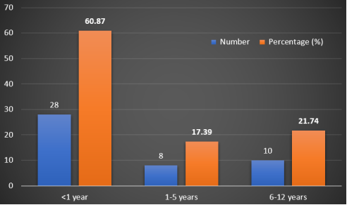
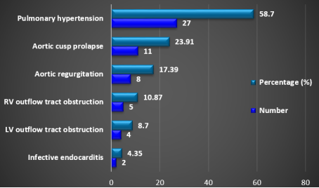
Ventricular septal defect (VSD) is the most common congenital heart defect in children, associated with important considerations for disease epidemiology, diagnosis, and long-term outcomes. VSD results from a deficiency in growth or the failure of alignment or fusion of the components of the ventricular septum. This study aimed to describe the epidemiological profile, diagnostic approach, and associated complications of VSD in a pediatric population. This cross-sectional study was conducted in the Department of Pediatric Cardiology, Combined Military Hospital, Dhaka, Bangladesh, from January 2018 to December 2018. This study included 46 pediatric patients less than 12 years old with isolated VSD. The mean age of the participants was 2.78 ± 3.44 years, with a slight male predominance (male-to-female ratio of 1.09:1). Most patients (60.87%) were under one year of age. A family history of congenital heart disease was reported in 13.04% of cases. Common presenting symptoms included feeding difficulty (69.57%), cough (60.87%), poor weight gain (54.35%), and head sweating (50%). Respiratory distress was the most frequently observed clinical sign (71.74%), followed by tachycardia (47.83%) and failure to thrive (41.30%). Echocardiography was the definitive diagnostic tool, performed in all cases. Other investigations included ECG (82.61%), chest X-ray (76.09%), and cardiac catheterization (8.70%). Perimembranous VSD was the most prevalent type (67.39%), and the majority of defects were small in size (58.70%). Pulmonary hypertension was the most common complication, noted in 58.70% of patients, followed by aortic cusp prolapse (23.91%) and aortic regurgitation (17.39%). Ventricular septal defect remains a significant pediatric cardiac condition, predominantly affecting infants. Early diagnosis, with the help of a clinical picture and widely and easily accessible diagnostic tools like echocardiography (ECHO), will help initiate timely treatment and decrease mortality. Pulmonary hypertension was the most frequent complication, followed by aortic cusp prolapse and aortic regurgitation.
| Published in | American Journal of Pediatrics (Volume 11, Issue 3) |
| DOI | 10.11648/j.ajp.20251103.18 |
| Page(s) | 166-172 |
| Creative Commons |
This is an Open Access article, distributed under the terms of the Creative Commons Attribution 4.0 International License (http://creativecommons.org/licenses/by/4.0/), which permits unrestricted use, distribution and reproduction in any medium or format, provided the original work is properly cited. |
| Copyright |
Copyright © The Author(s), 2025. Published by Science Publishing Group |
Ventricular Septal Defect, Echocardiography, Pulmonary Hypertension
Variable | Number | Percentage (%) | |
|---|---|---|---|
Age (Mean ±SD, years) | 2.78 ± 3.44 | ||
Sex | |||
Male | 24 | 52.17 | |
Female | 22 | 47.83 | |
Residence | |||
Urban | 28 | 60.87 | |
Rural | 18 | 39.13 | |
Family History of CHD | |||
Present | 6 | 13.04 | |
Absent | 40 | 86.96 | |
Baseline Findings | Number | Percentage (%) | |
|---|---|---|---|
Symptoms | |||
Head sweating | 23 | 50.00 | |
Dyspnea on exertion | 22 | 47.83 | |
Cough | 28 | 60.87 | |
Feeding difficulty | 32 | 69.57 | |
Poor weight gain | 25 | 54.35 | |
Asymptomatic | 14 | 30.43 | |
Signs | |||
Respiratory distress | 33 | 71.74 | |
Tachycardia | 22 | 47.83 | |
Fever | 11 | 23.91 | |
Failure to thrive (FTT) | 19 | 41.30 | |
Precordium Findings | |||
Bulge of the precordium | 17 | 36.96 | |
Cardiomegaly | 16 | 34.78 | |
Thrill | 33 | 71.74 | |
P2 palpable | 5 | 10.87 | |
Loud P2 | 7 | 15.22 | |
Pansystolic murmur | 32 | 69.57 | |
Ejection systolic murmur | 13 | 28.26 | |
Diagnostic Tool | Number | Percentage (%) |
|---|---|---|
Chest X-ray | 35 | 76.09 |
ECG | 38 | 82.61 |
Echocardiography | 46 | 100.00 |
Cardiac Catheterization | 4 | 8.70 |
Type | Number | Percentage (%) | |
|---|---|---|---|
Perimembranous | 31 | 67.39 | |
Muscular | 8 | 17.39 | |
Inlet | 4 | 8.70 | |
Outlet | 3 | 6.52 | |
Size | |||
Small (<5 mm) | 27 | 58.70 | |
Moderate (5-10 mm) | 15 | 32.61 | |
Large (>10 mm) | 4 | 8.70 | |
CHD | Congenital Heart Disease |
VSD | Ventricular Septal Defect |
ECHO | Echocardiography |
ECG | Electrocardiogram |
| [1] | Tantchou Tchoumi JC, Butera G, Giamberti A, Ambassa JC, Sadeu JC. Occurrence and pattern of congenital heart diseases in a rural area of sub-Saharan Africa. Cardiovasc J Afr. 2011; 22(2): 63-6. |
| [2] | Van Der Linde D, Konings EEM, Slager MA, Witsenburg M, Helbing WA, Takkenberg JJ, Roos-Hesselink JW. Birth prevalence of congenital heart disease worldwide: A systematic review and meta-analysis. J Am Coll Cardiol. 2011; 58(21): 2241-7. |
| [3] | Penny DJ, Vick GW III. Ventricular septal defect. Lancet. 2011; 377(9771): 1103-12. |
| [4] | Shafer K, Brickner ME. Ventricular septal defect. In: Braunwald’s heart disease: Essential echocardiography: a companion. Elsevier; 2018. p. 435-9. |
| [5] | Sarwar S, Shabana, Ehsan F, Tahir A, Jamil M, Shahid SU, Hasnain S, Khan A, Hyder SN. Hematological and demographic profile of Pakistani children with isolated ventricular septal defects (VSDs). Egypt J Med Hum Genet. 2020; 21: 1-8. |
| [6] | Rhodes LA, Keane JF, Keane JP, Fellows KE, Jonas RA, Castaneda AR. Long follow-up (43 years) of ventricular septal defect with audible aortic regurgitation. Am J Cardiol. 1990; 66: 340-5. |
| [7] | Sadiq M, Roshan B, Latif F, Bashir I, Sheikh SA, Khan A. Pattern of paediatric heart disease in Pakistan. J Coll Physicians Surg Pak. 2002; 12: 149-53. |
| [8] | Kazmi U, Sadiq M, Hyder SN. Pattern of ventricular septal defects and associated complications. J Coll Physicians Surg Pak. 2009 Jun; 19(6): 342-5. |
| [9] | Soto B, Becker AE, Moulaert AJ, Lie JT, Anderson RH. Classification of ventricular septal defects. Br Heart J. 1980; 43: 332-43. |
| [10] | Wickramasinghe P, Lamabadusuriya SP, Narenthiran S. Prospective study of congenital heart disease in children. Ceylon Med J. 2001; 46: 96-8. |
| [11] | Wu MH, Wu JM, Chang CI, et al. Implication of aneurysmal transformation in isolated perimembranous ventricular septal defect. Am J Cardiol. 1993; 72: 596-601. |
| [12] | Hrahshah AS, Hijazi IS. Natural and modified history of ventricular septal defect in infants. Pak J Med Sci. 2006; 22: 136-40. |
| [13] | McDaniel NL, Gutgesell HP. Ventricular septal defect. In: Allen HD, Driscoll DJ, Sheddy RE, Feltes TF, editors. Moss and Adams’ Heart Disease in Infants, Children, and Adolescents. 7th ed. Philadelphia: Lippincott Williams & Wilkins; 2008. p. 667-82. |
| [14] | Rao PS. Diagnosis and Management of Ventricular Septal Defects. Reviews in Cardiovascular Medicine. 2024 Nov 20; 25(11): 411. |
| [15] | Syamasundar Rao P. Diagnosis and management of acyanotic heart disease: part II– left-to-right shunt lesions. Indian Journal of Pediatrics. 2005; 72: 503–512. |
| [16] | Rao PS, Harris AD. Recent advances in managing septal defects: ventricular septal defects and atrioventricular septal defects. F1000Research. 2018; 7: F1000 Faculty Rev-498. |
| [17] | Rao PS. Congenital heart defects– A review. In: Rao PS, editor. Congenital Heart Disease: Selected Aspects. Rijeka (Croatia): InTech; 2011. p. 3–44. |
| [18] | Rees AH, Rao PS, Rigby JJ, Miller MD. Echocardiographic estimation of a left-to-right shunt in isolated ventricular septal defects. European Journal of Cardiology. 1978; 7: 25–33. |
| [19] | Rao PS. Other acyanotic heart defects presenting in the neonate. In: Rao PS, Vidyasagar D, editors. A Multidisciplinary Approach to Perinatal Cardiology. Vol. 2. New Castle upon Tyne (UK): Cambridge Scholars Publishing; 2021. p. 615–654. |
| [20] | Lue HC, Sung TC, Hou SH, Wu MH, Cheng SJ, Chu SH, et al. Ventricular septal defect in Chinese with aortic valve prolapse and aortic regurgitation. Heart Vessels. 1981; 2: 111-6. |
| [21] | Kobayashi J, Koike K, Senzaki H, Kobayashi T, Trunemoto M, Ishizawa A, et al. Correlation of anatomic and hemodynamic features with aortic valve leaflet deformity in doubly committed subarterial ventricular septal defect. Heart Vessels. 1999; 14: 240-5. |
| [22] | Chiu SN, Wang JK, Lin MT, Wu ET, Lu FL, Chang CI, et al. Aortic valve prolapse associated with outlet-type ventricular septal defect. Ann Thorac Surg. 2005; 79: 1366-71. |
| [23] | Chaudhry TA, Younas M, Baig A. Ventricular septal defect and associated complications. JPMA. 2011; 61(10): 1001-4. |
| [24] | Zielinsky P, Rossi M, Haertel JC. Subaortic fibrous ridge and ventricular septal defect: role of septal malalignment. Circulation. 1987; 75: 1124-9. |
| [25] | Kliegman RM, Jenson HB, Stanton NF. The cardiovascular system. In: Behrman RE, editor. Nelson Textbook of Pediatrics. 20th ed. Philadelphia: Saunders; 2016. p. 2194-7. |
| [26] | Fauci SA, Kasper DL, Longo DL, Braunwald E, Hauser SL, Jameson JL, et al. Disorders of cardiovascular system. In: Elliott M, Antman MD, editors. Harrison’s Principles of Medicine. 19th ed. USA: McGraw-Hill Companies; 2016. p. 1556-8. |
| [27] | Frontera-Izquierdo P, Cabezuelo-Huerta G, Cabezuelo-Huerta G. Natural and modified history of isolated ventricular septal defect: A 17-year study. Arch Dis Child. 1999 Nov; 81(5): 413-6. |
| [28] | Meryl S, Lopez L. Ventricular septal defect. In: Hugh D, Robert, Daniel J, Timothy F, Frank C, Howard PG, editors. Moss and Adams Heart Disease in Infants, Children, and Adolescents. 9th ed. Philadelphia: Lippincott Williams & Wilkins; 2016. p. 1450-60. |
| [29] | Aziz K. Ventricular septal defects. In: Heart Disease in Children. 2nd ed. Karachi: The Authors; 2000. p. 254-77. |
| [30] | Layangool T, Kirawittaya T, Sangtawesin C, Kojaranjit V, Makarapong P, Pechdamrongsakul A, Intasorn Y, Noisang P. Natural aortic valve complications of ventricular septal defect: A prospective cohort study. J Med Assoc Thai. 2008; 91: 53-9. |
| [31] | Rafiq I, Freeman L, Orzalkiewicz M, et al. Natural history of repaired and unrepaired VSD. Heart. 2015; 101: A82-A83. |
| [32] | Brauner R, Birk E, Blieden L, Sahar G, Vidne BA. Surgical management of ventricular septal defect with aortic valve prolapse: Clinical considerations and results. Eur J Cardiothorac Surg. 1995; 9: 315-9. |
| [33] | Ando M, Takao A. Pathological anatomy of ventricular septal defect associated with aortic valve prolapse and regurgitation. Heart Vessels. 1986; 2: 117-26. |
| [34] | Somanath HS, Gupta SK, Reddy KN, Murthy JS, Rao AS, Abraham KA. Ventricular septal defect with aortic regurgitation: A hemodynamic and angiographic profile in Indian subjects. Indian Heart J. 1990; 42: 113-6. |
| [35] | Hori Y, Yoshimura N, Kimura M. Right coronary cusp prolapse, evaluation by MRI. RSNA presentation Dec 2004 Hall D, Lakeside Centre. |
| [36] | Webb GD. Challenges in the care of adult patients with congenital heart defects. Heart. 2003; 89: 465-9. |
| [37] | Yetkin G, Refile B. An unusual case of chrysomonas luteala in infective endocarditis in a patient with ventricular septal defect. Indus Universatisi Tip Facultesi Dergisi. 2005; 12: 193-5. |
| [38] | Glen S, Burns J, Bloomfield P. Prevalence and development of additional cardiac abnormalities in 1448 patients with congenital ventricular septal defects. Heart. 2004; 90: 1321-5. |
APA Style
Moniruzzaman, M. (2025). Epidemiological Profile, Diagnostic Approach and Associated Complications of Ventricular Septal Defect in a Pediatric Population. American Journal of Pediatrics, 11(3), 166-172. https://doi.org/10.11648/j.ajp.20251103.18
ACS Style
Moniruzzaman, M. Epidemiological Profile, Diagnostic Approach and Associated Complications of Ventricular Septal Defect in a Pediatric Population. Am. J. Pediatr. 2025, 11(3), 166-172. doi: 10.11648/j.ajp.20251103.18
@article{10.11648/j.ajp.20251103.18,
  author = {Mohammad Moniruzzaman},
  title = {Epidemiological Profile, Diagnostic Approach and Associated Complications of Ventricular Septal Defect in a Pediatric Population
},
  journal = {American Journal of Pediatrics},
  volume = {11},
  number = {3},
  pages = {166-172},
  doi = {10.11648/j.ajp.20251103.18},
  url = {https://doi.org/10.11648/j.ajp.20251103.18},
  eprint = {https://article.sciencepublishinggroup.com/pdf/10.11648.j.ajp.20251103.18},
  abstract = {Ventricular septal defect (VSD) is the most common congenital heart defect in children, associated with important considerations for disease epidemiology, diagnosis, and long-term outcomes. VSD results from a deficiency in growth or the failure of alignment or fusion of the components of the ventricular septum. This study aimed to describe the epidemiological profile, diagnostic approach, and associated complications of VSD in a pediatric population. This cross-sectional study was conducted in the Department of Pediatric Cardiology, Combined Military Hospital, Dhaka, Bangladesh, from January 2018 to December 2018. This study included 46 pediatric patients less than 12 years old with isolated VSD. The mean age of the participants was 2.78 ± 3.44 years, with a slight male predominance (male-to-female ratio of 1.09:1). Most patients (60.87%) were under one year of age. A family history of congenital heart disease was reported in 13.04% of cases. Common presenting symptoms included feeding difficulty (69.57%), cough (60.87%), poor weight gain (54.35%), and head sweating (50%). Respiratory distress was the most frequently observed clinical sign (71.74%), followed by tachycardia (47.83%) and failure to thrive (41.30%). Echocardiography was the definitive diagnostic tool, performed in all cases. Other investigations included ECG (82.61%), chest X-ray (76.09%), and cardiac catheterization (8.70%). Perimembranous VSD was the most prevalent type (67.39%), and the majority of defects were small in size (58.70%). Pulmonary hypertension was the most common complication, noted in 58.70% of patients, followed by aortic cusp prolapse (23.91%) and aortic regurgitation (17.39%). Ventricular septal defect remains a significant pediatric cardiac condition, predominantly affecting infants. Early diagnosis, with the help of a clinical picture and widely and easily accessible diagnostic tools like echocardiography (ECHO), will help initiate timely treatment and decrease mortality. Pulmonary hypertension was the most frequent complication, followed by aortic cusp prolapse and aortic regurgitation.},
 year = {2025}
}
TY  - JOUR
T1  - Epidemiological Profile, Diagnostic Approach and Associated Complications of Ventricular Septal Defect in a Pediatric Population

AU  - Mohammad Moniruzzaman
Y1  - 2025/07/28
PY  - 2025
N1  - https://doi.org/10.11648/j.ajp.20251103.18
DO  - 10.11648/j.ajp.20251103.18
T2  - American Journal of Pediatrics
JF  - American Journal of Pediatrics
JO  - American Journal of Pediatrics
SP  - 166
EP  - 172
PB  - Science Publishing Group
SN  - 2472-0909
UR  - https://doi.org/10.11648/j.ajp.20251103.18
AB  - Ventricular septal defect (VSD) is the most common congenital heart defect in children, associated with important considerations for disease epidemiology, diagnosis, and long-term outcomes. VSD results from a deficiency in growth or the failure of alignment or fusion of the components of the ventricular septum. This study aimed to describe the epidemiological profile, diagnostic approach, and associated complications of VSD in a pediatric population. This cross-sectional study was conducted in the Department of Pediatric Cardiology, Combined Military Hospital, Dhaka, Bangladesh, from January 2018 to December 2018. This study included 46 pediatric patients less than 12 years old with isolated VSD. The mean age of the participants was 2.78 ± 3.44 years, with a slight male predominance (male-to-female ratio of 1.09:1). Most patients (60.87%) were under one year of age. A family history of congenital heart disease was reported in 13.04% of cases. Common presenting symptoms included feeding difficulty (69.57%), cough (60.87%), poor weight gain (54.35%), and head sweating (50%). Respiratory distress was the most frequently observed clinical sign (71.74%), followed by tachycardia (47.83%) and failure to thrive (41.30%). Echocardiography was the definitive diagnostic tool, performed in all cases. Other investigations included ECG (82.61%), chest X-ray (76.09%), and cardiac catheterization (8.70%). Perimembranous VSD was the most prevalent type (67.39%), and the majority of defects were small in size (58.70%). Pulmonary hypertension was the most common complication, noted in 58.70% of patients, followed by aortic cusp prolapse (23.91%) and aortic regurgitation (17.39%). Ventricular septal defect remains a significant pediatric cardiac condition, predominantly affecting infants. Early diagnosis, with the help of a clinical picture and widely and easily accessible diagnostic tools like echocardiography (ECHO), will help initiate timely treatment and decrease mortality. Pulmonary hypertension was the most frequent complication, followed by aortic cusp prolapse and aortic regurgitation.
VL  - 11
IS  - 3
ER  -
Department of Pediatrics, Combined Military Hospital, Dhaka, Bangladesh
Information