
 ,
Khalid Al-Shamrani,
Faisal Al-Shamrani,
Yousef Alturki,
Mohammed Alturki,
Maryam A. Dabbour
,
Khalid Al-Shamrani,
Faisal Al-Shamrani,
Yousef Alturki,
Mohammed Alturki,
Maryam A. Dabbour
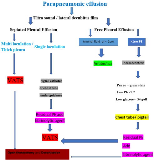
Background: Parapneumonic effusion (PPE) and empyema remain serious complications of childhood pneumonia associated with significant morbidity despite advances in antimicrobial therapy and surgical techniques. Accurate disease staging and timely stage-directed intervention are fundamental to optimal outcomes. Objective: To provide a current, evidence-based synthesis of the definition, etiology, diagnosis, and management of complicated pneumonia in children, with particular focus on areas of ongoing clinical controversy. Methods: A narrative review incorporating randomized controlled trials, systematic reviews, meta-analyses, Cochrane reviews, and international clinical guidelines published between 2016 and 2026 was conducted. PubMed was searched through May 2026 using terms including pneumonia, complicated pneumonia, parapneumonic effusion, empyema, VATS, fibrinolysis, decortication, and pediatric. Results: Chest radiography remains the initial imaging modality for suspected complicated pneumonia, with lateral decubitus views needed to assess pleural fluid mobility. However, chest ultrasonography is the preferred imaging modality for evaluating parapneumonic effusions and empyema because of its superior diagnostic accuracy, ability to characterize pleural collections, and absence of ionizing radiation. Management is stage-dependent, combining targeted antibiotic therapy with procedural intervention when indicated. Image-guided pigtail catheter drainage with intrapleural fibrinolytic therapy is effective for most patients. VATS demonstrates particular utility during the fibrinopurulent stage. No single fibrinolytic agent has demonstrated clear superiority; however, tissue plasminogen activator is most widely employed. Open decortication remains reserved for refractory or advanced-stage disease. Conclusion: Early diagnosis, appropriate antibiotics, and prompt intervention or surgery are all important for optimal care. The optimal outcomes require a multidisciplinary, individualized, stage-directed approach. Interventional radiology is appropriate for single loculations, while early VATS is favored for thick pleural rind or multiple loculations. Further high-quality prospective research is needed to establish definitive treatment algorithms.
| Published in | American Journal of Pediatrics (Volume 12, Issue 3) |
| DOI | 10.11648/j.ajp.20261203.11 |
| Page(s) | 87-102 |
| Creative Commons |
This is an Open Access article, distributed under the terms of the Creative Commons Attribution 4.0 International License (http://creativecommons.org/licenses/by/4.0/), which permits unrestricted use, distribution and reproduction in any medium or format, provided the original work is properly cited. |
| Copyright |
Copyright © The Author(s), 2026. Published by Science Publishing Group |
Parapneumonic Effusion, Empyema, Complicated Pneumonia, Children, Video Assisted Thoracoscopy (VATS), Fibrinolysis, Chest Ultrasonography, Pediatric Pleural Infection
Stage | Name | Pathophysiology | Pleural Fluid Characteristics | Ultrasound Appearance | Typical Management |
|---|---|---|---|---|---|
I | Exudative | Increased capillary permeability; protein-rich fluid accumulates; sterile or minimally contaminated | pH >7.3; glucose >3.4 mmol/L; LDH <500 IU/L; clear/straw-colored | Anechoic; freely mobile; no septations | IV antibiotics ± diagnostic thoracentesis; no routine drainage |
II | Fibrinopurulent | Fibrin deposition, bacterial invasion, and neutrophil infiltration; loculation begins | pH 7.0-7.2; glucose 1.6-3.4 mmol/L; LDH 500-1,000 IU/L; turbid/purulent | Echogenic fluid; fibrinous strands; septations; partial loculation | IV antibiotics + chest tube/pigtail drainage + intrapleural fibrinolytics; VATS if complex or failing |
III | Organizing | Fibroblast infiltration; collagen deposition; rigid fibrous pleural peel; trapped lung | pH <7.0; glucose <1.6 mmol/L; LDH >1,000 IU/L; thick pus/caseous | Thick hyperechoic pleural rind; dense loculations; reduced lung movement | VATS decortication (early); open thoracotomy + formal decortication (advanced/refractory) |
Clinical Feature | Infants (<12 months) | Toddlers/Preschool (1-5 years) | School-Age/Adolescents (>5 years) |
|---|---|---|---|
Fever | High, persistent | High, persistent >48-72h despite antibiotics | High, persistent; often with rigors |
Respiratory symptoms | Tachypnea; grunting; poor feeding | Tachypnea; retractions; reduced activity | Tachypnea; dyspnea; pleuritic chest pain |
Pain | Irritability; nonspecific distress | Abdominal pain (lower lobe); shoulder pain | Pleuritic chest pain; referred shoulder pain |
Constitutional | Poor feeding; lethargy | Reduced oral intake; malaise | Malaise; anorexia; weight loss |
Physical signs | Reduced chest expansion; dullness on percussion | Reduced breath sounds; dullness | Stony dullness; absent breath sounds; friction rub |
Oxygen saturation | May drop precipitously | SpO2 <92% indicates severe disease | SpO2 <92% requires urgent intervention |
Atypical features | Sepsis-like presentation; apnea | Mimics acute abdomen | Scoliosis; mediastinal shift in large effusions |
Investigation | Indication | Key Findings Suggesting Complicated PPE | Sensitivity/Specificity | Clinical Utility | Limitations |
|---|---|---|---|---|---|
Complete blood count | All patients | WBC >15×109/L; neutrophilia; elevated bands | Moderate | Baseline assessment; treatment monitoring | Nonspecific; may be normal in early disease |
C-reactive protein | All patients | CRP >60-100 mg/L | Moderate-High | Distinguishes bacterial vs viral; monitors response | Nonspecific; elevated in any inflammation |
Procalcitonin | All patients | PCT >0.5-2.0 ng/mL suggests bacterial etiology | Moderate | Antibiotic stewardship guidance | Limited stage-specific utility |
Blood cultures | All patients before antibiotics | Positive in 10-20% of cases | Low-Moderate | Directs targeted therapy; identifies bacteremia | Low yield; often negative with prior antibiotics |
Chest X-ray (AP/Lateral) | Initial imaging all patients | Blunted costophrenic angle; homogeneous opacity; mediastinal shift | Moderate | Confirms effusion presence; baseline | Cannot characterize fluid; misses small effusions |
Chest ultrasound | First-line for all suspected PPE | Anechoic/echogenic fluid; septations; pleural thickening; guides drainage | High (>90% for effusion) | Stage characterization; drainage guidance; no radiation | Operator-dependent |
CT thorax | Suspected NP; lung abscess; BPF; failure to improve | Necrotizing foci; abscess; complex loculations; peel thickness | Very High | Surgical planning defines anatomy | Radiation exposure; requires sedation in young children |
Pleural fluid pH | At time of drainage | pH <7.2 indicates drainage required | High | Determines need for drainage | Requires anaerobic sample; affected by air contamination |
Pleural fluid glucose | At time of drainage | Glucose <2.2 mmol/L indicates drainage | Moderate-High | Complements pH and LDH | Less reliable in isolation |
Pleural fluid LDH | At time of drainage | LDH >1,000 IU/L indicates complicated PPE | Moderate | Staging tool | Overlap between stages |
Pleural fluid culture | All drained fluid | Positive in 30-50% of empyemas | Low-Moderate | Guides antibiotic rationalization | Markedly reduced with prior antibiotics |
16S rRNA PCR | Culture-negative empyema | Identifies pathogen DNA | Higher than culture | Improves etiological diagnosis | Limited availability; cost |
Parameter | Transudate (Simple PPE Stage I) | Exudate (Complex PPE Stage II) | Empyema (Stage II-III) | Frank Empyema (Stage III) |
|---|---|---|---|---|
Appearance | Clear/straw-colored | Turbid/yellow | Turbid/purulent | Thick pus; frank empyema |
pH | >7.3 | 7.2-7.3 | 7.0-7.2 | <7.0 |
Glucose (mmol/L) | >3.4 | 2.2-3.4 | 1.6-2.2 | <1.6 |
LDH (IU/L) | <500 | 500-700 | 700-1,000 | >1,000 |
Protein (pleural/serum ratio) | <0.5 | >0.5 | >0.5 | >0.5 |
WBC (cells/mm3) | <1,000 | 1,000-5,000 | 5,000-50,000 | >50,000 |
Predominant cell type | Mononuclear | Mixed | Neutrophil predominant | Neutrophil predominant |
Gram stain/Culture | Negative | Usually, negative | Often positive | Positive or visibly purulent |
Management implication | Antibiotics ± observation | Antibiotics + monitor closely | Drainage + fibrinolytics | Drainage + fibrinolytics / VATS |
Pathogen | Frequency | Age Group | Clinical Features | Antibiotic of Choice | Alternative | Special Considerations |
|---|---|---|---|---|---|---|
S. pneumoniae | Most common (40-60%) | All ages; peak 2-5 years | Acute onset; rapid progression; single loculation common | Amoxicillin-clavulanate IV; Ceftriaxone IV | Penicillin G (sensitive strains) | Serotype shift post-PCV13; non-vaccine serotypes 3, 19A increasing |
S. aureus (MSSA) | 10-20% | All ages; infants at risk | Rapidly progressive; necrotizing features; multiple loculations; high surgical rate | Flucloxacillin IV (UK); Nafcillin IV (US) | Cefazolin IV | Associated with necrotizing pneumonia, lung abscess; pneumatocele |
S. aureus (CA-MRSA) | 5-15% (regional variation) | All ages; school-age | Very severe; necrotizing pneumonia; sepsis; high VATS rate | Vancomycin IV; Clindamycin (sensitive strains) | Linezolid | Higher prevalence in North America; monitor local epidemiology |
S. pyogenes | 5-10% | School-age | Rapid evolution; can be fulminant | Benzylpenicillin IV + Clindamycin | Amoxicillin-clavulanate | Clindamycin added for toxin suppression |
Mycoplasma pneumoniae | 5-10% | School-age; adolescents | Subacute onset; bilateral infiltrates; atypical features | Azithromycin PO/IV; Clarithromycin | Doxycycline (>8 years) | PCR pleural fluid often positive; culture-negative |
Gram-negative bacilli | <5% (immunocompetent); higher in immunocompromised | All ages; neonates | Nosocomial or underlying disease; severe course | Piperacillin-tazobactam IV; Meropenem (severe) | Cefotaxime + aminoglycoside | Consider in immunocompromised, neonates, aspiration |
Anaerobes | <5%; underdiagnosed | All ages | Subacute; foul-smelling fluid; aspiration history | Amoxicillin-clavulanate; Metronidazole + beta-lactam | Clindamycin | Oral flora; aspiration pneumonia; dental disease |
Culture-negative | 30-50% of drained empyemas | All ages | Prior antibiotic use most common explanation | Broaden empirically; consider molecular diagnostics | — | 16S rRNA PCR and multiplex PCR improve yield |
Clinical Scenario | Stage | First-Line Management | Second-Line (if no improvement 48-72h) | Third-Line (refractory) | Evidence Level |
|---|---|---|---|---|---|
Small free-flowing PPE; child clinically stable; SpO2 >95% | I Exudative | IV antibiotics; close monitoring; repeat US at 48-72h | Add diagnostic thoracentesis; consider drainage if increasing | Chest tube + fibrinolytics | Grade B; Expert consensus |
Moderate-large PPE; child unwell; SpO2 <95%; pH 7.2-7.3 | I-II Transitional | IV antibiotics + pigtail catheter drainage under US guidance | Add intrapleural fibrinolytics (tPA) | VATS if failing or loculating | Grade A; RCT/Meta-analysis |
Complex/loculated PPE; multiple septations on US; pH <7.2 | II Fibrinopurulent | IV antibiotics + pigtail/chest tube + intrapleural tPA | VATS (primary or rescue within 72h) | Open decortication (rare) | Grade A; RCT/Meta-analysis |
Thick pleural rind on US/CT; multiple dense loculations | II-III Early organizing | IV antibiotics + early VATS | Open thoracotomy if VATS incomplete | Formal decortication | Grade B; Cohort studies |
Established fibrous pleural peel; trapped lung; failed VATS | III Organizing | Open thoracotomy + formal surgical decortication | Prolonged IV antibiotics post-operatively | — | Grade C; Expert consensus |
Necrotizing pneumonia with PPE | II-III | IV antibiotics (include MRSA coverage) + drainage; CT for assessment | VATS; avoid early decortication of necrotic lung | Lobectomy (rare; last resort) | Grade C; Cohort series |
Suspected bronchopleural fistula | II-III | IV antibiotics + chest tube; CT to confirm; thoracic surgery consultation | VATS repair; water-seal monitoring | Open repair | Grade C; Expert consensus |
Agent | Mechanism | Pediatric Dosing (Typical) | Dwell Time | Frequency | Evidence Base | Success Rate | Advantages | Limitations |
|---|---|---|---|---|---|---|---|---|
Tissue Plasminogen Activator (tPA / Alteplase) | Converts plasminogen to plasmin; lyses fibrin clots and septations | 4 mg in 40 mL normal saline (weight-based: 0.1 mg/kg, max 4 mg) | 1-4 hours | 2-3× daily for 3 days | Most widely used in pediatric practice; multiple case series; extrapolation from adult MIST2 trial | 70-85% avoid surgery | Most evidence base; widely available; rapid action | Hemorrhagic risk; chest pain during instillation; limited dedicated pediatric RCT data |
Urokinase | Directly activates plasminogen; lyses fibrin | 40,000 IU in 40 mL normal saline (weight-based protocols vary: 10,000-40,000 IU) | 4 hours | Once or twice daily for 3-5 days | Multiple pediatric RCTs including MIST1 equivalent pediatric studies; Cochrane reviews | 65-80% avoid surgery | Established safety profile in children; lower cost | Less potent fibrin lysis than tPA; no longer available in some countries |
Streptokinase | Binds plasminogen forming activator complex; indirect fibrinolysis | 15,000-25,000 IU/kg in 50 mL normal saline | 4 hours | Once or twice daily for 3-5 days | Older RCT evidence; largely superseded | 60-75% avoid surgery | Low cost; widely available in low-resource settings | Allergic reactions; limited use in streptococcal infection; largely replaced by tPA |
tPA + DNase (combined) | tPA lyses fibrin; DNase degrades DNA, reducing fluid viscosity | tPA 4 mg + DNase 5 mg alternating doses | 1-4 hours | Alternating doses twice daily | MIST2 trial (adults; N=210); pediatric data limited to case reports and small series | Adult data: superior to either alone | Theoretically superior combined mechanism; adult evidence strong | No dedicated pediatric RCT; cost; DNase availability variable |
Outcome Measure | VATS | Fibrinolytic Therapy + Drain | Open Decortication | Statistical Significance | Evidence Quality |
|---|---|---|---|---|---|
Hospital length of stay (days) | 7-12 | 10-18 | 14-21 | VATS significantly shorter vs fibrinolytics (MD −3.2 days; 95% CI −5.1 to −1.3; p<0.001) | Meta-analysis; RCTs |
Duration of fever post-procedure (days) | 2-4 | 4-7 | 4-8 | VATS significantly shorter (MD −2.1 days; p=0.002) | Meta-analysis |
Treatment failure rate | 5-10% | 15-30% | <5% (definitive) | VATS superior to fibrinolytics (RR 0.42; 95% CI 0.26-0.68) | Meta-analysis |
Requirement for further intervention | 5-8% | 20-35% (require escalation to VATS) | <5% | Fibrinolytics significantly higher escalation rate | RCT/Cohort data |
Surgical complications | 3-8% (air leak; wound infection; port site complications) | 2-5% (hemorrhage; drain displacement) | 8-15% (prolonged air leak; wound complications; longer ICU stay) | Open decortication highest complication rate | Cohort studies |
Mortality | <1% | <1% | <2% | No significant difference across groups | Meta-analysis |
Time to radiological resolution (months) | 1-3 | 2-4 | 2-5 | VATS fastest; open decortication longest | Cohort studies |
Long-term pulmonary function | Normal or near-normal in >90% at 6 months | Normal or near-normal in >85% at 6 months | Normal or near-normal in >85% at 12 months | No significant difference at 12 months | Prospective cohort |
Cost (relative) | High (OR time; equipment) | Moderate | Highest (prolonged hospitalization; OR time) | Context and setting dependent | Economic analyses |
Patient/family preference | Favoured for shorter stay | Favoured for less invasive initial approach | Least preferred; reserved for refractory disease | — | Survey/qualitative data |
Study | Year | Design | N | Population | Intervention | Key Findings | Evidence Level |
|---|---|---|---|---|---|---|---|
Ricciardi et al. [13] | 2022 | Systematic review & meta-analysis | 1,847 | Children <18 years with empyema | VATS vs non-surgical management | VATS: shorter LOS (MD −3.2d), faster fever resolution, lower failure rate (RR 0.42) | Level I |
Sola et al. [21] | 2022 | Prospective multicenter | 312 | Children with fibrinopurulent empyema | VATS vs fibrinolytics | Comparable outcomes when case-selected appropriately; VATS is superior for multiple loculations | Level II |
Piccolo et al. [20] | 2021 | Systematic review | 847 | Adults and pediatric pleural infection | tPA vs tPA+DNase vs urokinase | tPA+DNase is superior in adults; pediatric data insufficient for conclusion | Level I |
Bhatt & Everard. [19] | 2018 | Narrative review | — | Pediatric empyema | Comprehensive management review | Pigtail equivalent to large-bore tube; US guidance mandatory; tPA first-line fibrinolytic | Level III |
Baranowski et al. [16] | 2021 | Guideline/Review | — | Pediatric pleural disease | Ultrasound guidance | US mandatory for all drainage procedures; morphological staging guides management | Level III |
Buckingham et al. [18] | 2019 | Retrospective multicenter | 423 | Children with necrotizing pneumonia/empyema | CA-MRSA epidemiology | CA-MRSA accounts for 15% of necrotizing pneumonia; vancomycin empirical coverage justified | Level III |
Krenke et al. [11] | 2017 | Prospective cohort | 186 | Children with PPE | Risk factors for local complications | pH <7.2, LDH >1,000, loculation on US independently predict need for procedural escalation | Level II |
Krenke et al. [14] | 2018 | Prospective follow-up cohort | 143 | Children post-empyema at 12-24 months | Long-term pulmonary function | 92% normal spirometry at 24 months; residual pleural thickening in 18% at 6 months | Level II |
Nenna et al. [22] | 2021 | Prospective cohort | 98 | Children post-empyema | Radiological and functional outcomes | Full radiological recovery in 87% by 6 months; restrictive defects in 13% post open decortication | Level II |
Mistry et al. [15] | 2019 | Multicenter retrospective | 521 | Children with culture-confirmed PPE | Microbiological trends | S. pneumoniae 52%; CA-MRSA 14%; non-vaccine serotypes in 67% of pneumococcal cases | Level III |
Al-Shamrani [23] | 2020 | Narrative review | - | Complicated pneumonia | Comprehensive management review | US helpful Decubitus film recommended Dornase alfa is the best fibrinolytic Earlier VATS is recommended | Level III |
Clinical or Laboratory Indicator | Threshold/Finding | Action Recommended | Urgency |
|---|---|---|---|
Persistent fever despite antibiotics | >48-72 hours | Repeat US; consider drainage | Urgent |
Oxygen saturation | SpO2 <92% on room air | Supplemental O2; assess for drainage | Immediate |
Pleural fluid pH | <7.2 | Drainage mandatory | Urgent |
Pleural fluid glucose | <2.2 mmol/L | Drainage strongly indicated | Urgent |
Pleural fluid LDH | >1,000 IU/L | Drainage strongly indicated | Urgent |
Frank pus on thoracentesis | Grossly purulent | Drainage mandatory | Immediate |
Positive Gram stain | Organisms on stain | Drainage mandatory; optimize antibiotics | Urgent |
Ultrasound: multiple loculations | ≥3 septations; dense partitioning | Consider early VATS over fibrinolytics | Urgent |
Ultrasound: thick pleural rind | Peel >5 mm; fixed lung | Early VATS or open decortication | Urgent |
Failure of fibrinolytic therapy | No improvement after 48-72h of tPA | Escalate to VATS | Urgent |
Suspected necrotizing pneumonia | Cavitation on CT; failure to improve | CT thorax; thoracic surgery consultation; broad-spectrum antibiotics | Immediate |
Hemodynamic instability/sepsis | SIRS criteria: vasopressor requirement | ICU admission; urgent source control drainage; broad-spectrum antibiotics | Emergency |
Suspected bronchopleural fistula | Air leak through drain; CT confirmation | Thoracic surgery consultation; water-seal drainage | Urgent |
PPE | Parapneumonic Effusion |
VATS | Video-assisted Thoracoscopic Surgery |
PCR | Polymerase Chain Reaction |
DNase | Deoxyribonuclease |
tPA | Tissue Plasminogen Activator |
CA-MRSA | Community-Associated Methicillin-resistant Staphylococcus Aureus |
PRISMA | Preferred Reporting Items for Systematic Reviews and Meta-Analyses |
LDH | Lactate Dehydrogenase |
rRNA | Ribosomal RNA |
RNA | Ribonucleic Acid |
MIST2 | Second Multicenter Intrapleural Sepsis Trial |
WBC | White Blood Cell Count |
PCT | Procalcitonin |
AP | Anteroposterior |
NP | Necrotizing Pneumonia |
BPF | Bronchopleural Fistula |
MSSA | Methicillin-sensitive S. Aureus |
PCV13 | 13-valent Pneumococcal Conjugate Vaccine |
IV | Intravenous |
PO | Oral |
US | Ultrasound |
CT | Computed Tomography |
RCT | Randomized Controlled Trial |
SpO2 | Oxygen Saturation |
IU | International Units |
MD | Mean Difference |
CI | Confidence Interval |
RR | Relative Risk |
ICU | Intensive Care Unit |
OR | Operating Room |
LOS | Length of Stay |
SIRS | Systemic Inflammatory Response Syndrome |
Evidence grade A | RCT/ Meta - Analysis |
Evidence grade B | Cohort/ Observational Evidence |
Evidence grade C | Expert Consensus/ Case Series |
| [1] | Shen KR, Bribri Esco A, Crabtree T, et al. The American Association for Thoracic Surgery consensus guidelines for the management of empyema. J Thorac Cardiovasc Surg. 2017; 153(6): e129-e146. |
| [2] | Bender JM, Ampofo K, Korgenski K, et al. Epidemiological shifts in pediatric empyema following the introduction of the pneumococcal conjugate vaccine. Pediatr Infect Dis J. 2020; 39(4): 289-295. |
| [3] | Jain S, Williams DJ, Arnold SR, et al. Community-acquired pneumonia requiring hospitalization among US children. N Engl J Med. 2015; 372(9): 835-845. |
| [4] | Elemraid MA, Rushton SP, Thomas MF, et al. Risk factors for pleural empyema in children. Pediatr Pulmonol. 2019; 54(4): 442-448. |
| [5] | Rodrigues F, Foster D, Caramelli B, et al. Pneumococcal serotypes causing empyema in European children following PCV13 introduction. Vaccine. 2020; 38(18): 3416-3423. |
| [6] | Principi N, Esposito S. Management of severe community-acquired pneumonia of children in developing and developed countries. Lancet Respir Med. 2016; 4(3): 232-245. |
| [7] | Wolfler A, Frassinelli P, Morandi A, et al. Management of complicated pneumonia in children: a clinical approach. Arch Dis Child. 2020; 105(6): 548-555. |
| [8] | Birkbak AC, Jensen JU, Gottlieb M. Diagnosis and management of pleural infection. Eur Clin Respir J. 2016; 3: 31320. |
| [9] | Tomà P, Bartoloni A, Mastria A, et al. Multidetector CT in children with complicated pneumonia. Pediatr Radiol. 2017; 47(10): 1271-1281. |
| [10] | Principi N, Esposito S. Management of parapneumonic pleural effusion and empyema in children: state of the art. J Infect. 2020; 80(2): 150-157. |
| [11] | Krenke K, Krawiec M, Kraj G, Peradzyńska J, Kulus M. Risk factors for local complications of parapneumonic effusion in children. Pediatr Pulmonol. 2017; 52(7): 930-936. |
| [12] | Harris M, Clark J, Coote N, et al. British Thoracic Society guidelines for the management of community-acquired pneumonia in children: update 2017. Thorax. 2017; 72(Suppl 2): ii1-ii39. |
| [13] | Ricciardi GL, Ciarlo L, Moretti A, et al. Surgical versus non-surgical management of empyema in children: a systematic review and meta-analysis. J Pediatr Surg. 2022; 57(4): 601-610. |
| [14] | Krenke K, Krawiec M, Kraj G, Kulus M. Long-term pulmonary outcomes following parapneumonic empyema in children. Pediatr Pulmonol. 2018; 53(9): 1210-1216. |
| [15] | Mistry RD, Gillespie SE, Doern GV, et al. Microbiological trends in pediatric complicated pneumonia. Pediatr Infect Dis J. 2019; 38(6): 612-618. |
| [16] | Baranowski E, Baranowski AM, Dore M, et al. Updated guidance on pleural ultrasonography in children. Eur Respir Rev. 2021; 30(161): 200224. |
| [17] | Lichtenstein DA, Mezière G, Biderman P, Gepner A. Current role of chest CT in assessment of pediatric complicated pneumonia. Chest. 2019; 156(4): 812-819. |
| [18] | Buckingham SC, McDougal LK, Cathey LD, et al. Emergence of community-associated MRSA in pediatric necrotizing pneumonia and empyema. Pediatr Infect Dis J. 2019; 38(11): 1082-1089. |
| [19] | Bhatt JM, Everard ML. Empyema thoracis in children. Paediatr Respir Rev. 2018; 28: 10-15. |
| [20] | Piccolo F, Popowicz N, Smith D, Lee YC. Intrapleural tPA and DNase therapy for pleural infection: an update. J Thorac Dis. 2021; 13(8): 5070-5081. |
| [21] | Redden MD, Chin TY, van Driel ML. Surgical versus non-surgical management for pleural empyema. Cochrane Database Syst Rev. 2017; 3: CD010651. |
| [22] | Nenna R, Mancino E, Petrarca L, et al. Long-term radiological and functional pulmonary outcomes following childhood empyema thoracis. Pediatr Pulmonol. 2021; 56(5): 1102-1108. |
| [23] | Al-Shamrani AS. Management of complicated pneumonia in children: evidence beyond guidelines. Am J Pediatr. 2020; 6(3): 240-252. |
| [24] | Esposito S, et al. Role of inflammatory markers in pediatric community-acquired pneumonia. Pediatr Pulmonol. 2020; 55(2): 265-275. |
| [25] | Briggs C, et al. The erythrocyte sedimentation rate and C-reactive protein measurements and their relevance in clinical medicine. Ann Clin Biochem. 2017; 54(1): 31-36. |
| [26] | Yao G, Ma C, Liu J, Sun Z, Wei B. Interleukin-6 serum levels are independently associated with severe adenovirus pneumonia in children: a cross-sectional study. Translational Pediatrics. 2022; 11(12): 2013-2024. |
| [27] | Zhang Y, et al. Cytokine profiles in children with severe pneumonia. Front Pediatr. 2021; 9: 663465. |
| [28] | Bennett TD, et al. Hyperferritinemia and outcomes in critically ill children with sepsis. Crit Care Med. 2019; 47(4): e330-e337. |
| [29] | Carcillo JA, et al. Ferritin as a diagnostic and prognostic marker of hyperinflammation in pediatric sepsis. Pediatr Crit Care Med. 2020; 21(5): e277-e285. |
| [30] | Yao Y, et al. D-dimer as a biomarker for disease severity and mortality in pneumonia. J Thromb Thrombolysis. 2020; 50(4): 804-810. |
| [31] | Lippi G, et al. D-dimer elevation in infectious diseases. Semin Thromb Hemost. 2021; 47(1): 43-49. |
| [32] | Krenke K, Urbankowska E, Urbankowski T, Lange J, Kulus M. Pleural effusion in children with pneumonia: diagnostic and management approach. Adv Respir Med. 2021; 89(1): 74-84. |
| [33] | Wang B, Zheng Y, Jia L. The diagnostic value of plasma procalcitonin (PCT) and serum amyloid A protein (SAA) in children with Mycoplasma pneumonia. J Med Biochem. 2025; 44(3): 534-543. |
| [34] | St. Peter SD, Ampofo K, Brogan T, et al. Clinical Practice Guideline by the Infectious Diseases Society of America and the Pediatric Infectious Diseases Society: 2026 Guideline Update on the Management of Community-Acquired Pneumonia in Infants and Children Older than 3 Months of Age. Clin Infect Dis. 2026. |
| [35] | Lodha R, Florin TA, Ambroggio L, et al. Community-acquired pneumonia in infants and children: clinical practice guideline by the Pediatric Infectious Diseases Society and the Infectious Diseases Society of America. Clin Infect Dis. 2026; 72(4): e1-e68. |
| [36] | Bradley JS, Byington CL, Shah SS, et al. The management of community-acquired pneumonia in infants and children older than 3 months of age: clinical practice guidelines. Clin Infect Dis. 2011; 53(7): e25-e76. |
| [37] | Zar HJ, et al. Induced sputum for the diagnosis of pneumonia in children. Lancet Respir Med. 2014; 2(5): 395-404. |
| [38] | Maskell NA, Batt S, Hedley EL, et al. The bacteriology of pleural infection by genetic and standard methods and its mortality significance. Am J Respir Crit Care Med. 2006; 174(7): 817-823. |
| [39] | Cohen E, Mahant S, Dell SD, et al. The long-term outcomes of pediatric pleural empyema: a prospective study. Arch Pediatr Adolesc Med. 2012; 166(11): 999-1004. |
| [40] | Krishna R, Antoine MH, Alahmadi MH, Rudrappa M. Pleural effusion. In: StatPearls [Internet]. Treasure Island (FL): StatPearls Publishing; 2024. Available from: |
| [41] | Felson B, Goodman LR. Felson’s principles of chest roentgenology: a programmed text. 4th ed. Philadelphia: Elsevier; 2015. |
| [42] | Chibuk TK, Cohen E, Robinson JL, Mahant S, Hartfield DS. Pediatric complicated pneumonia: diagnosis and management of empyema. Paediatr Child Health. 2011; 16(7): 425-429. |
| [43] | Bloise S, et al. Point-of-care thoracic ultrasound in children. Diagnostics (Basel). 2023; 13(10): 1765. |
| [44] | Balfour-Lynn IM, Abrahamson E, Cohen G, et al. Pediatric pleural infections: current concepts and controversies. Lancet Child Adolesc Health. 2021; 5(6): 427-436. |
| [45] | Kuo PH, Wu HD, Lin YH. Management of parapneumonic effusion and empyema in children. J Thorac Dis. 2021; 13(6): 3842-3853. |
| [46] | Kuo PH, et al. Imaging of pleural disease. Radiol Clin North Am. 2020; 58(2): 287-303. |
| [47] | Tanase, E., Marusca, L., Marusca, L., Monica, S., Razvan, S., Horhat, R., Dinu, S., Dragomir, T. L., & Tanasescu, S. (2024). Assessing the Impact of Vitamin D Supplementation on Respiratory Infections in Children and Adolescents: A Cross-Sectional Study. Nutrients, 16(22), 3953. |
| [48] | Light RW. Parapneumonic effusions and empyema. Proc Am Thorac Soc. 2006; 3(1): 75-80. |
| [49] | Bender JM, et al. Molecular diagnostics for pediatric parapneumonic effusions and empyema. Clin Infect Dis. 2018; 66(10): 1478-1485. |
| [50] | Blaschke AJ, et al. Molecular detection of pathogens in pediatric parapneumonic effusions. Pediatr Infect Dis J. 2011; 30(4): 289-294. |
| [51] | Strachan RE, et al. Pediatric empyema: current management and outcomes. Pediatr Respir Rev. 2018; 26: 15-21. |
| [52] | Li STT, et al. Advances in the diagnosis and management of pediatric parapneumonic effusion. Front Pediatr. 2024. |
| [53] | Elsheikh A, McKeon DJ, Rahman NM. Diagnosis and management of pleural infection. Breathe (Sheff). 2023; 19(4): 230146. |
| [54] | Sagoo P, Chiu CY, Argent A. Pleural empyema in children: a comprehensive review of management and outcomes. Children (Basel). 2024; 11(2): 198. |
| [55] | Erlichman I, Breuer O, Shoseyov D, Cohen-Cymberknoh M, Koplewitz BZ, Averbuch D, Kerem E, Blau H. Complicated pneumonia in children: factors associated with treatment failure and the need for surgical intervention. Pediatr Pulmonol. 2020; 55(9): 2415-2423. |
| [56] | Tzoni G, Dimopoulou A, Dimopoulou D, et al. A systematic review of randomized controlled trials comparing surgical and non-surgical management of pediatric empyema. J Pediatr Surg. 2025; 60(1): 161969. |
| [57] | Anderson D, Jensen E, Karmarkar A, et al. Comprehensive review of chest tube management. JAMA Surg. 2022; 157(3): 269-274. |
| [58] | Buonsenso D, et al. Parapneumonic empyema in children: a scoping review of the literature. Ital J Pediatr. 2024; 50: 136. |
| [59] | Mahant S, Cohen E, Weinstein M, Wadhwa A. Use of intrapleural tissue plasminogen activator and dornase alfa in pediatric empyema. J Pediatr Surg. 2020; 55(6): 1068-1072. |
| [60] | Livingston MH, Mahant S, Connolly B, MacLusky I, Laberge S, Giglia L, et al. Effectiveness of intrapleural tissue plasminogen activator and dornase alfa vs tissue plasminogen activator alone in children with pleural empyema: a randomized clinical trial. JAMA Pediatr. 2020; 174(4): 332-340. |
| [61] | Garwan YM, Alsalloum MA, Thompson AJ, Morrisette T, Chessman KH. Use of intrapleural alteplase in the treatment of parapneumonic effusion in children: a report of a 10-year experience. J Pediatr Pharmacol Ther. 2025; 30(4): 484-493. |
| [62] | Fernandez Elviro C, Longcroft-Harris B, Allin E, Leache L, Woo K, Bone JN, et al. Conservative and surgical modalities in the management of pediatric parapneumonic effusion and empyema: a living systematic review and network meta-analysis. Chest. 2023; 164(5): 1125-1138. |
| [63] | Rahman NM, Maskell NA, West A, et al. Intrapleural use of tissue plasminogen activator and DNase in pleural infection. N Engl J Med. 2011; 365(6): 518-526. |
APA Style
Al-Shamrani, A., Al-Shamrani, K., Al-Shamrani, F., Alturki, Y., Alturki, M., et al. (2026). Current Evidence and Clinical Controversies in the Management of Complicated Pneumonia in Children. American Journal of Pediatrics, 12(3), 87-102. https://doi.org/10.11648/j.ajp.20261203.11
ACS Style
Al-Shamrani, A.; Al-Shamrani, K.; Al-Shamrani, F.; Alturki, Y.; Alturki, M., et al. Current Evidence and Clinical Controversies in the Management of Complicated Pneumonia in Children. Am. J. Pediatr. 2026, 12(3), 87-102. doi: 10.11648/j.ajp.20261203.11
@article{10.11648/j.ajp.20261203.11,
  author = {Abdullah Al-Shamrani and Khalid Al-Shamrani and Faisal Al-Shamrani and Yousef Alturki and Mohammed Alturki and Maryam A. Dabbour},
  title = {Current Evidence and Clinical Controversies in the Management of Complicated Pneumonia in Children},
  journal = {American Journal of Pediatrics},
  volume = {12},
  number = {3},
  pages = {87-102},
  doi = {10.11648/j.ajp.20261203.11},
  url = {https://doi.org/10.11648/j.ajp.20261203.11},
  eprint = {https://article.sciencepublishinggroup.com/pdf/10.11648.j.ajp.20261203.11},
  abstract = {Background: Parapneumonic effusion (PPE) and empyema remain serious complications of childhood pneumonia associated with significant morbidity despite advances in antimicrobial therapy and surgical techniques. Accurate disease staging and timely stage-directed intervention are fundamental to optimal outcomes. Objective: To provide a current, evidence-based synthesis of the definition, etiology, diagnosis, and management of complicated pneumonia in children, with particular focus on areas of ongoing clinical controversy. Methods: A narrative review incorporating randomized controlled trials, systematic reviews, meta-analyses, Cochrane reviews, and international clinical guidelines published between 2016 and 2026 was conducted. PubMed was searched through May 2026 using terms including pneumonia, complicated pneumonia, parapneumonic effusion, empyema, VATS, fibrinolysis, decortication, and pediatric. Results: Chest radiography remains the initial imaging modality for suspected complicated pneumonia, with lateral decubitus views needed to assess pleural fluid mobility. However, chest ultrasonography is the preferred imaging modality for evaluating parapneumonic effusions and empyema because of its superior diagnostic accuracy, ability to characterize pleural collections, and absence of ionizing radiation. Management is stage-dependent, combining targeted antibiotic therapy with procedural intervention when indicated. Image-guided pigtail catheter drainage with intrapleural fibrinolytic therapy is effective for most patients. VATS demonstrates particular utility during the fibrinopurulent stage. No single fibrinolytic agent has demonstrated clear superiority; however, tissue plasminogen activator is most widely employed. Open decortication remains reserved for refractory or advanced-stage disease. Conclusion: Early diagnosis, appropriate antibiotics, and prompt intervention or surgery are all important for optimal care. The optimal outcomes require a multidisciplinary, individualized, stage-directed approach. Interventional radiology is appropriate for single loculations, while early VATS is favored for thick pleural rind or multiple loculations. Further high-quality prospective research is needed to establish definitive treatment algorithms.},
 year = {2026}
}
TY  - JOUR
T1  - Current Evidence and Clinical Controversies in the Management of Complicated Pneumonia in Children
AU  - Abdullah Al-Shamrani
AU  - Khalid Al-Shamrani
AU  - Faisal Al-Shamrani
AU  - Yousef Alturki
AU  - Mohammed Alturki
AU  - Maryam A. Dabbour
Y1  - 2026/07/22
PY  - 2026
N1  - https://doi.org/10.11648/j.ajp.20261203.11
DO  - 10.11648/j.ajp.20261203.11
T2  - American Journal of Pediatrics
JF  - American Journal of Pediatrics
JO  - American Journal of Pediatrics
SP  - 87
EP  - 102
PB  - Science Publishing Group
SN  - 2472-0909
UR  - https://doi.org/10.11648/j.ajp.20261203.11
AB  - Background: Parapneumonic effusion (PPE) and empyema remain serious complications of childhood pneumonia associated with significant morbidity despite advances in antimicrobial therapy and surgical techniques. Accurate disease staging and timely stage-directed intervention are fundamental to optimal outcomes. Objective: To provide a current, evidence-based synthesis of the definition, etiology, diagnosis, and management of complicated pneumonia in children, with particular focus on areas of ongoing clinical controversy. Methods: A narrative review incorporating randomized controlled trials, systematic reviews, meta-analyses, Cochrane reviews, and international clinical guidelines published between 2016 and 2026 was conducted. PubMed was searched through May 2026 using terms including pneumonia, complicated pneumonia, parapneumonic effusion, empyema, VATS, fibrinolysis, decortication, and pediatric. Results: Chest radiography remains the initial imaging modality for suspected complicated pneumonia, with lateral decubitus views needed to assess pleural fluid mobility. However, chest ultrasonography is the preferred imaging modality for evaluating parapneumonic effusions and empyema because of its superior diagnostic accuracy, ability to characterize pleural collections, and absence of ionizing radiation. Management is stage-dependent, combining targeted antibiotic therapy with procedural intervention when indicated. Image-guided pigtail catheter drainage with intrapleural fibrinolytic therapy is effective for most patients. VATS demonstrates particular utility during the fibrinopurulent stage. No single fibrinolytic agent has demonstrated clear superiority; however, tissue plasminogen activator is most widely employed. Open decortication remains reserved for refractory or advanced-stage disease. Conclusion: Early diagnosis, appropriate antibiotics, and prompt intervention or surgery are all important for optimal care. The optimal outcomes require a multidisciplinary, individualized, stage-directed approach. Interventional radiology is appropriate for single loculations, while early VATS is favored for thick pleural rind or multiple loculations. Further high-quality prospective research is needed to establish definitive treatment algorithms.
VL  - 12
IS  - 3
ER  -
Department of Pediatrics, Prince Sultan Military Medical City, Alfaisal University, Saudi Arabia
College of Medicine, Almaarefa University, Riyadh, Saudi Arabia
College of Medicine, Alfaisal University, Riyadh, Saudi ArabiaA
College of Medicine, Alfaisal University, Riyadh, Saudi ArabiaA
Royal College of Surgeons, Dublin, Ireland
Department of Pediatrics, Security Forces Hospital, Riyadh, Saudi Arabia

Information


 Share This Article
 Twitter
 Linked In
 Facebook
 Copy
Copy Download