Research Article | | Peer-Reviewed

Assessing the Validity and Reliability of the Comet Assay in Detecting EMS-induced Genotoxicity

Received: 26 June 2026     Accepted: 11 July 2026     Published: 28 July 2026
Views:       Downloads:
Abstract

The widespread use of industrial chemicals has raised concerns about their potential to damage deoxyribonucleic acid (DNA), a process linked to cancer and other serious diseases. To explore this risk, we examined the genotoxic and cytotoxic effects of ethyl methane sulfonate (EMS), a known mutagen, in mice using the comet assay. Saline and corn oil served as vehicle controls. Body weight (BW) measurements showed no major differences between controls and treated groups over two days, although higher EMS doses were associated with slight reductions and greater variability, suggesting systemic stress. In contrast, DNA damage was strikingly dose-dependent. Duodenum, stomach, and liver tissues all showed significant increases in DNA strand breaks after EMS exposure, with duodenal cells in females appearing particularly sensitive. Cell viability declined progressively with increasing EMS doses across all tissues, while ghost cell frequency, a marker of cytotoxicity, rose in parallel. Importantly, vehicle controls remained stable, confirming that observed effects were due to EMS rather than solvents. Together, these findings demonstrate that EMS induces clear, dose-dependent DNA damage and cell stress in gastrointestinal and hepatic tissues. The comet assay proved to be a sensitive and reliable tool for detecting such genotoxic effects, reinforcing its value in toxicity testing. By highlighting tissue-specific and sex-related responses, this study underscores the importance of considering biological variability when assessing chemical hazards.

Published in International Journal of Genetics and Genomics (Volume 14, Issue 3)
DOI 10.11648/j.ijgg.20261403.11
Page(s) 99-112
Creative Commons

This is an Open Access article, distributed under the terms of the Creative Commons Attribution 4.0 International License (http://creativecommons.org/licenses/by/4.0/), which permits unrestricted use, distribution and reproduction in any medium or format, provided the original work is properly cited.

Copyright

Copyright © The Author(s), 2026. Published by Science Publishing Group

Keywords

Genotoxicity, Comet Assay, Ethyl Methanesulfonate, DNA Damage

1. Introduction
The extraordinary growth in the chemical industry during the second half of the twentieth century has led to the emergence of thousands of new products in nature every year, many of which have significant genetic-level effects. Therefore, it's important to identify substances that might harm or alter human deoxyribonucleic acid (DNA). To meet this demand, the potential genetic hazards to human somatic and germ cells arising from chemical exposure are assessed using a genetic toxicity test recognised globally by regulatory bodies .
This test may identify a wide range of genotoxic effects in vitro and in vivo, including chromosomal abnormalities, point mutations, and genetic harm caused by various causes . In addition to DNA damage, genotoxic chemicals and ionizing radiation can cause a variety of undesirable reactions in mammalian cells, including lipid and protein degradation. Several disorders, such as Huntington's, Parkinson's, Alzheimer's, and acquired immune deficiency syndrome, have genetic mutations, cancer development, and progression linked to DNA damage . Another cause of DNA damage involves ionizing radiation, which is likewise often employed in clinical settings to treat tumors . In this sense, having reliable techniques or tests to evaluate DNA strand breaks caused by chemical agents by identifying the resulting damage to DNA within cells is essential. This involves measuring DNA damage through single-cell gel electrophoresis (comet assay, such as alkaline and neutral), identifying micronuclei in cells with the micronucleus assay, measuring fragmented DNA using capillary or gel electrophoresis, and assessing a variety of oxidatively modified DNA molecules using gas chromatography-mass spectrometry (GC-MS) and liquid chromatography-mass spectrometry (LC-MS).
The comet assay has garnered significant attention lately because it can detect a wide range of DNA damage, requires only a tiny number of cells for analysis, and can evaluate damage in nondividing cells, allowing the assessment of DNA damage in almost any tissue. Quantifying breaks in the DNA chains of plants and animals provides a straightforward method of assessing the harm inflicted by mutagenic and carcinogenic agents. . Because of these features, the Comet assay can be easily incorporated into conventional rodent toxicity testing.
Alkaline (pH > 13) comet assays detect DNA damage, including apurinic sites, alkali-labile covalently bound DNA adducts, DNA strand breaks, and a variety of other DNA alterations caused by reactive oxygen/lipid peroxidation species . Neutral comet assays are another type.
Because the alkaline version may detect both single and double-strand breaks. The benefit of an in vivo comet test is that it may be used to evaluate genotoxicity in a range of organs. The comet assay is a common technique for detecting genotoxicity both in vitro and in vivo. In vivo comet tests are often conducted in the stomach and liver. This is especially true since the stomach is the initial point of entry following oral exposure , and the liver is the primary organ involved in the metabolic activation of chemicals, as well as one of the usual organs selected for carcinogenicity .
To achieve this, we've employed the comet test to compare two dosages of ethyl methane sulfonate (EMS) with 0.9% saline and corn oil. By substituting nucleotides in DNA, EMS is a possibly cancer-causing substance that results in point mutations. EMS's ethyl group forms O6-ethylguanine via alkylation of guanine bases. DNA polymerases subsequently replace the cytosine opposite the O6-ethylguanine with thymine. This causes the initial G: C base pair to change into an A: T mutation while replication .
In recent years, the comet test has been used for a variety of species, situations, and genotoxicology evaluation methods. Therefore, the main goal of this research is to critically analyze the comet assay findings utilizing EMS in comparison to 0.9% saline and corn oil in the framework of genotoxicology. We assessed and interpreted the data, including the percentage of tail DNA (tail intensity) of duodenum, stomach, and liver tissue cells, which was based on the intensity of the DNA fragment in the tail. We also analyzed the body weight (BW) findings for both male and female mice on days 1 and 2, as well as the average percentage of viability and ghost cells for each group.
2. Materials and Methods
2.1. Chemicals
Chemicals were purchased from the following suppliers: Ethyl Methane Sulfonate (EMS) (CAS #62-50-0) from Tokyo Chemical Industry Co. Ltd., Japan, which was dissolved in physiological 0.9% Saline (Batch #A3753246) and Corn Oil (CAS #8001-30-7), was ordered from Abaris Healthcare Pvt. Ltd., Delhi, India, and ANJ Biomedicals, Bordentown, NJ, USA. Low-melting agarose (CAS #9012-36-6) and Triton X-100 (CAS #9002-93-1) from SRL Pvt. Ltd., Mumbai, India, and dimethyl sulfoxide (DMSO) (CAS #67-68-5) were purchased from Himedia Laboratories, Maharashtra, India. Dulbecco's phosphate-buffered saline (DPBS) (LOT #3144480) and Hanks balanced salt mixture (HBSS) (LOT #2639071) from Gibco™, Thermo Fisher Scientific, India, and Fetal bovine serum (FBS) were purchased from MP Biomedicals, India. Ethylene diamine tetra-acetic acid (EDTA) (6381-92-6) from SRL Pvt. Ltd, Mumbai, India; Tris base (CAS #77-86-1) from TM Media, Delhi, India; and SYBR Green (LOT #0000667963) were purchased from Himedia Laboratories, Maharashtra, India.
2.2. Animals
Specific pathogen-free Swiss albino male and female mice (All females will be nulliparous and non-pregnant), approximately 6-10 weeks of age, were purchased from Spring Labs, Vasantha Narasapura, Industrial Area, Tumakuru, Karnataka, India.
This study was performed at Vipragen Biosciences Private Limited, a CCSEA-approved facility under the registration number 1683/PO/RcBiBt/S/13/CPCSEA, following all ethical practices as laid down in the guideline/s for animal care and accredited by AAALAC International, USA (Refer NRC Guide 2011 and Annexure 1). This study has been approved (VIP-IAEC-476-2024) by the Institutional Animals Ethics Committee (IAEC) of the test facility.
2.3. Animal Housing, Bedding and Feeding Conditions
Animals were acclimatized for 10 days under laboratory conditions (temperature, air changes per hour and humidity) and were observed for clinical signs daily, and weighed during the randomization, before the dose administration, and before necropsy. Animals of the same sex and group were housed with artificial vegetative form in a maximum of five animals per cage in standard polycarbonate cages (L290 x W220 x H140mm). Autoclaved corncob (Rowan Agro Nature Private Limited - Lot No. 17) was used as bedding material and changed along the cage at the time of permanent individual animal number marking.
The cages were fitted with stainless steel mesh top grills with facilities for holding pelleted food (Purina Lab Diet 5L79 Rat and Mouse 18%-, Hylasco Biotechnology Pvt. Ltd, India) and drinking water in polycarbonate bottles with stainless steel sipper tubes. Reverse osmosis water was provided ad libitum via water bottles throughout the experiment period. Animals were fed with the rodent feed (Purina Lab Diet 5L79 Rat and Mouse 18%-Hylasco Biotechnology (India) Pvt. Ltd., Batch No.: 08AUG20241. Feed was analyzed for proximate, contaminant, and microbial analysis.
2.4. Environmental Conditions
Animals were housed under controlled environmental conditions in a room with an adequate fresh air supply (12.8 air changes per hour), room temperature 21.3 - 22.8 ºC, relative humidity 53 - 60% and a lighting cycle of 12 hrs light and dark. The animal husbandry conditions, such as temperature and relative humidity, were recorded once a day, in the morning. The maximum and minimum temperatures and relative humidity were also recorded.
3. Experimental Design
3.1. Randomization, Animal Grouping, and Allocation
In the main study, animals were allocated to groups manually. A total of 40 healthy mice were grouped and allocated to their respective treatment groups using a BW-based stratified randomization using MS Excel spreadsheets. It was ensured that the mean BW of each group before the start of the treatment did not exceed ± 20% of the mean BW in each sex.
3.2. Dose Formulations
The doses selected for the main study were 100 and 200 mg/kg bw in male and female animals. EMS was formulated as soluble in 0.9% Saline. The dose formulations were prepared shortly before each dosing. In this study, the quantities of EMS 120.05 mg and 120.07 mg were weighed in a labelled container on days 1 and 2, respectively, and covered with aluminium foil until formulation preparation. EMS was mixed with the vehicle. Then the formulation was transferred to a measuring cylinder and made up to the required volume by the addition of vehicle. The concentrations of 10.00 and 20.00 mg/mL were prepared for the main study.
3.3. Animal Treatment
The dose volume was 10 mL/kg bw/day for all treated animal groups. The volume of formulation administered was adjusted based on the recent BW of individual mice. Preferably, two-day dosing using vehicle and EMS was performed at 24-hour intervals and administered at 24-hour intervals. EMS formulations and control vehicles (0.9% Saline and Corn oil) were administered through the oral route by gavage. The oral route was selected as it is the intended route of administration in humans. Animal feed was removed 1 hour before dosing and provided after half an hour of the dosing. Once daily for two days, animals were euthanized by carbon dioxide inhalation at 3 hr after the final administration, followed by abdominal exsanguination, and the duodenum, stomach and liver were removed according to the In-vivo Mammalian Alkaline Comet Assay test guideline (OECD, 2016).
3.4. Comet Assay Method
Animals were euthanized, consistent with effective animal welfare legislation, at the appropriate time(s) after the last treatment with EMS. Following exsanguination, samples of the duodenum, glandular stomach and liver were collected for analysis and processed as follows. For liver, a portion of the left lateral lobe of the liver was removed and washed in the cold mincing buffer until as much blood as possible is removed; thereafter tissue was placed in 3mL mincing buffer (ice-cold Hank’s Balanced Salt Solution (HBSS) containing 20 mM EDTA and 10% DMSO) and minced with a pair of fine scissors to release the cells.
The entire stomach was removed, cut open, and washed to free from food materials using cold mincing buffer (ice-cold Hank’s Balanced Salt Solution containing 20 mM EDTA and 10% DMSO). The fore-stomach was removed, and the remaining part was discarded. The glandular stomach was then placed into ~3mL of cold mincing buffer and incubated on ice. After the incubation, the surface epithelia were gently scraped about two times using a scalpel blade. This layer was discarded, and the gastric mucosa was rinsed with cold mincing buffer. Thereafter, the stomach epithelium was carefully scraped 4-5 times with a scalpel blade or minced with scissors to get single cells.
The duodenum was removed, cut open, and rinsed out using cold mincing buffer (ice-cold Hank’s Balanced Salt Solution containing 20 mM EDTA and 10% DMSO). Thereafter, the duodenum was placed into cold mincing buffer and incubated on ice. After incubation, the surface epithelia were gently scraped using forceps or minced with scissors to get single cells.
The duodenum, stomach, and liver cell suspensions were strained into pre-labeled conical polypropylene tubes through a cell strainer and were kept on ice during the preparation of the comet slides.
3.5. Comet Assay Slide Preparation
For analysis of fresh tissues, the samples were kept on wet ice until slide preparation (slides were made within 1 hr after single cell preparation). The samples were partially thawed at room temperature and then placed on ice until slide preparation. Contents of the tubes were mixed gently by inversion, and then placed upright on ice for 15-30 seconds to allow large cell clumps to settle; the supernatant was used to prepare comet slides. Fully frosted microscope slides were coated with a volume of 200 µL containing a layer of 1% normal melting agarose (NMA) and left on the ice for solidification. Subsequently, 20 µL of tissue cells were mixed with 100 µL of 0.5% low-melting agarose (LMA), and this suspension was pipetted onto the pre-coated (1% NMA) layer of the slides and covered with a cover slip. The preparation was kept for solidification for 5 min in the absence of light, and after solidification of the suspension, the cover slip was removed.
Slides with cells embedded in LMA were submerged in a chilled alkaline (~4°C) lysis solution (2.5 M NaCl, 100 mM Na2EDTA, 10 mM Tris-base pH 10, 1% Triton X-100, 10% DMSO) at pH 10 and maintained at about 2-8°C temperature overnight in the refrigerator. Further, the slides were placed in an alkaline electrophoresis buffer of pH >13 (1 mM Na2EDTA / 300 mM NaOH) for 15-20 minutes to induce unwinding of DNA strands. Electrophoresis was performed at a constant voltage of about 20-30 Volt/cm and an electric current of about 250-300 mA for 20 ± 5 min in chilled electrophoresis buffer or at ~ 2-8°C temperature in the refrigerator. Following electrophoresis, the samples were neutralized by incubation of slides in the 0.4 M Tris, pH 7.4, for 5 min at room temperature and allowed to air-dry. Slides were stored at room temperature in a desiccator at a relative humidity of 60% until stained and scored.
3.6. Staining and Visualization
DNA was stained by using 20-30 µL SYBR green (2 μg/mL working solution, from 1mg/mL stock) fluorescent dye on the agarose, which is then covered with a cover slip and incubated for 5 min in the dark. From the time of placing the suspended cells on the slides through the electrophoresis, the cells were protected from additional DNA damage resulting from direct exposure to visible light by performing all steps in the absence of light. The DNA damage was visualized by observing the cells under 10x/20x objective magnification of a fluorescent microscope equipped with an excitation filter of 510-560 nm and an emission filter of 590 nm. A total of 150 cells was recorded for each sample (2-4 slides, 150 cells from total prepared slides).
3.7. Statistical Analysis
Statistical analysis was performed on the data of the mean% tail DNA GraphPad Prism (version 10.6.1). Descriptive statistics were calculated for each observation. For comparisons between the vehicle control and EMS groups, Statistical analyses were performed with GraphPad Prism (version 10.6.1). Two-Way ANOVA, Tukey's multiple comparison test was used to determine the data.
4. Result
4.1. BW Analysis
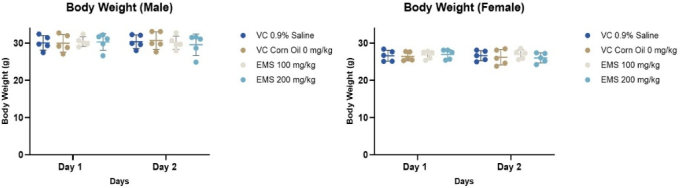
Male and female mice BW over time under various treatment circumstances are compared in the Figure 1. Using a two-way ANOVA with Tukey's multiple comparison test, the objective is to determine if administering 0.9% saline, corn oil 0 mg/kg, EMS 100 mg/kg BW, and EMS 200 mg/kg BW resulted in detectable changes in BW and whether these outcomes vary by sex and time point.
Figure 1. Body Weight Assessment in Male and Female Mice Treated with Vehicle Control (0.9% Saline and Corn Oil) and EMS (100 and 200 mg/kg) Over Two Consecutive Days: Individual and mean body weights were recorded for male and female groups receiving VC (0.9% saline), VC (corn oil, 0 mg/kg), and EMS at 100 or 200 mg/kg on Days 1 and 2. In both sexes, body weights were comparable between treatment and control groups at all time points. No statistically or biologically significant alterations were noted following EMS administration.
On day 1, the BW values of male mice were similar in all treatment groups. On day 2, there were no noticeable, significant differences in BW between the vehicle control, or EMS-induced groups in comparison to baseline, illustrated in Table 1. There was overlap across treatment conditions, and the distribution of individual measures showed comparable BW profiles across dosages. On day 1, female individuals showed similar distributions of BW in all groups. There were no noticeable changes in BW between day 1 and day 2 measurements. Although there was individual variation within each group, the general trend held for both vehicle and EMS-induced animals. While BW distributions varied between males and females at both time periods, as would be predicted given sex-related variances, BW seemed to be similar within each sex at both treatment conditions and time points.
Table 1. Summary of BW Measurements in Male and Female Mice Across Control and EMS Treatment Groups.

Group

Sex

No. of mice

Average BW (gm)

Day 1

Day 2

G1-VC 0.9% Saline (0 mg/kg BW)

Male

05

30.06

30.36

Female

05

26.58

26.62

G2 Corn Oil (0 mg/kg BW)

Male

05

29.938

30.684

Female

05

26.41

26.168

G3 EMS (100 mg/kg BW)

Male

05

30.38

30.096

Female

05

26.814

26.964

G4 EMS (200 mg/kg BW)

Male

05

30.278

29.53

Female

05

26.954

26.00

When combined, the closely clustered body-weight measurements of the control groups showed typical development trends and physiological consistency. The body-weight distributions of low-dose groups are similar to those of controls. On the other hand, both the low- and high-dose groups exhibited a decrease in mean BW in comparison to controls, along with a wider range of individual values. A dose-related change in BW increase is indicated by this pattern, which might be the result of systemic toxicity, decreased food intake, metabolic disturbance, or overall stress brought on by increasing exposure levels. Reduced mean BW and increased variability indicate a definite treatment-related detrimental impact, with the most noticeable effects seen in the highest dosage groups.
4.2. Effect of % Tail DNA
4.2.1. Duodenum Cells
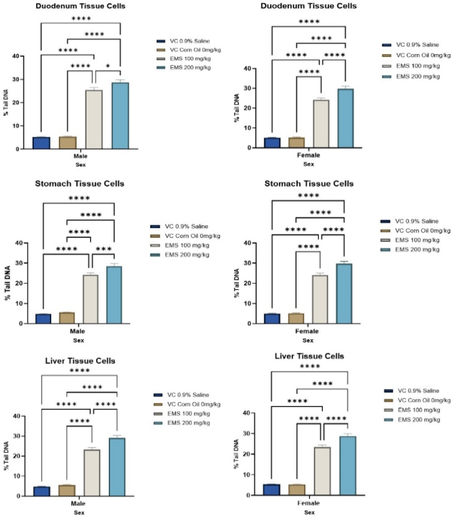
Figure 2. Ethyl Methanesulfonate (EMS)-Induced DNA Damage in Duodenum, Stomach, and Liver Cells of Male and Female Mice Assessed by% Tail DNA: % tail DNA was quantified in duodenal, stomach, and liver cells collected from male and female mice treated with vehicle controls (0.9% saline and corn oil) or EMS at 100 and 200 mg/kg. Data are presented as mean ± SD, and statistical comparisons are indicated (*p < 0.05; ****p < 0.0001). In both sexes, EMS administration resulted in a marked and statistically significant increase in DNA damage across all examined tissues compared with vehicle controls. A clear dose-dependent elevation in% tail DNA was observed, with the 200 mg/kg group consistently showing the highest level of genotoxicity.
DNA damage in duodenal cells was measured using the alkaline Comet assay, and the results were exhibited as mean ± SEM for typical comet features (e.g.,% tail DNA), shown in Table 2. Using two-way ANOVA and Tukey's multiple comparison tests, the main consequences of sex (male and female) and dosage (0.9% saline 0 mg/kg, corn oil 0 mg/kg, EMS 100 mg/kg BW, and EMS 200 mg/kg BW) were statistically evaluated in Figure 2. A two-way ANOVA revealed that the EMS dose significantly affected all DNA damage (***p < 0.001), indicating that the incidence of DNA damage in duodenal cells is dose dependent.
Table 2. DNA Damage (% Tail DNA) in Duodenum, Stomach, and Liver Cells of Male and Female Mice Following EMS Exposure.

Group

Sex

Average cells counted

Duodenum Tissue Cells

Stomach Tissue Cells

Liver Tissue Cells

Mean

Mean

Mean

% Tail DNA

% Tail DNA

% Tail DNA

VC 0.9% Saline 0 mg/kg

Male

150

4.75 ± 2.50

4.77 ± 2.68

4.75 ± 2.50

Female

150

5.31 ± 3.05

5.01 ± 2.95

5.31 ± 3.05

VC Corn Oil 0 mg/kg

Male

150

5.54 ± 3.08

5.54 ± 3.12

5.54 ± 3.08

Female

150

5.25 ± 2.87

5.11 ± 3.01

5.25 ± 2.87

EMS 100 mg/kg

Male

150

23.33 ± 11.89

24.26 ± 10.88

23.33 ± 11.89

Female

150

23.44 ± 12.05

24.08 ± 11.59

23.44 ± 12.05

EMS 200 mg/kg

Male

150

29.11 ± 14.20

28.52 ± 14.77

29.11 ± 14.20

Female

150

28.81 ± 14.81

29.73 ± 14.55

28.81 ± 14.81

According to Tukey's analysis, EMS exposure at 100 mg/kg BW significantly increased DNA damage when compared to both control groups (*p < 0.05). At 200 mg/kg BW, however, EMS generated a very significant elevation (***p <0.001) in comet values, which indicated substantial breakage in DNA strands.
Across matching EMS-induced groups, female duodenal cells showed greater amounts of DNA damage than males, indicating a significant main impact of sex (*p <0.05). At the highest EMS dose (200 mg/kg BW), Tukey's comparisons revealed that these sex-based differences were most noticeable, with females exhibiting very high statistical significance (***p <0.001) in comparison to males, while no significant differences were found between the male and female groups in the saline and corn oil controls (p >0.05).
In conclusion, the Comet test results shown that EMS causes dose-dependent DNA damage in duodenal cells, with females showing higher sensitivity. This emphasizes the significance of taking sex into account as a biological variable when evaluating genotoxicity.
4.2.2. Stomach Cells
The comet assay was used to assess the degree of DNA damage in stomach cells in male and female mice subjected to various vehicle groups, such as 0.9% saline, 0 mg/kg, corn oil 0 mg/kg, and EMS-treated groups (100 and 200 mg/kg BW). Tukey's multiple comparison tests were then performed (Figure 2 and Table 2).
There was very little DNA damage in mice treated with 0.9% saline and corn oil in both sexes. There was no statistically significant difference between these two control groups (p >0.05), demonstrating the lack of solvent-related genotoxic effects and validating their use as vehicle controls. Male and female stomach cells exposed to 100 mg/kg BW of EMS showed a statistically significant increase in DNA damage (p < 0.05) when compared to both control groups. This rise reveals the genotoxic potential of EMS at this dosage and validates the comet assay's sensitivity in identifying breaks in DNA strands.
When comparing stomach cells of both sexes to vehicle controls and the EMS 100 mg/kg BW group, a substantial increase in DNA damage was seen at the higher dosage, EMS 200 mg/kg BW (p < 0.01 and p < 0.001). This result suggests that DNA damage increases with increasing EMS concentration. Female mice at similar EMS dosages had a somewhat greater mean DNA damage response than male mice when comparing sex-based differences; however, this difference was not statistically significant (p >0.05). This implies that, despite possible small differences, sex had no discernible impact on the EMS-induced genotoxicity in stomach cells during the trial.
Overall, the statistical analysis showed that EMS causes substantial, dose-dependent DNA damage to stomach cells, but that treatments with saline and corn oil were not associated with any genotoxic consequences. The lack of discernible sex-related variations suggested that the genotoxic reaction in male and female animals is similar.
4.2.3. Liver Cells
DNA damage clearly increased after EMS exposure in both male and female mice, according to statistical evaluation of DNA damage in liver cells. There were no statistically significant differences between the two control groups (p >0.05) in the baseline levels of DNA damage seen in 0.9% saline and corn oil, suggesting that there were no genotoxic effects, shown in Figure 2 and Table 2.
On the other hand, in both males and females, treatment with EMS at 100 mg/kg BW led to a substantial increase in DNA damage when compared to both vehicle control groups (*p <0.05). This rise implies that at this dosage, detectable DNA strand breaks are induced in liver cells. When compared to controls and the EMS 100 mg/kg BW group, a statistically significant rise in DNA damage was seen at the higher EMS dose (200 mg/kg BW) (***p <0.001), indicating a strong dose-response connection. Male and female rats showed comparable patterns in every treatment group when sexes were compared. However, in contrast to male liver cells, female liver cells displayed a somewhat greater increase in DNA damage parameters at both EMS dosages. The statistical comparison between the sexes within the same dose group showed that the sex-related variation was either not significant or only slightly significant, even though this difference was clearly descriptive. This suggests that EMS-induced genotoxicity in liver cells is primarily dose-dependent rather than sex-dependent.
In summary, the statistical results demonstrate that EMS significantly and dose-dependently causes genotoxic effects in liver cells. While the same responses from male and female mice suggest that both sexes are equally vulnerable to EMS-induced DNA damage under the study's settings, the lack of significant differences between saline and vehicle controls confirms the experimental setup.
4.3. Effect Of% Viability
4.3.1. Duodenum Tissue Cells
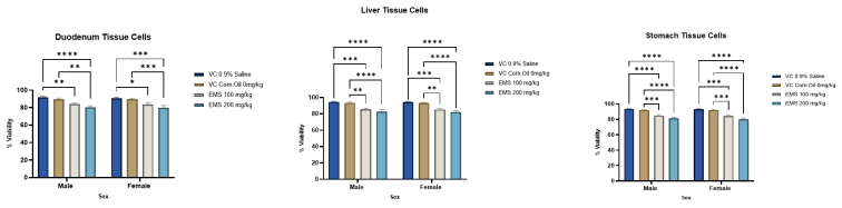
The comet test analysis of duodenal tissue cells revealed consistent patterns in both the male and female groups, as well as a significant dose-dependent decrease in the percentage of cell viability after EMS exposure (Table 3). Using two-way ANOVA and post-hoc multiple comparison tests, statistical comparisons were performed between the EMS-treated groups and the vehicle controls (0.9% saline and corn oil), as well as across dosages within each sex. P-values were used to interpret significance (*p < 0.05), as mentioned in Figure 3.
Figure 3. Effect of Ethyl Methanesulfonate (EMS) on Cell Viability in Duodenum, Liver, and Stomach Tissues of Male and Female Mice: % Cell viability was evaluated in duodenal, hepatic, and gastric tissues collected from male and female mice following treatment with vehicle controls (0.9% saline and corn oil) or EMS at doses of 100 and 200 mg/kg. Data are presented as mean ± SD. Compared with vehicle controls, EMS exposure resulted in a statistically significant reduction in cell viability across all examined tissues. The decrease was more pronounced at 200 mg/kg, demonstrating a dose-related effect. Comparable responses were observed in both male and female animals, suggesting minimal sex-related differences in EMS-induced cytotoxicity.
Table 3. Comparative analysis of relative cell viability in duodenum, glandular stomach, and liver tissues of male and female groups exposed to VC, corn oil, and EMS.

Group

Sex

Duodenum

G. Stomach

Liver

Average% viability per group

Relative viability compared to vehicle control

Average% viability per group

Relative viability compared to vehicle control

Average% viability per group

Relative viability compared to vehicle control

G1-VC 0.9% Saline (0 mg/kg BW)

Male

91.73

0.00

93.58

0.00

94.52

0.00

Female

90.83

0.00

93.01

0.00

94.21

0.00

G2 Corn Oil (0 mg/kg BW)

Male

89.65

0.98

92.20

0.99

93.58

0.99

Female

89.43

0.98

92.15

0.99

93.30

0.99

G3 EMS (100 mg/kg BW)

Male

84.02

0.92

84.71

0.91

85.61

0.91

Female

83.66

0.92

84.47

0.91

85.28

0.91

G4 EMS (200 mg/kg BW)

Male

80.40

0.88

81.48

0.87

82.73

0.88

Female

79.76

0.88

80.15

0.86

82.15

0.87

The highest levels of cell survival were viewed in the male duodenal cells of mice given 0.9% saline and corn oil (0 mg/kg), which may indicate that there was no cytotoxicity or genotoxic stress associated with the vehicle. When compared to the vehicle controls, the exposure to EMS at a dose of 100 mg/kg BW resulted in a significant decrease in cell viability (**p < 0.01), suggesting the possible production of DNA damage and a loss in cellular integrity. An even more notable decrease was seen at the 200 mg/kg BW EMS dosage, which was highly significant when compared to the 100 mg/kg group and the vehicle controls (****p < 0.0001), showing a strong dose-responsive genotoxic impact of EMS in male duodenal cells.
A similar pattern appeared in the female duodenum's cells. Cell viability in the 0.9% saline and corn oil groups did not change, indicating that the vehicles did not have any sex-specific effects. Cell viability significantly decreased after treatment with EMS at a dose of 100 mg/kg BW in comparison to the control group (*p < 0.05), whereas exposure to EMS at a dose of 200 mg/kg BW resulted in a considerable and very noticeable decrease (***p < 0.001). A clear dose-response connection in the female duodenal cells is highlighted by the progressive decline in viability with higher EMS dosages.
Males and females both showed comparable baseline viability under control settings in the framework of sex-related comparisons, indicating that there was no intrinsic difference in duodenal cell viability between the sexes. The general reaction pattern was the same for both sexes, although when exposed to EMS, females displayed a stronger sensitivity at the higher dosage. While the effects of EMS were significant in both sexes, there was no statistically significant interaction between sex and treatment at control levels. In conclusion, the statistical findings (shown by *, **, ***, and ****, which stand for p < 0.05, p < 0.01, p < 0.001, and p < 0.0001, respectively) confirm that EMS has notable, dose-dependent cytotoxic and genotoxic effects on duodenal tissue cells, irrespective of sex. These findings support the comet assay as a reliable technique for detecting DNA damage-related reductions in cell viability within gastrointestinal tissues and highlight the duodenum's susceptibility to alkylating chemicals like EMS.
4.3.2. Liver Tissue Cells
The lack of any vehicle-related toxicity was confirmed by the elevated levels and comparable percentage of cell viability in male liver cells from the vehicle control groups (0.9% saline and corn oil, 0 mg/kg), illustrated in Figure 3 and Table 3. No difference in significance was seen between them (p > 0.05). Cell viability significantly decreased after exposure to EMS at a dose of 100 mg/kg BW in comparison to both vehicle controls (**p < 0.01 to ***p < 0.001), indicating the start of the destruction of DNA and a deterioration in cellular integrity. A more considerable reduction was seen at EMS 200 mg/kg BW, where the percentage viability was significantly lower than that of EMS 100 mg/kg and both control groups (***p < 0.001 to ****p < 0.0001). This finding supports a strong dose-response connection in the liver tissue of males.
Similarly, as compared to EMS-treated groups, female liver cells showed that both vehicle controls maintained a far greater proportion of cell viability, with no discernible difference between saline and corn oil (p > 0.05). When contrasted to the controls, the viability decreased statistically significantly when EMS was administered at 100 mg/kg BW (**p < 0.01 - ***p < 0.001), while the percentage of cell viability decreased significantly and significantly when EMS was administered at 200 mg/kg BW (****p < 0.0001). A clear dose-dependent toxic impact was confirmed by the statistical significance of the gradual decline from EMS 100 to 200 mg/kg.
Both male and female liver cells showed comparable response patterns when sexes were compared, and neither the magnitude of EMS-induced decrease nor baseline viability showed any discernible sex-specific variations (p > 0.05 for sex interaction). This suggests that, under the examined conditions, the genotoxic effects of EMS in liver tissue are mostly sex-independent. In the conclusion, the statistical results and p-value distributions clearly showed that EMS exposure significantly reduced liver cell viability and DNA integrity in a dose-dependent way, as shown by the comet test, but vehicle treatments didn't trigger genotoxicity.
4.3.3. Stomach Tissue Cell
From a statistical standpoint, the impact of EMS exposure on stomach tissue cell viability was thoroughly assessed, taking into account treatment dose (0.9% saline, corn oil vehicle control, EMS 100 mg/kg BW, and EMS 200 mg/kg BW) as well as sex (male and female). Two-way ANOVA was used to analyze the data, and the findings were expressed as mean ± SEM after pertinent post-hoc multiple comparison tests. At *p < 0.05, statistical significance was established (Figure 3 and Table 3).
Both vehicle control groups, 0.9% saline and corn oil, showed good and similar cell viability inside the male stomach tissue cells; no statistically significant variation was observed between them (p > 0.05). This outcome confirms that the vehicles did not cause any cytotoxicity. On the other hand, EMS exposure resulted in a decrease in cell viability that varied according to the dosage. Thus, compared to both saline and corn oil controls, administration of EMS at 100 mg/kg BW significantly lowered cell viability (***p < 0.001). The EMS 200 mg/kg BW therapy caused a further and more noticeable drop, which was substantially reduced compared to the EMS 100 mg/kg BW treatment (***p < 0.001) and extremely significant when compared to the controls (****p < 0.0001).
Similarly, the control groups failed to demonstrate any significant differences between the saline and corn oil in the setting of female stomach tissue cells (p > 0.05). A statistically significant decrease in cell viability was observed when EMS exposure at 100 mg/kg BW was compared to both vehicle controls (***p < 0.001). Even more significant decline was seen in the EMS 200 mg/kg BW group, which was significantly less than the EMS 100 mg/kg BW group (**p < 0.001) and extremely significant versus the controls (****p < 0.0001).
The stomach tissue cells of males and females in the control and EMS-treated groups did not vary significantly when sex-based comparisons were performed at matching dosages (p > 0.05). This result suggests that both sexes experienced a similar cytotoxic reaction to EMS. The thorough statistical analysis showed that EMS has a clear dose-dependent cytotoxic effect on stomach tissue cells, with significant reductions in cell viability at both EMS dosages delivered, whereas the vehicle controls showed no toxicity at all. Importantly, the observed DNA damage may be mainly attributed to the genotoxic characteristics of EMS rather than an overwhelming cytotoxic response because cell viability remained above the allowable level for DNA damage assessment.
4.4. Effect of % Ghost Cell
4.4.1. Duodenum Tissue Cells
In order to facilitate the interpretation of comet assay data, the percentages of ghost cells in duodenal tissue were assessed as a standard for cytotoxicity, as mentioned in Table 4. Treatment doses (0.9% saline, corn oil vehicle, EMS 100 mg/kg BW, and EMS 200 mg/kg BW) and sex (male versus female) were determined to be experimental factors (Figure 4).
Table 4. Comparative distribution of ghost cells in gastrointestinal and hepatic tissues with relative ratios to control across treatment groups.

Group

Sex

Duodenum

G. Stomach

Liver

% Ghost cells per group

Relative ratio of ghost cells compared to the control

% Ghost cells/ group

Relative ratio of ghost cells compared to the control

% Ghost cells/ group

Relative ratio of ghost cells compared to the control

G1-VC 0.9% Saline (0 mg/kg BW)

Male

4.93

0.00

5.20

0.00

4.00

0.00

Female

5.07

0.00

5.33

0.00

4.27

0.00

G2 Corn Oil (0 mg/kg BW)

Male

4.93

1.00

5.87

1.13

4.40

1.10

Female

5.20

1.03

6.13

1.15

4.67

1.09

G3 EMS (100 mg/kg BW)

Male

9.47

1.92

11.20

2.15

8.80

2.20

Female

9.87

1.95

10.93

2.05

8.93

2.09

G4 EMS (200 mg/kg BW)

Male

11.47

2.32

13.07

2.51

11.07

2.77

Female

12.00

2.37

13.33

2.50

11.33

2.66

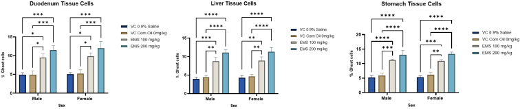
Figure 4. Induction of Ghost Cells in Duodenum, Liver, and Stomach Tissues Following EMS Exposure in Male and Female Mice: The percentage of ghost cells, indicative of apoptotic or severely damaged cells, was quantified in duodenal, hepatic, and gastric tissues from male and female mice treated with vehicle controls (0.9% saline and corn oil) and ethyl methanesulfonate (EMS) at 100 and 200 mg/kg. Data are expressed as mean ± SD. EMS administration produced a statistically significant increase in the proportion of ghost cells across all examined tissues compared with both vehicle controls (*p < 0.05 to ****p < 0.0001). A clear dose-dependent trend was observed, with the highest percentages detected in the 200 mg/kg groups.
Ghost cell extents of the vehicle control groups (0.9% saline and corn oil) were baseline and relatively low (~5%) in both male and female animals. The absence of treatment-related cytotoxicity under control circumstances is shown by the fact that no significant variations were found between the two vehicle controls. Both sexes had the same initial scores, demonstrating that there is no innate sex-dependent variation in the cytotoxicity of the vehicle in the duodenum.
For both sexes, administration of EMS has led to a noticeable and statistically significant increase in the percentage of ghost cells in a dose-dependent fashion. When as opposed to saline and corn oil controls, EMS at a dose of 100 mg/kg BW significantly raised the number of ghost cells in male participants (*p < 0.05). Furthermore, compared to the controls and the 100 mg/kg BW group, EMS at 200 mg/kg BW caused an even larger and more significant rise (***p < 0.001). Similarly, in females, EMS at 100 mg/kg BW markedly boosted the frequency of ghost cells compared to vehicle controls (*p < 0.05), while EMS at 200 mg/kg BW produced a considerable and extremely significant increase (**p < 0.001).
The number of ghost cells has been identified as very similar across the sexes when they were given the same dosages. At EMS 200 mg/kg BW, females exhibited a little greater mean sensitivity than males, but this distinction was not significant when considering the powerful main impact of EMS. Therefore, the two-way ANOVA showed that the treatment dosage had a substantial main effect (***p < 0.001), but the main effects of sex and the interaction between sex and dose were negligible in comparison to the dose effect. Overall, the findings show that EMS causes a dose-dependent cytotoxic impact on duodenal tissue cells, as demonstrated by an increase in the creation of ghost cells in both male and female participants. Particularly, the degree of cytotoxicity remained within a range appropriate for comet test assessment, indicating that the DNA damage seen at high EMS dosages represents real genotoxic effects rather than just an excess of cell death.
4.4.2. Liver Tissue Cells
EMS treatment led to a considerable and significant increase in the proportion of ghost cells in liver tissue cells, with dosage-dependent effects. When EMS was administered to male participants at a dose of 100 mg/kg BW, the proportion of ghost cells was considerably higher than in both vehicle controls (**p < 0.01 - ***p < 0.001). Comparing 200 mg/kg BW to vehicle controls and the lower EMS dosage revealed an additional significant increase that was very significant (****p < 0.0001). Similar results were shown in female individuals, where EMS at 100 mg/kg BW significantly increased the frequency of ghost cells in comparison to controls (**p < 0.01). Additionally, EMS at 200 mg/kg BW produced the greatest numbers of ghost cells, indicating extremely significant differences from EMS at 100 mg/kg BW and vehicle controls (****p < 0.0001) (Figure 4 and Table 4).
Males and females responded to EMS therapy identically, according to a comprehensive sex-to-sex comparison, with no discernible variations in the overall cytotoxicity profile. Although the main impact was dosage-driven rather than sex-specific, females showed a slightly greater mean percentage of ghost cells at the maximal EMS dose, suggesting a possible tendency towards increased sensitivity. Overall, the statistical results show that, whereas vehicle treatments refused to compromise cell integrity, EMS significantly and dose-dependently cytotoxically damages liver tissue cells, as seen by the increase in ghost cell production. The conclusion that the detected DNA damage is primarily genotoxic rather than an artifact of excessive cytotoxicity is supported by the fact that, despite a rise in ghost cell frequencies at higher EMS dosages, these frequencies stayed within an accessible limit for the comet test.
4.4.3. Stomach Tissue Cells
It appeared that there was no statistically significant difference between the two vehicles (p > 0.05), and the vehicle control groups (0.9% saline and corn oil, 0 mg/kg) had comparably low levels of ghost cells (around 5-6%) in both males and females, shown in Figure 4 and Table 4. The regularity of ghost cells clearly increased following EMS treatment, and this increase was dose-related. A substantial increase was observed when EMS was administered at a dose of 100 mg/kg, compared with both vehicle controls (males: ***p < 0.001; females: **p < 0.01 to ***p < 0.001). The proportion of ghost cells was considerably larger at 200 mg/kg than at 100 mg/kg and in all control groups, further amplifying this impact (****p < 0.0001).
However, there was a minor trend for females to have a larger mean at the highest EMS concentration, but no statistically significant differences were seen when comparing the sexes within each treatment group (p > 0.05). When considered collectively, these results show that there are no discernible sex-related variations in the robust, dose-dependent cytotoxic response to EMS in stomach tissue, as seen by enhanced ghost cell appearance. In addition to supporting the accurate interpretation of DNA damage results in the comet assay, the degree of ghost cell formation validates treatment-associated cytotoxicity.
5. Discussion
The Comet assay has been used to identify prospective genotoxic chemical agents' mechanisms of action in tumor target tissues or tissues exhibiting illness, to evaluate the in-vivo significance of positive results from in-vitro genotoxicity studies, and to look into suspected genotoxicity at the region of interaction. In this context, the comet assay has been widely employed to examine how genotoxic chemicals affect the breaking of DNA strands in mammalian cells.
In this work, we assessed whether EMS causes destruction of DNA in the duodenum, liver, and stomach tissue cells of mice using the in vivo comet test. To determine genotoxicity, three specific tissues were chosen: the liver, a frequently examined organ in rodent cancer bioassays, a tissue that is advised for assessing DNA damage in ICH S2 (R1), and a tissue being studied in the context of the JaCVAM international Comet assay validation endeavor . The glandular stomach, a tissue being investigated in the JaCVAM validation endeavor and the duodenum, a common region of tumors in the United States and other Western populations, making it a potential tissue of interest due to its crucial role in food digestion, first-pass metabolism, and as an initial site of exposure of toxicant contact with the body.
In a validation study of an in vivo comet assay, where EMS was employed as a positive control chemical in the stomach and liver, the results eligibility requirements demanded that the percentage of tail DNA be 1) statistically significantly greater than the control, 2) increased by more than 5% to the control, and 3) increased by 2-fold or more, relative to the control. The study's rise in the percentage of tail DNA by EMS satisfied all of the requirements for data acceptance for the stomach, duodenum, and liver tissues mentioned above. Since an increase in tail DNA must be taken into account for both genotoxic and cytotoxic effects . According to the aforementioned findings, the genotoxic impacts of EMS on the mice liver, duodenum, and stomach were the cause of the rise in EMS-induced percentage tail DNA.
EMS, a familiar genotoxic chemical, damages DNA in a variety of ways, including depurination , DNA adducts , and DNA alkylation processes . At the phospho-triester spine of the DNA helix and at nearly every purine or pyrimidine nucleophilic site, EMS is capable of producing DNA adducts . Furthermore, it has been documented that EMS damages DNA in the stomach, bladder, and bone marrow and causes cancers in the liver, kidneys, lungs, brain, mammary glands, and uterus . The Comet test can therefore identify a range of DNA damages (DNA adducts, apurinic sites) that may be caused by EMS.
We observed that the ghost cell population doubles at both low and high concentrations of EMS in comparison to 0.9% saline and corn oil in the tissues of the liver, duodenum, and stomach, resulting in a loss of cellular viability of less than 5% (low concentration) and 10% (high concentration). Despite its high treatment concentration, EMS, which was the most successful at inducing strand breaks (almost 90%), did not result in a discernible rise in DNA base lesions. In conclusion, EMS appears to be a highly helpful positive control substance for multi-organ comet assays in mice, specifically targeting the bone marrow, thyroid gland, liver, stomach, intestine, and bladder.
6. Conclusion
Overall, EMS administration satisfied established in vivo comet test data acceptance requirements by causing statistically significant and clinically relevant increases in the percentage of tail DNA in the liver, duodenum, and stomach tissues. When compared to controls, the extent of DNA damage was greater than the two-fold change and 5% absolute increase thresholds. Crucially, very mild increases in the frequency of ghost cells and negligible decreases in cell viability suggest that genotoxic rather than cytotoxic effects were mostly responsible for the observed DNA movement. The results further verify EMS as a suitable and trustworthy positive control molecule for regulatory genotoxicity testing in mice and validate the sensitivity and robustness of the multi-organ comet assay.
7. Future Scope
In the future, the utilization of sophisticated 3D (three-dimensional)-reconstructed tissues, such as intestine organoids, gastric mucosa equivalents, and liver spheroids, shows promise. Tissue architecture, metabolic capacity, and exposure gradients are all closely modelled by these models. Assessment of first-pass metabolism, local tissue vulnerability, and DNA repair capacity in site-of-contact tissues would be possible by performing comet assay studies in these 3D systems following EMS exposure. These investigations may help predict the risk of gastrointestinal and hepatic cancer and shed light on the genotoxic pathways unique to particular organs. Another avenue for the future is to combine two-dimensional (2D) in-vitro assays with integrated testing methodologies. According to the 3Rs framework, demonstrating agreement between 2D in-vitro comet assay data and in-vivo results could encourage the regulatory adoption of substitute techniques, minimize the use of animals, and facilitate more predictive, mechanism-based genotoxicity screening for pharmaceuticals and environmental chemicals. To improve mechanistic knowledge, human risk prediction, and ethical toxicological testing, this model should move away from only animal-based comet assay research and toward human-relevant in-vitro systems, sophisticated 3D organoid models, 2D in-vitro procedures, and integrated testing strategies.
Abbreviations

EMS

Ethyl Methane Sulfonate

DNA

Deoxyribonucleic Acid

DMSO

Dimethyl Sulfoxide

DPBS

Dulbecco's Phosphate-buffered Saline

FBS

Fetal Bovine Serum

EDTA

Ethylene Diamine Tetra-acetic Acid

CAS

Chemical Abstracts Service

HBSS

Hanks Balanced Salt Mixture

OECD

Organisation for Economic Co-operation and Development

NMA

Normal Melting Agarose

LMA

Low Melting Agarose

BW

Body Weight

MG

Milligram

ML

Milliliter

mM

Millimolar

KG

Kilogram

VC

Vehicle Control

µL

Microliter

%

Percentage

3D

Three-diamensional

2D

Two-diamensional

Acknowledgments
We acknowledge the support of Vipragen Biosciences Private Limited, Hootagalli, Mysore, Karnataka, for providing resources.
Author Contributions
Samit Kadam: Conceptualization, Data curation, Formal Analysis, Investigation, Methodology, Supervision, Validation, Visualization, Writing – original draft, Writing – review & editing
Aditya Dipakrao Hajare: Data curation, Formal Analysis, Validation, Visualization, Writing – original draft, Writing – review & editing
Bhumika Nataraj: Formal Analysis, Validation, Visualization
Data Availability Statement
No new data were generated or analysed in this study. All data referenced in this review are available in the cited publications.
Conflicts of Interest
The authors declare no conflicts of interest.
References
[1] International Conference on Harmonisation of Technical Requirements for Registrations of Pharmaceuticals in Human Use (ICH). 1996. S2A: Guidance on Specific Aspects of Regulatory Genotoxicity Tests for Pharmaceuticals.
[2] International Conference for Harmonisation of Technical Requirements for Registration of Pharmaceuticals for Human Use (ICH). 1997. S2B: Genotoxicity: A Standard Battery for Genotoxicity Testing for Pharmaceuticals.
[3] Eastmond DA, Hartwig A, Anderson D, Anwar WA, Cimino MC, Dobrev I, Douglas GR, Nohmi T, Phillips DH, Vickers C. 2009. Mutagenicity testing for chemical risk assessment: Update on the WHO/OPCS Harmonized Scheme. Mutagenesis 24:341-349.
[4] B. Halliwell and J. M. C. Gutteridge, Free Radicals in Biology and Medicine, Oxford University Press, New York USA, 4th edition, 2007.
[5] N. T. Henthorn, J. W. Warmenhoven, M. Sotiropoulos et al., “Clinically relevant nanodosimetric simulation of DNA dam age complexity from photons and protons,” RSC Advances, vol. 9, no. 12, pp. 6845-6858, 2019.
[6] de Lapuente, Joaquín, Joana Lourenço, Sónia A. Mendo, Miquel Borràs, Marta G. Martins, Pedro M. Costa, and Mário Pacheco. "The Comet Assay and its applications in the field of ecotoxicology: a mature tool that continues to expand its perspectives." Frontiers in genetics 6 (2015): 180.
[7] Tice RR, Agurell E, Anderson D, Burlinson B, Hartmann A, Kobayashi H, Miyamae Y, Rojas E, Ryu JC, Sasaki YK. 2000. Single cell gel/comet assay: Guidelines for in vitro genetic toxicology test ing. Environ Mol Mutagen 35:206-221.
[8] Burlinson B, Tice RR, Speit G, Agurell E, Brendler-Schwaab SY, Col lins AR, Escobar, P, Honma M, Kumaravel, TS, Nakajima M, Sasaki YF, Thybaud V, Uno Y, Vasquez M, Hartmann A. 2007. Fourth International Workgroup on Genotoxicity testing: Results of the in vivo Comet assay workgroup. Mutat Res 627:31-35.
[9] Uno, Y., Kojima, H., Omori, T., Corvi, R., Honma, M., Schechtman, M. L., Tice, R. R., Burlinson, B., Escobar, P. A., Kraynak, A. R., Nakagawa, Y., Nakajima, M., Pant, K., Asano, N., Lovell, D., Morita, T., Ohno, Y. and Hayashi, M. (2015a): JaCVAM-organ- ized international validation study of the in vivo rodent alkaline comet assay for the detection of genotoxic carcinogens: I. Sum- mary of pre-validation study results. Mutat. Res. Genet. Toxicol. Environ. Mutagen., 786-788, 3-13.
[10] Susanne Brendler-Schwaab, Andreas Hartmann, Stefan Pfuhler, Günter Speit, The in vivo comet assay: use and status in genotoxicity testing, Mutagenesis, Volume 20, Issue 4, July 2005, Pages 245-254,
[11] Bunček, M. Griffiths, A. J. F., Miller, J. H., Suzuki, D. T., Lewontin, R., Gelbart, W. M.: An Introduction to Genetic Analysis. Biologia Plantarum 45, 50 (2002).
[12] OECD (2016), Test No. 489: In Vivo Mammalian Alkaline Comet Assay, OECD Guidelines for the Testing of Chemicals, Section 4, OECD Publishing, Paris.
[13] Recio L, Hobbs C, Caspary W, Witt KL. 2010. Dose-response assess ment of four genotoxic chemicals in a combined mouse and rat micronucleus (MN) and comet assay protocol. J Toxicol Sci 35:149-162.
[14] Witt KL, Malarkey DE, Hobbs CA, Davis JP, Kissling GE, Casp ary W, Travelos G, Recio L. 2010. No increases in bio markers of genetic damage or pathological changes in heart and brain tissues in male rats administered methylphenidate hydrochloride (Ritalin) for 28 days. Environ Mol Mutagen 51:80-88
[15] Elmar, G., Heinrich, B., Lutz, M. and Thomas, P. (2009): Literature review on the genotoxicity, reproductive toxicity, and carcino- genicity of ethyl methanesulfonate. Toxicol. Lett., 190, 254-265.
[16] Jones GD, Le Pla RC, Farmer PB. 2010 Phosphotriester adducts (PTEs): DNA’s overlooked lesion. Mutagenesis 25:3-16.
[17] Kitamoto, S., Matsuyama, R., Uematsu, Y., Ogata, K., Ota, M., Yamada, T., Miyata, K., Funabashi, H. and Saito, K. (2015): Opti- mal dose selection of N-methyl-N-nitrosourea for the rat com- et assay to evaluate DNA damage in organs with different sus- ceptibility to cytotoxicity. Mutat. Res. Genet. Toxicol. Environ. Mutagen., 786-788, 129-136.
[18] Kraynak, A. R., Barnum, J. E., Cunningham, C. L., Ng, A., Ykoruk, B. A., Bennet, B., Stoffregen, D., Merschman, M., Freeland, E. and Galloway, S. M. (2015): Alkaline comet assay in liv- er and stomach, and micronucleus assay in bone marrow, from rats treated with 2-acetylaminofluorene, azidothymidine, cispl- atin, or isobutyraldehyde. Mutat. Res. Genet. Toxicol. Environ. Mutagen., 786-788, 77-86.
Cite This Article
  • APA Style

    Kadam, S., Hajare, A. D., Nataraj, B. (2026). Assessing the Validity and Reliability of the Comet Assay in Detecting EMS-induced Genotoxicity. International Journal of Genetics and Genomics, 14(3), 99-112. https://doi.org/10.11648/j.ijgg.20261403.11

    Copy | Download

    ACS Style

    Kadam, S.; Hajare, A. D.; Nataraj, B. Assessing the Validity and Reliability of the Comet Assay in Detecting EMS-induced Genotoxicity. Int. J. Genet. Genomics 2026, 14(3), 99-112. doi: 10.11648/j.ijgg.20261403.11

    Copy | Download

    AMA Style

    Kadam S, Hajare AD, Nataraj B. Assessing the Validity and Reliability of the Comet Assay in Detecting EMS-induced Genotoxicity. Int J Genet Genomics. 2026;14(3):99-112. doi: 10.11648/j.ijgg.20261403.11

    Copy | Download

  • @article{10.11648/j.ijgg.20261403.11,
      author = {Samit Kadam and Aditya Dipakrao Hajare and Bhumika Nataraj},
      title = {Assessing the Validity and Reliability of the Comet Assay in Detecting EMS-induced Genotoxicity},
      journal = {International Journal of Genetics and Genomics},
      volume = {14},
      number = {3},
      pages = {99-112},
      doi = {10.11648/j.ijgg.20261403.11},
      url = {https://doi.org/10.11648/j.ijgg.20261403.11},
      eprint = {https://article.sciencepublishinggroup.com/pdf/10.11648.j.ijgg.20261403.11},
      abstract = {The widespread use of industrial chemicals has raised concerns about their potential to damage deoxyribonucleic acid (DNA), a process linked to cancer and other serious diseases. To explore this risk, we examined the genotoxic and cytotoxic effects of ethyl methane sulfonate (EMS), a known mutagen, in mice using the comet assay. Saline and corn oil served as vehicle controls. Body weight (BW) measurements showed no major differences between controls and treated groups over two days, although higher EMS doses were associated with slight reductions and greater variability, suggesting systemic stress. In contrast, DNA damage was strikingly dose-dependent. Duodenum, stomach, and liver tissues all showed significant increases in DNA strand breaks after EMS exposure, with duodenal cells in females appearing particularly sensitive. Cell viability declined progressively with increasing EMS doses across all tissues, while ghost cell frequency, a marker of cytotoxicity, rose in parallel. Importantly, vehicle controls remained stable, confirming that observed effects were due to EMS rather than solvents. Together, these findings demonstrate that EMS induces clear, dose-dependent DNA damage and cell stress in gastrointestinal and hepatic tissues. The comet assay proved to be a sensitive and reliable tool for detecting such genotoxic effects, reinforcing its value in toxicity testing. By highlighting tissue-specific and sex-related responses, this study underscores the importance of considering biological variability when assessing chemical hazards.},
     year = {2026}
    }
    

    Copy | Download

  • TY  - JOUR
    T1  - Assessing the Validity and Reliability of the Comet Assay in Detecting EMS-induced Genotoxicity
    AU  - Samit Kadam
    AU  - Aditya Dipakrao Hajare
    AU  - Bhumika Nataraj
    Y1  - 2026/07/28
    PY  - 2026
    N1  - https://doi.org/10.11648/j.ijgg.20261403.11
    DO  - 10.11648/j.ijgg.20261403.11
    T2  - International Journal of Genetics and Genomics
    JF  - International Journal of Genetics and Genomics
    JO  - International Journal of Genetics and Genomics
    SP  - 99
    EP  - 112
    PB  - Science Publishing Group
    SN  - 2376-7359
    UR  - https://doi.org/10.11648/j.ijgg.20261403.11
    AB  - The widespread use of industrial chemicals has raised concerns about their potential to damage deoxyribonucleic acid (DNA), a process linked to cancer and other serious diseases. To explore this risk, we examined the genotoxic and cytotoxic effects of ethyl methane sulfonate (EMS), a known mutagen, in mice using the comet assay. Saline and corn oil served as vehicle controls. Body weight (BW) measurements showed no major differences between controls and treated groups over two days, although higher EMS doses were associated with slight reductions and greater variability, suggesting systemic stress. In contrast, DNA damage was strikingly dose-dependent. Duodenum, stomach, and liver tissues all showed significant increases in DNA strand breaks after EMS exposure, with duodenal cells in females appearing particularly sensitive. Cell viability declined progressively with increasing EMS doses across all tissues, while ghost cell frequency, a marker of cytotoxicity, rose in parallel. Importantly, vehicle controls remained stable, confirming that observed effects were due to EMS rather than solvents. Together, these findings demonstrate that EMS induces clear, dose-dependent DNA damage and cell stress in gastrointestinal and hepatic tissues. The comet assay proved to be a sensitive and reliable tool for detecting such genotoxic effects, reinforcing its value in toxicity testing. By highlighting tissue-specific and sex-related responses, this study underscores the importance of considering biological variability when assessing chemical hazards.
    VL  - 14
    IS  - 3
    ER  - 

    Copy | Download

Author Information
  • Vipragen Biosciences Private Limited, Mysuru, India
  • Vipragen Biosciences Private Limited, Mysuru, India

  • Vipragen Biosciences Private Limited, Mysuru, India

  • Abstract
  • Keywords
  • Document Sections

    1. 1. Introduction
    2. 2. Materials and Methods
    3. 3. Experimental Design
    4. 4. Result
    5. 5. Discussion
    6. 6. Conclusion
    7. 7. Future Scope
    Show Full Outline
  • Abbreviations
  • Acknowledgments
  • Author Contributions
  • Data Availability Statement
  • Conflicts of Interest
  • References
  • Cite This Article
  • Author Information