Limb-Body Wall Complex (LBWC) is a rare and typically lethal congenital anomaly characterized by severe defects of the ventral body wall, limbs, and neural tube. It is considered one of the most severe malformation syndromes. Its etiology remains uncertain, with proposed mechanisms including abnormal embryonic folding, vascular disruption, and early amnion rupture. We report a stillborn neonate delivered at Alem Ketema Enat Hospital, North Shoa, Amhara, Ethiopia, to a 27-year-old multiparous woman with no antenatal care. The neonate presented with multiple congenital anomalies consistent with the caudal variant of LBWC, including a large abdominal wall defect with evisceration of viscera, spinal dysraphism, and limb deformities. Unique to this case was the coexistence of a well-defined skin-covered sacrococcygeal mass distinct from the spinal defect, absence of external genitalia, imperforate anus, and unilateral clubfoot—an unusual constellation of findings rarely documented together in previous reports. The umbilical cord was notably short, further supporting the diagnosis. This case expands the phenotypic spectrum of the caudal variant of LBWC and highlights the diagnostic challenges in low-resource settings where routine prenatal ultrasonography is unavailable. The combination of these caudal defects suggests a failure of early embryonic folding in this region. Early antenatal screening and access to skilled sonography are essential for the timely identification of lethal congenital malformations, appropriate counseling, and informed pregnancy management in such environments. This report serves as the first from Ethiopia documenting this specific presentation, underscoring the urgent need to strengthen anomaly detection programs.
This is an Open Access article, distributed under the terms of the Creative Commons Attribution 4.0 International License (http://creativecommons.org/licenses/by/4.0/), which permits unrestricted use, distribution and reproduction in any medium or format, provided the original work is properly cited.
Limb-Body Wall Complex (LBWC) is a rare and usually lethal congenital disorder characterized by multiple major anomalies, including defects of the ventral body wall, limbs, and neural tube. Historically, the diagnosis was established primarily through comprehensive postmortem examinations; however, with advances in imaging, LBWC can now be identified during the first or second trimester using prenatal ultrasonography
[1]
Thomas MA, Crawford S, Bedard T. A population based study of limb body wall complex with proposed features for prenatal diagnosis. Am J Med Genet A. 2025; 197(11): e64140.
LBWC is typically classified into thoracoabdominal (classic) and caudal variants, with the latter predominantly affecting the lower abdomen, pelvis, sacrum, and lower limbs
[2]
Van Allen MI, Curry C, Gallagher L. Limb body wall complex: diagnostic criteria and clinical spectrum. Am J Med Genet. 1987; 28(3): 529-539.
. Classically, three major anomalies are associated with LBWC: 1. Exencephaly or encephalocele, often accompanied by facial clefts 2. Thoraco- and/or abdominoschisis, involving defects of the thoracic and/or abdominal wall 3. Limb defects, which may include clubfoot, limb reduction, or other malformations
[1]
Thomas MA, Crawford S, Bedard T. A population based study of limb body wall complex with proposed features for prenatal diagnosis. Am J Med Genet A. 2025; 197(11): e64140.
Cases of gastroschisis associated with additional anomalies, such as limb defects, kyphosis, talipes equinovarus, imperforate anus, and absence of external genitalia and nipples, are extremely rare
[4]
Kar A, Kar T, Nayak GD, et al. Gastroschisis with multiple skeletal deformities, imperforate anus, and absent genitalia: a rare presentation. Case Rep. 2019.
There is no universal consensus regarding definitive diagnostic criteria for LBWC. Its incidence is estimated at 1 in 14,000 to 1 in 31,000 pregnancies, and it is frequently associated with intrauterine fetal demise
[1]
Thomas MA, Crawford S, Bedard T. A population based study of limb body wall complex with proposed features for prenatal diagnosis. Am J Med Genet A. 2025; 197(11): e64140.
The etiology of LBWC remains unclear. Several theories have been proposed: - Abnormal embryonic folding (germinal disc defect theory): defective folding of the trilaminar germ disc results in maldevelopment of the ventral body wall and associated structures
[1]
Thomas MA, Crawford S, Bedard T. A population based study of limb body wall complex with proposed features for prenatal diagnosis. Am J Med Genet A. 2025; 197(11): e64140.
- Amnion rupture theory: early rupture of the amniotic sac causes mechanical entanglement of the developing embryo by amniotic bands, disrupting normal organogenesis
[5]
Torpin R. Amniochorionic mesoblastic fibrous strings and amniotic bands: associated constricting fetal malformations or fetal death. Am J Obstet Gynecol. 1965; 91: 65-75.
We report a rare case of LBWC - caudal variant in a term stillborn neonate presenting with a large gastroschisis, isolated sacral mass, spinal defect, unilateral limb deformity, and absent external genitalia and anal canal. This case highlights the variable spectrum of LBWC, emphasizes the presence of an isolated sacral mass separate from the spinal defect, and underscores the importance of early prenatal detection for counseling and pregnancy management.
2. Case Presentation
Maternal History: A 27-year-old Gravida 2, Para 2 woman with no antenatal care delivered a term stillborn neonate at a rural Ethiopian hospital. There was no history of consanguinity, teratogenic exposure, abortion, or previous stillbirth.
Ultrasound Findings: Bedside ultrasound revealed a gestational age of 38 weeks, a large anterior abdominal wall defect, and a solid sacral mass (5 × 5 cm) overlying a lumbosacral spinal defect. Fetal cardiac activity was absent.
Delivery Outcome: A stillborn neonate weighing 3.2 kg was delivered via spontaneous vaginal delivery after four hours.
Clinical Findings at Birth:
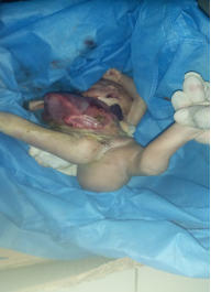
Abdominal wall defect: Large Gastroschisis with evisceration of intestines and part of the liver; short umbilical cord (10 cm).
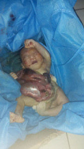
Spinal and limb anomalies: Sacrococcygeal mass in the postero-inferior perineum; dorsally turned lower limbs with malrotated feet; unilateral clubfoot and femoral deformity (Figures 1, 2).
Urogenital anomalies: Absent external genitalia and imperforate anus (Figure 2).
Ethical approval was obtained from the Institutional Review Board of Debre Berhan University, Ethiopia. Written informed consent for publication of this case and accompanying images was obtained from the patient’s parents.
4. Discussion
Limb-Body Wall Complex (LBWC) is one of the most severe congenital malformation syndromes, typically characterized by major defects of the ventral body wall, limbs, and neural tube. The condition is usually incompatible with postnatal life, with most affected fetuses resulting in intrauterine fetal demise or early neonatal death
[1]
Thomas MA, Crawford S, Bedard T. A population based study of limb body wall complex with proposed features for prenatal diagnosis. Am J Med Genet A. 2025; 197(11): e64140.
Van Bennekom CM, Mitchell AA, Moore CA, et al. Vasoactive exposures during pregnancy and risk of microtia. Birth Defects Res A Clin Mol Teratol. 2013; 97(12): 833-840.
The etiology of LBWC remains uncertain, with several hypotheses proposed. The amnion rupture theory, first described by Torpin
[7]
Guzmán Huerta ME, Muro Barragán SA, Acevedo Gallegos S, et al. Amniotic band sequence: prenatal diagnosis, phenotype descriptions, and a proposal of a new classification based on morphologic findings. Rev Invest Clin. 2013; 65(4): 300-306.
, suggests that early rupture of the amniotic sac causes entanglement of the developing embryo by amniotic bands, resulting in mechanical disruption of organogenesis. The vascular disruption theory attributes anomalies to ischemic injury during embryogenesis
[2]
Van Allen MI, Curry C, Gallagher L. Limb body wall complex: diagnostic criteria and clinical spectrum. Am J Med Genet. 1987; 28(3): 529-539.
. The abnormal embryonic folding (germinal disc defect) theory posits that defective folding of the trilaminar germ disc leads to maldevelopment of the ventral body wall and associated structures
[7]
Guzmán Huerta ME, Muro Barragán SA, Acevedo Gallegos S, et al. Amniotic band sequence: prenatal diagnosis, phenotype descriptions, and a proposal of a new classification based on morphologic findings. Rev Invest Clin. 2013; 65(4): 300-306.
Our case, presenting with gastroschisis, spina bifida, unilateral clubfoot, absent external genitalia, and imperforate anus, aligns most closely with the embryonic folding theory. The presence of multiple midline and caudal anomalies suggests a primary developmental defect rather than secondary mechanical damage, although overlap with amniotic band-related disruptions cannot be excluded.
Diagnostic criteria for LBWC remain debated. Van Allen et al. proposed that the presence of at least two of the following anomalies is sufficient for diagnosis: (1) exencephaly or encephalocele with facial clefts, (2) thoraco- or abdominoschisis, and (3) limb defects
[9]
Daskalakis G, Sebire NJ, Jurkovic D, et al. Body stalk anomaly at 10-14 weeks of gestation. Ultrasound Obstet Gynecol. 1997; 10: 405-410.
. Our patient fulfilled at least two criteria—abdominal wall defect and limb deformity—along with a neural tube defect, supporting the diagnosis.
Prenatal diagnosis is crucial but often missed in low-resource settings. LBWC can typically be identified by first-trimester ultrasonography, which may reveal severe body wall disruption, fixed fetal posture, and absence of umbilical cord visualization due to a short or absent cord
[9]
Daskalakis G, Sebire NJ, Jurkovic D, et al. Body stalk anomaly at 10-14 weeks of gestation. Ultrasound Obstet Gynecol. 1997; 10: 405-410.
. In this case, lack of antenatal care and absence of early ultrasound prevented timely detection, highlighting a common challenge in rural Ethiopia.
The differential diagnosis includes omphalocele, gastroschisis, amniotic band syndrome (ABS), and caudal regression syndrome (CRS). Gastroschisis is usually isolated without associated limb or neural anomalies, while ABS often presents with asymmetric limb amputations or constriction rings without major ventral body wall defects
[10]
Bergman JEH, Barišić I, Addor MC, et al. Amniotic band syndrome and limb body wall complex in Europe 1980-2019. Am J Med Genet A. 2023; 191(12): 3047-3063.
. CRS may show imperforate anus and urogenital anomalies but lacks extensive body wall and limb abnormalities characteristic of LBWC, confirming the diagnosis in this case
[11]
Mian DB, Nguessan KLP, Aissi G, et al. Amniotic band syndrome (ABS): can something be done during pregnancy in African poor countries? Clin Exp Obstet Gynecol. 2014; 41(4): 439-444.
[11]
.
Specific Features of This Case.
This case exhibits several phenotypic features rarely reported together in LBWC:
Skin-covered sacrococcygeal mass clearly separated from the spinal defect, unlike the typical open neural tube lesions described in most LBWC cases
[12]
Baruah P, Choudhury PR. Limb Body Wall Complex with sacrococcygeal mass and agenesis of external genitalia. Case Rep Med. 2013.
Adeleke OA, Ogunbiyi AO, et al. Rare presentation of limb body wall complex in a neonate: sacrococcygeal mass and imperforate anus without kyphoscoliosis. J Matern Fetal Neonatal Med. 2022; 35(24): 5000-5005.
Moerman P, Fryns JP, Vandenberghe K, et al. Pathogenesis of body stalk anomaly: disruption, vascular compromise, or early maldevelopment? Am J Med Genet. 1992; 42(4): 433-437.
Russo R, D’Armiento M, Angrisani P, Vecchione R. Limb body wall complex: a critical review and a nosological proposal. Am J Med Genet. 1993; 47(6): 893-900.
Adeleke OA, Ogunbiyi AO, et al. Rare presentation of limb body wall complex in a neonate: sacrococcygeal mass and imperforate anus without kyphoscoliosis. J Matern Fetal Neonatal Med. 2022; 35(24): 5000-5005.
Unilateral femoral deformity with clubfoot, whereas limb reduction or bilateral defects are more typical, highlighting phenotypic variability
[14]
Moerman P, Fryns JP, Vandenberghe K, et al. Pathogenesis of body stalk anomaly: disruption, vascular compromise, or early maldevelopment? Am J Med Genet. 1992; 42(4): 433-437.
Russo R, D’Armiento M, Angrisani P, Vecchione R. Limb body wall complex: a critical review and a nosological proposal. Am J Med Genet. 1993; 47(6): 893-900.
Absence of kyphoscoliosis or craniofacial anomalies, features usually observed in the thoracoabdominal (classic) variant, confirming a pure caudal phenotype.
First Ethiopian case combining gastroschisis, sacrococcygeal mass, and absent external genitalia, expanding the regional spectrum of LBWC and emphasizing diagnostic challenges in low-resource settings.
These features contribute new insights into the variability of caudal LBWC and support the notion that embryonic folding failure may occur at different axial levels, producing diverse malformations.
From a clinical and public health perspective, this report underscores the urgent need for improved antenatal ultrasound coverage and training for anomaly detection in resource-limited regions. Early recognition of lethal congenital anomalies allows for informed parental counseling, ethical decision-making, and optimal planning of obstetric care.
5. Conclusion
Limb-Body Wall Complex (LBWC) is a rare and typically lethal congenital malformation involving severe defects of the body wall, limbs, and neural tube. This case demonstrates a classic presentation with gastroschisis, spina bifida, limb deformities, absent external genitalia, and imperforate anus, highlighting the broad spectrum of anomalies associated with LBWC.
Early prenatal diagnosis through first- or second-trimester ultrasonography is essential for timely identification, parental counseling, and pregnancy management, particularly in resource-limited settings where routine antenatal care may be lacking. This report emphasizes the importance of raising awareness among healthcare providers regarding the complex presentation of LBWC and underscores the need to strengthen antenatal screening programs to improve early detection of lethal congenital anomalies.
Thomas MA, Crawford S, Bedard T. A population based study of limb body wall complex with proposed features for prenatal diagnosis. Am J Med Genet A. 2025; 197(11): e64140.
Kar A, Kar T, Nayak GD, et al. Gastroschisis with multiple skeletal deformities, imperforate anus, and absent genitalia: a rare presentation. Case Rep. 2019.
Van Bennekom CM, Mitchell AA, Moore CA, et al. Vasoactive exposures during pregnancy and risk of microtia. Birth Defects Res A Clin Mol Teratol. 2013; 97(12): 833-840.
Guzmán Huerta ME, Muro Barragán SA, Acevedo Gallegos S, et al. Amniotic band sequence: prenatal diagnosis, phenotype descriptions, and a proposal of a new classification based on morphologic findings. Rev Invest Clin. 2013; 65(4): 300-306.
Bergman JEH, Barišić I, Addor MC, et al. Amniotic band syndrome and limb body wall complex in Europe 1980-2019. Am J Med Genet A. 2023; 191(12): 3047-3063.
Mian DB, Nguessan KLP, Aissi G, et al. Amniotic band syndrome (ABS): can something be done during pregnancy in African poor countries? Clin Exp Obstet Gynecol. 2014; 41(4): 439-444.
[12]
Baruah P, Choudhury PR. Limb Body Wall Complex with sacrococcygeal mass and agenesis of external genitalia. Case Rep Med. 2013.
Adeleke OA, Ogunbiyi AO, et al. Rare presentation of limb body wall complex in a neonate: sacrococcygeal mass and imperforate anus without kyphoscoliosis. J Matern Fetal Neonatal Med. 2022; 35(24): 5000-5005.
Moerman P, Fryns JP, Vandenberghe K, et al. Pathogenesis of body stalk anomaly: disruption, vascular compromise, or early maldevelopment? Am J Med Genet. 1992; 42(4): 433-437.
Russo R, D’Armiento M, Angrisani P, Vecchione R. Limb body wall complex: a critical review and a nosological proposal. Am J Med Genet. 1993; 47(6): 893-900.
Beyazn, S., Belete, A. (2025). Caudal Variant of Limb-Body Wall Complex with Sacrococcygeal Mass, Absent Genitalia, and Imperforate Anus: A Case Report from Ethiopia. Medicine and Health Sciences, 1(1), 29-32. https://doi.org/10.11648/j.mhs.20250101.15
Beyazn, S.; Belete, A. Caudal Variant of Limb-Body Wall Complex with Sacrococcygeal Mass, Absent Genitalia, and Imperforate Anus: A Case Report from Ethiopia. Med. Health Sci.2025, 1(1), 29-32. doi: 10.11648/j.mhs.20250101.15
Beyazn S, Belete A. Caudal Variant of Limb-Body Wall Complex with Sacrococcygeal Mass, Absent Genitalia, and Imperforate Anus: A Case Report from Ethiopia. Med Health Sci. 2025;1(1):29-32. doi: 10.11648/j.mhs.20250101.15
@article{10.11648/j.mhs.20250101.15,
  author = {Sisay Beyazn and Awoke Belete},
  title = {Caudal Variant of Limb-Body Wall Complex with Sacrococcygeal Mass, Absent Genitalia, and Imperforate Anus: A Case Report from Ethiopia},
  journal = {Medicine and Health Sciences},
  volume = {1},
  number = {1},
  pages = {29-32},
  doi = {10.11648/j.mhs.20250101.15},
  url = {https://doi.org/10.11648/j.mhs.20250101.15},
  eprint = {https://article.sciencepublishinggroup.com/pdf/10.11648.j.mhs.20250101.15},
  abstract = {Limb-Body Wall Complex (LBWC) is a rare and typically lethal congenital anomaly characterized by severe defects of the ventral body wall, limbs, and neural tube. It is considered one of the most severe malformation syndromes. Its etiology remains uncertain, with proposed mechanisms including abnormal embryonic folding, vascular disruption, and early amnion rupture. We report a stillborn neonate delivered at Alem Ketema Enat Hospital, North Shoa, Amhara, Ethiopia, to a 27-year-old multiparous woman with no antenatal care. The neonate presented with multiple congenital anomalies consistent with the caudal variant of LBWC, including a large abdominal wall defect with evisceration of viscera, spinal dysraphism, and limb deformities. Unique to this case was the coexistence of a well-defined skin-covered sacrococcygeal mass distinct from the spinal defect, absence of external genitalia, imperforate anus, and unilateral clubfoot—an unusual constellation of findings rarely documented together in previous reports. The umbilical cord was notably short, further supporting the diagnosis. This case expands the phenotypic spectrum of the caudal variant of LBWC and highlights the diagnostic challenges in low-resource settings where routine prenatal ultrasonography is unavailable. The combination of these caudal defects suggests a failure of early embryonic folding in this region. Early antenatal screening and access to skilled sonography are essential for the timely identification of lethal congenital malformations, appropriate counseling, and informed pregnancy management in such environments. This report serves as the first from Ethiopia documenting this specific presentation, underscoring the urgent need to strengthen anomaly detection programs.},
 year = {2025}
}
TY  - JOUR
T1  - Caudal Variant of Limb-Body Wall Complex with Sacrococcygeal Mass, Absent Genitalia, and Imperforate Anus: A Case Report from Ethiopia
AU  - Sisay Beyazn
AU  - Awoke Belete
Y1  - 2025/12/24
PY  - 2025
N1  - https://doi.org/10.11648/j.mhs.20250101.15
DO  - 10.11648/j.mhs.20250101.15
T2  - Medicine and Health Sciences
JF  - Medicine and Health Sciences
JO  - Medicine and Health Sciences
SP  - 29
EP  - 32
PB  - Science Publishing Group
UR  - https://doi.org/10.11648/j.mhs.20250101.15
AB  - Limb-Body Wall Complex (LBWC) is a rare and typically lethal congenital anomaly characterized by severe defects of the ventral body wall, limbs, and neural tube. It is considered one of the most severe malformation syndromes. Its etiology remains uncertain, with proposed mechanisms including abnormal embryonic folding, vascular disruption, and early amnion rupture. We report a stillborn neonate delivered at Alem Ketema Enat Hospital, North Shoa, Amhara, Ethiopia, to a 27-year-old multiparous woman with no antenatal care. The neonate presented with multiple congenital anomalies consistent with the caudal variant of LBWC, including a large abdominal wall defect with evisceration of viscera, spinal dysraphism, and limb deformities. Unique to this case was the coexistence of a well-defined skin-covered sacrococcygeal mass distinct from the spinal defect, absence of external genitalia, imperforate anus, and unilateral clubfoot—an unusual constellation of findings rarely documented together in previous reports. The umbilical cord was notably short, further supporting the diagnosis. This case expands the phenotypic spectrum of the caudal variant of LBWC and highlights the diagnostic challenges in low-resource settings where routine prenatal ultrasonography is unavailable. The combination of these caudal defects suggests a failure of early embryonic folding in this region. Early antenatal screening and access to skilled sonography are essential for the timely identification of lethal congenital malformations, appropriate counseling, and informed pregnancy management in such environments. This report serves as the first from Ethiopia documenting this specific presentation, underscoring the urgent need to strengthen anomaly detection programs.
VL  - 1
IS  - 1
ER  -