The study describes morphometric changes in the adenohypophysis during sudden cardiac death. The study investigated the morphometric parameters of pituitary gland tissues obtained from 75 individuals who died of sudden cardiac death, categorized according to the WHO age classification into cohorts of 18–44, 45–59, 60–74, and 75–90 years and older. The specimens were examined at the Andijan branch of the Republican Scientific and Practical Center for Forensic Medical Examination during the period of 2015–2024. In acute cardiac death associated with ischemic heart disease (IHD), age-specific features revealed that in the 18–44 and 45–59 age groups, the diameters of acidophilic cells, thyrotroph cells, corticotroph cells, and gonadotroph cells significantly increased; these indicators explained the enhancement of metabolic processes and morphofunctional activity in them. In the 60–74 and 75–90 age groups, a statistically significant decrease in the diameters of acidophilic cells, thyrotroph cells, corticotroph cells, and gonadotroph cells was observed, which manifested a decline in cellular metabolic processes. A decrease in the area occupied by the adenohypophyseal parenchyma accompanied by an increase in the stromal area occurred in parallel with advancing age, demonstrating a decline in the gland's morphofunctional activity. These changes were explained by the fact that age-related atrophic and sclerotic alterations of the pituitary gland in these individuals were accelerated against the background of IHD, and atrophic changes accelerated the aging processes.
This is an Open Access article, distributed under the terms of the Creative Commons Attribution 4.0 International License (http://creativecommons.org/licenses/by/4.0/), which permits unrestricted use, distribution and reproduction in any medium or format, provided the original work is properly cited.
Relevance: Studying the structure, underlying causes, and pathomorphological criteria of sudden cardiac death (SCD) remains one of the most complex tasks in pathological anatomy and forensic medicine. Statistical indices of SCD serve as critical metrics reflecting a nation's demographic, economic, social, and political development
[1]
Boytsov, S. A., Nikulina, N. N., Yakushin, S. S., et al. Sudden cardiac death in patients with coronary heart disease: prevalence, detection rates, and problems of statistical accounting. Russian Journal of Cardiology; 2011, 2, 59–64.
[2]
Bockeria, O. L., Biniashvili, M. B. Sudden cardiac death and ischemic heart disease. Annals of Arrhythmology; 2013, 10(2), 69–79.
Ventricular fibrillation stands as the dominant mechanism of SCD, driven and maintained by various electrophysiological mechanisms during tachycardia and fibrillation events
[3]
Camm, A. J., Lüscher, T. F., Serruys, P. W., eds. Diseases of the Heart and Vessels: European Society of Cardiology Guidelines. Moscow: GEOTAR-Media; 2011, pp. 114–120.
[4]
Khalikov, A. A., et al. Forensic medical aspects of sudden autopsy-negative cardiac death. Forensic Medical Examination; 2021, 64(3), 59–63.
. Among extracardiac factors, a leading regulatory role belongs to dysfunctions within the hypothalamus-pituitary-adrenal axis (HPAA), which exerts the highest level of control over internal organ operations
[5]
Khakimov, Z. K. The role of the hypothalamic-pituitary system in sudden coronary death. Economics and Society; 2022, 11(102)-1, 961–964. Available from:
The accurate analysis of morphological alterations during diseases and pathological processes requires a systematic approach, combining qualitative descriptive assessments with objective quantitative evaluations.
Study Objectives and Tasks: To investigate the age-related morphometric changes and morphofunctional status of the pituitary gland in individuals of various age cohorts who succumbed to sudden cardiac death.
2. Materials and Methods
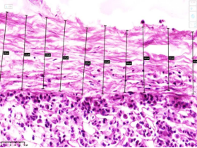
The study objects comprised pituitary gland tissues collected from 75 individuals who died of sudden cardiac death, classified into age groups based on the WHO guidelines: 18–44, 45–59, 60–74, and 75–90 years and older. Autopsy materials were registered and processed at the Andijan branch of the Republican Scientific and Practical Center for Forensic Medical Examination during the period of 2015–2024. For morphometric evaluations, tissue sections measuring 5–7 µm in thickness were scanned using a NanoZoomer digital slide scanner at 20× magnification. Digital micro-images were processed to quantify cellular structures in micrometers (µm) and area percentages (%). An increase in the average thickness of the pituitary gland capsule and disorganization of fibrous structures were noted.
Data regarding the designated microscopic fields, their occupied volume share within a specific grid area of µm2, and cellular dimensions were extracted and evaluated using the specialized QuPath-0.5.0 (console) + ImageJ software platforms. This process was carried out under automated artificial intelligence control without human investigator bias.
Statistical data management and arithmetic mean calculations were performed on a Pentium-4 personal computer using the Microsoft Excel-2019 software suite.
Morphometric data capturing the alterations occurring within the parenchymal and mesenchymal tissue components of the anterior adenohypophysis were analyzed in arithmetic means and presented in tables (Tables 1-2). Cellular volume alterations, total occupied surface area, parenchyma-to-stroma ratios, blood vessel density, and other criteria were extracted, taking into account the direct age-specific shifts in the pituitary gland under acute ischemic heart disease (IHD) and physiological aging trajectories.
3. Results and Discussion
In the analysis of the data presented in Table 1, under acute cardiac death associated with IHD, age-specific features revealed that in the 18–44 age group, the diameter of acidophilic cells reached 12.59 ± 0.22 µm compared to 11.2 ± 0.31 µm in the control group. This represents a statistically significant increase of 12.5% relative to the control group. Regarding thyrotroph cell dimensions, they measured 26.14 ± 2.09 µm within the 18–44 cohort, rising by 25% over the control baseline, indicating a hyper-metabolic response
[7]
Gupta, S., Sharma, R., & Verma, A. Morphometric and histopathological evaluation of the human pituitary gland across various age groups: An autopsy-based study. Journal of Clinical Endocrinology & Metabolism, 2023, 108(4), e112–e120.
. Morphologically, this reflects an accelerated synthesis of thyroid hormones under acute coronary syndromes
[8]
Smith, J. D., & Taylor, K. L. Pathomorphological indicators of acute neuroendocrine stress responses during sudden cardiac arrest. Forensic Science International, 2023, 345, 111620.
. Corticotroph cell sizes in the pituitary gland of the 18–44 cohort reached 29.91 ± 1.11 µm compared to 24.97 ± 1.11 µm in the control group, establishing a statistically significant increase of 20%.
Specifically, this volumetric expansion of corticotroph cells serves as a distinct morphological substrate reflecting hyperproductive morphofunctional activity
[12]
Anderson, C. R., & White, T. M. Acute ischemic changes of the hypothalamic-pituitary axis in sudden unexpected cardiovascular death. Cardiovascular Pathology, 2024, 69, 107601.
. Gonadotroph cells averaged 26.51 ± 1.22 µm compared to 21.94 ± 1.41 µm in controls, representing a 21% inflation, which matches an upregulated testosterone synthesis and cholesterol-driven steroidogenesis within acute cardiac stress states
[14]
Zhao, H., Wang, J., & Sun, L. Morphofunctional activity of thyrotroph and corticotroph cells under acute cardiovascular stress conditions. BMC Endocrine Disorders, 2025, 25(1), 42.
Figure 1. The average thickness of the pituitary gland capsule is presented. This micro-image was scanned using a NanoZoomer digital slide scanner. Digital values were extracted via NDP. view 2 software. H&E staining. Magnification: 10×10.
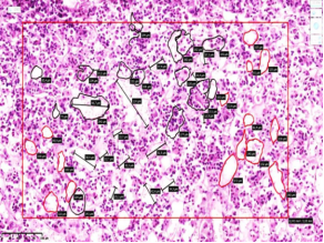
Figure 2. The area of acidophilic cell clusters within the pituitary gland structure is presented. This micro-image was scanned using a NanoZoomer digital slide scanner. Digital values were extracted via NDP.view 2 software. H&E staining. Magnification: 10×10.
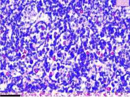
Figure 3. The cellular composition, total occupied area, and nucleocytoplasmic ratio within the pituitary gland parenchyma are presented. This micro-image was scanned using a NanoZoomer digital slide scanner. Digital values were extracted via QuPath-0.5.0 + NDP.view 2 software applications. H&E staining. Magnification: 10×10.

In the 45–59 age bracket, the diameter of acidophilic cells reached 14.32 ± 0.23 µm compared to 13.41 ± 0.12 µm in controls. Comparative analysis revealed a minor 7% increase, which lacked definitive statistical significance, pointing toward relative physiological adaptations that were morphologically mirrored across examinations
[11]
Patel, R., & Kumar, S. Quantitative immunohistochemical profiling of acidophilic and basophilic cells in the aging adenohypophysis. Endocrine Pathology, 2024, 35(1), 88–97.
. Thyrotroph cell sizes within the 45–59 cohort measured 28.76 ± 2.61 µm, reflecting an increase of 25% over controls and confirming a persistent hyperproductive functional curve
[16]
Kim, D. H., & Park, J. S. Post-mortem pituitary morphometry as a forensic marker for terminal stress duration in acute ischemic heart disease. Legal Medicine, 2025, 74, 102415.
Corticotroph cell diameters among the 45–59 cohort dying of IHD reached 34.36 ± 1.62 µm against 27.71 ± 1.31 µm in age-matched controls. This indicates a 24% volumetric cellular inflation, predominantly driven by the dense accumulation of granular inclusions within the cellular cytoplasm. Gonadotroph diameters within this cohort averaged 26.51 ± 1.09 µm versus 23.05 ± 1.63 µm in controls. Comparative data showed a 15% increase, indicating relative gonadal activity advantages matching prolonged hypercholesterolemia trends during chronic coronary pathology courses.
In the third investigated group (60–74 years), the average diameter of acidophilic cells fell to 12.66 ± 0.49 µm against 11.62 ± 0.14 µm in controls. This reduction signals cellular hypofunction regarding local growth hormone processing, and with a localized 8% variance line, it lacked strong statistical significance.
Thyrotroph cell diameters within the 60–74 age bracket dying of IHD averaged 16.05 ± 1.59 µm compared to 18.88 ± 1.88 µm in controls. Chronic IHD backgrounds noticeably accelerated senescent atrophic and sclerotic transformations within the pituitary, generating an average cellular shrinkage of 15%. Such structural atrophic shifts are tightly associated with an accelerated systemic aging cascade
[9]
Zhang, Y., Chen, X., & Liu, M. Age-dependent structural degeneration and sclerotic alterations in the anterior pituitary: Implications for metabolic homeostasis. Aging Cell, 2024, 23(2), e14055.
Corticotroph cell dimensions within the 60–74 cohort fell to 19.09 ± 0.84 µm versus 22.47 ± 0.99 µm in controls. Comparative data analysis established a 15% cell boundaries contraction, validating that basophilic cells maintain steady involutionary rates during ischemic cardiac pathways. These specific parameters of cellular shrinkage confirmed parallel hypofunctional rates unfolding within synchronous organs of the systemic sympathoadrenal cascade
[10]
Miller, P. A., & Davis, H. E. Adenohypophyseal parenchyma volume reduction and stromal fibrosis in elderly post-mortem cases. Pathology - Research and Practice, 2024, 220, 154812.
Gonadotroph cell boundaries within the 60–74 cohort averaged 16.78 ± 1.08 µm against 19.75 ± 1.27 µm in control segments, certifying a statistically visible 15–17% cellular shrinkage. In the 60–74 age bracket, this structural decline mirrors a sharp reduction in sexual function and a pronounced progression of systemic atherosclerosis dynamics under an IHD background, where vascular lipidosis processes accumulate vital precursor metabolic resources
[15]
Martinez, E., & Fernandez, G. Age-related vascular involution and interstitial sclerosis in human pituitary autopsy tissue. Neuroendocrinology, 2025, 115(3), 205–218.
In the final investigated group (75–90 years), biological senescence across all visceral and endocrine frameworks combined with advanced coronary pathology. Age-dependent morphologic shifts within the pituitary gland under IHD patterns revealed that acidophilic cells averaged 10.01 ± 0.12 µm compared to 11.06 ± 0.31 µm in controls, accelerating senescent involution via a 10–12% cellular contraction
[13]
Rossi, L., & Bianchi, F. Histomorphometric changes of endocrine glands in sudden cardiac death vs. traumatic death controls: A comparative post-mortem evaluation. International Journal of Legal Medicine, 2025, 139(2), 311–320.
Thyrotroph cellular dimensions in those dying of IHD in the 75–90 age bracket reached 15.02 ± 1.21 µm, while the control lines registered 12.76 ± 1.02 µm. These parameters confirmed that structural atrophic and sclerotic pituitary renovations are significantly accelerated by ischemic artery backgrounds, resulting in a 15% cellular boundary reduction that speeds up involutionary mechanisms
[17]
Brown, L. M., & Wilson, E. J. Cellular metabolic downregulation in the adenohypophysis during senile atrophy: A stereological post-mortem analysis. Acta Neuropathologica Communications, 2026, 14(1), 12.
Corticotroph cells dropped to an average diameter of 18.07 ± 0.41 µm versus 15.36 ± 0.71 µm in controls. This 12–15% cellular shrinkage directly represents advanced functional downregulation, confirmed by parallel atrophic and sclerotic changes documented in the adrenal glands of the same decedents.
Gonadotroph cellular dimensions within the 75–90 cohort demonstrated a critical volumetric collapse, falling to 9.67 ± 1.34 µm against an average of 12.91 ± 1.69 µm in control subjects, confirming a 33% statistical reduction. In the 75–90 age bracket, this decline reflects a profound loss of reproductive axis activity, coinciding with extensive fibro-sclerotic alterations across the testes and prostate gland, pushing atrophic pituitary indices to critical senescent limits
[18]
Tanaka, K., & Sato, T. Histological remodeling of pituitary cellular subpopulations in elderly subjects with chronic ischemic cardiovascular pathologies. Frontiers in Endocrinology, 2026, 17, 1359012.
During the assessment of the area allocated to individual structural tissue parameters within the adenohypophysis parenchyma (% per 64,000µm2, Table 2), the 45–59, 60–74, and 75–90 age cohorts demonstrated a progressive increase in capillary blood vessel diameters and connective tissue stroma surfaces, while the functional areas occupied by basophilic and eosinophilic cells significantly decreased.
Table 1. Morphometric dimensions of parenchymal cells in the pituitary gland (diameter, µm).
No.
Group Profile
Acidophilic Cells
Thyrotroph Cells
Corticotroph Cells
Gonadotroph Cells
P≤0.05 / P≤0.01
1
Control Group 18–44 years (n=8)
11.2 ± 0.31
20.91 ± 2.09
24.97 ± 1.11
21.94 ± 1.41
0.05
2
Group 2: 18–44 years (n=58)
12.59 ± 0.22
26.14 ± 2.09
29.91 ± 1.11
26.51 ± 1.22
0.01
3
Control Group 45–59 years (n=7)
13.41 ± 0.12
23.01 ± 1.79
27.71 ± 1.31
23.05 ± 1.63
0.01
4
Group 3: 45–59 years (n=30)
14.32 ± 0.23
28.76 ± 2.61
34.36 ± 1.62
26.51 ± 1.09
0.01
5
Control Group 60–74 years (n=8)
11.62 ± 0.14
18.88 ± 1.88
22.47 ± 0.99
19.75 ± 1.27
0.01
6
Group 4: 60–74 years (n=7)
12.66 ± 0.49
16.05 ± 1.59
19.09 ± 0.84
16.78 ± 1.08
0.05
7
Control Group 75–90 years (n=7)
11.06 ± 0.31
15.02 ± 1.21
18.07 ± 0.41
12.91 ± 1.69
0.05
8
Group 5: 75–90 years (n=11)
10.01 ± 0.12
12.76 ± 1.02
15.36 ± 0.71
9.67 ± 1.34
0.05
Table 2. Area occupied by structural components in the adenohypophysis parenchyma (%per 64,000µm2area).
No.
Group Profile
Blood Capillaries
Connective Tissue
Basophilic Cells
Acidophilic Cells
P≤0.05 / P≤0.01
1
Control Group 18–44 years (n=8)
33920.22 ± 5.95
7040.31 ± 1.79
13438.21 ± 1.26
9598.35 ± 1.49
0.05
2
Group 2: 18–44 years (n=58)
36480.12 ± 4.17
8317.13 ± 2.01
14715.39 ± 1.19
4480.11 ± 1.13
0.01
3
Control Group 45–59 years (n=7)
39008.25 ± 4.14
8096.23 ± 1.13
14453.94 ± 1.41
11038.12 ± 1.91
0.01
4
Group 3: 45–59 years (n=30)
30069.31 ± 3.71
14715.12 ± 1.89
14055.16 ± 2.59
5120.23 ± 1.65
0.01
5
Control Group 60–74 years (n=8)
30528.19 ± 4.14
6336.27 ± 1.79
12094.34 ± 1.09
8638.5 ± 1.13
0.01
6
Group 4: 60–74 years (n=7)
26861.16 ± 2.39
22375.31 ± 1.19
10045.55 ± 2.54
4672.11 ± 1.33
0.05
7
Control Group 75–90 years (n=7)
27136.17 ± 2.61
5632.11 ± 1.09
10750.56 ± 1.02
7678.68 ± 1.08
0.05
8
Group 5: 75–90 years (n=11)
24312.16 ± 3.01
28781.16 ± 1.66
7478.29 ± 1.11
3392.12 ± 1.65
0.05
4. Conclusions
In acute cardiac death linked to ischemic heart disease, age-specific pituitary assessment reveals distinct pathomorphological criteria. In the 18–44 and 45–59 age segments, a statistically significant increase in the diameters of acidophilic cells, thyrotroph cells, corticotroph cells, and gonadotroph cells represents a reactive hyperproductive state; these indicators serve as the morphofunctional substrate establishing elevated cellular metabolic output during terminal crises.
Conversely, in the 60–74 and 75–90 age cohorts, a uniform contraction in the diameters of acidophilic cells, thyrotroph cells, corticotroph cells, and gonadotroph cells manifests pronounced metabolic downregulation. The age-parallel contraction of the total area occupied by functional adenohypophyseal parenchyma and the widening of the fibrous stromal framework confirm that localized atrophic and sclerotic transformations are prominently accelerated by underlying coronary artery pathology, noticeably precipitating senescent degradation paths via central endocrine mechanisms.
Abbreviations
SCD
Sudden Cardiac Death
IHD
Ischemic Heart Disease
HPAA
Hypothalamus-Pituitary-Adrenal Axis
WHO
World Health Organization
Acknowledgments
The authors express their gratitude to the Republican Scientific and Practical Center for Forensic Medical Examination, Andijan Branch, and the Republican Center of Pathological Anatomy for providing technical assistance and organizational aid during the data collection phases of this research.
Author Contributions
Imomov Khojiakbar: Conceptualization, Data curation, Methodology, Writing – original draft
The data supporting the outcome of this research work has been reported in this manuscript.
Conflicts of Interest
The authors declare no conflicts of interest.
References
[1]
Boytsov, S. A., Nikulina, N. N., Yakushin, S. S., et al. Sudden cardiac death in patients with coronary heart disease: prevalence, detection rates, and problems of statistical accounting. Russian Journal of Cardiology; 2011, 2, 59–64.
[2]
Bockeria, O. L., Biniashvili, M. B. Sudden cardiac death and ischemic heart disease. Annals of Arrhythmology; 2013, 10(2), 69–79.
Camm, A. J., Lüscher, T. F., Serruys, P. W., eds. Diseases of the Heart and Vessels: European Society of Cardiology Guidelines. Moscow: GEOTAR-Media; 2011, pp. 114–120.
[4]
Khalikov, A. A., et al. Forensic medical aspects of sudden autopsy-negative cardiac death. Forensic Medical Examination; 2021, 64(3), 59–63.
Khakimov, Z. K. The role of the hypothalamic-pituitary system in sudden coronary death. Economics and Society; 2022, 11(102)-1, 961–964. Available from:
Gupta, S., Sharma, R., & Verma, A. Morphometric and histopathological evaluation of the human pituitary gland across various age groups: An autopsy-based study. Journal of Clinical Endocrinology & Metabolism, 2023, 108(4), e112–e120.
Smith, J. D., & Taylor, K. L. Pathomorphological indicators of acute neuroendocrine stress responses during sudden cardiac arrest. Forensic Science International, 2023, 345, 111620.
Miller, P. A., & Davis, H. E. Adenohypophyseal parenchyma volume reduction and stromal fibrosis in elderly post-mortem cases. Pathology - Research and Practice, 2024, 220, 154812.
Patel, R., & Kumar, S. Quantitative immunohistochemical profiling of acidophilic and basophilic cells in the aging adenohypophysis. Endocrine Pathology, 2024, 35(1), 88–97.
Anderson, C. R., & White, T. M. Acute ischemic changes of the hypothalamic-pituitary axis in sudden unexpected cardiovascular death. Cardiovascular Pathology, 2024, 69, 107601.
Rossi, L., & Bianchi, F. Histomorphometric changes of endocrine glands in sudden cardiac death vs. traumatic death controls: A comparative post-mortem evaluation. International Journal of Legal Medicine, 2025, 139(2), 311–320.
Martinez, E., & Fernandez, G. Age-related vascular involution and interstitial sclerosis in human pituitary autopsy tissue. Neuroendocrinology, 2025, 115(3), 205–218.
Kim, D. H., & Park, J. S. Post-mortem pituitary morphometry as a forensic marker for terminal stress duration in acute ischemic heart disease. Legal Medicine, 2025, 74, 102415.
Brown, L. M., & Wilson, E. J. Cellular metabolic downregulation in the adenohypophysis during senile atrophy: A stereological post-mortem analysis. Acta Neuropathologica Communications, 2026, 14(1), 12.
Tanaka, K., & Sato, T. Histological remodeling of pituitary cellular subpopulations in elderly subjects with chronic ischemic cardiovascular pathologies. Frontiers in Endocrinology, 2026, 17, 1359012.
Khojiakbar, I., Avazbek, M., Rokhatoy, F., Madina, A. (2026). Age-Related Morphometric Changes of the Pituitary Gland in Sudden Cardiac Death. American Journal of Clinical and Experimental Medicine, 14(4), 86-90. https://doi.org/10.11648/j.ajcem.20261404.15
Khojiakbar, I.; Avazbek, M.; Rokhatoy, F.; Madina, A. Age-Related Morphometric Changes of the Pituitary Gland in Sudden Cardiac Death. Am. J. Clin. Exp. Med.2026, 14(4), 86-90. doi: 10.11648/j.ajcem.20261404.15
Khojiakbar I, Avazbek M, Rokhatoy F, Madina A. Age-Related Morphometric Changes of the Pituitary Gland in Sudden Cardiac Death. Am J Clin Exp Med. 2026;14(4):86-90. doi: 10.11648/j.ajcem.20261404.15
@article{10.11648/j.ajcem.20261404.15,
author = {Imomov Khojiakbar and Mamataliyev Avazbek and Fazlitdinova Rokhatoy and Ahmedova Madina},
title = {Age-Related Morphometric Changes of the Pituitary Gland in Sudden Cardiac Death},
journal = {American Journal of Clinical and Experimental Medicine},
volume = {14},
number = {4},
pages = {86-90},
doi = {10.11648/j.ajcem.20261404.15},
url = {https://doi.org/10.11648/j.ajcem.20261404.15},
eprint = {https://article.sciencepublishinggroup.com/pdf/10.11648.j.ajcem.20261404.15},
abstract = {The study describes morphometric changes in the adenohypophysis during sudden cardiac death. The study investigated the morphometric parameters of pituitary gland tissues obtained from 75 individuals who died of sudden cardiac death, categorized according to the WHO age classification into cohorts of 18–44, 45–59, 60–74, and 75–90 years and older. The specimens were examined at the Andijan branch of the Republican Scientific and Practical Center for Forensic Medical Examination during the period of 2015–2024. In acute cardiac death associated with ischemic heart disease (IHD), age-specific features revealed that in the 18–44 and 45–59 age groups, the diameters of acidophilic cells, thyrotroph cells, corticotroph cells, and gonadotroph cells significantly increased; these indicators explained the enhancement of metabolic processes and morphofunctional activity in them. In the 60–74 and 75–90 age groups, a statistically significant decrease in the diameters of acidophilic cells, thyrotroph cells, corticotroph cells, and gonadotroph cells was observed, which manifested a decline in cellular metabolic processes. A decrease in the area occupied by the adenohypophyseal parenchyma accompanied by an increase in the stromal area occurred in parallel with advancing age, demonstrating a decline in the gland's morphofunctional activity. These changes were explained by the fact that age-related atrophic and sclerotic alterations of the pituitary gland in these individuals were accelerated against the background of IHD, and atrophic changes accelerated the aging processes.},
year = {2026}
}
TY - JOUR
T1 - Age-Related Morphometric Changes of the Pituitary Gland in Sudden Cardiac Death
AU - Imomov Khojiakbar
AU - Mamataliyev Avazbek
AU - Fazlitdinova Rokhatoy
AU - Ahmedova Madina
Y1 - 2026/08/26
PY - 2026
N1 - https://doi.org/10.11648/j.ajcem.20261404.15
DO - 10.11648/j.ajcem.20261404.15
T2 - American Journal of Clinical and Experimental Medicine
JF - American Journal of Clinical and Experimental Medicine
JO - American Journal of Clinical and Experimental Medicine
SP - 86
EP - 90
PB - Science Publishing Group
SN - 2330-8133
UR - https://doi.org/10.11648/j.ajcem.20261404.15
AB - The study describes morphometric changes in the adenohypophysis during sudden cardiac death. The study investigated the morphometric parameters of pituitary gland tissues obtained from 75 individuals who died of sudden cardiac death, categorized according to the WHO age classification into cohorts of 18–44, 45–59, 60–74, and 75–90 years and older. The specimens were examined at the Andijan branch of the Republican Scientific and Practical Center for Forensic Medical Examination during the period of 2015–2024. In acute cardiac death associated with ischemic heart disease (IHD), age-specific features revealed that in the 18–44 and 45–59 age groups, the diameters of acidophilic cells, thyrotroph cells, corticotroph cells, and gonadotroph cells significantly increased; these indicators explained the enhancement of metabolic processes and morphofunctional activity in them. In the 60–74 and 75–90 age groups, a statistically significant decrease in the diameters of acidophilic cells, thyrotroph cells, corticotroph cells, and gonadotroph cells was observed, which manifested a decline in cellular metabolic processes. A decrease in the area occupied by the adenohypophyseal parenchyma accompanied by an increase in the stromal area occurred in parallel with advancing age, demonstrating a decline in the gland's morphofunctional activity. These changes were explained by the fact that age-related atrophic and sclerotic alterations of the pituitary gland in these individuals were accelerated against the background of IHD, and atrophic changes accelerated the aging processes.
VL - 14
IS - 4
ER -
Khojiakbar, I., Avazbek, M., Rokhatoy, F., Madina, A. (2026). Age-Related Morphometric Changes of the Pituitary Gland in Sudden Cardiac Death. American Journal of Clinical and Experimental Medicine, 14(4), 86-90. https://doi.org/10.11648/j.ajcem.20261404.15
Khojiakbar, I.; Avazbek, M.; Rokhatoy, F.; Madina, A. Age-Related Morphometric Changes of the Pituitary Gland in Sudden Cardiac Death. Am. J. Clin. Exp. Med.2026, 14(4), 86-90. doi: 10.11648/j.ajcem.20261404.15
Khojiakbar I, Avazbek M, Rokhatoy F, Madina A. Age-Related Morphometric Changes of the Pituitary Gland in Sudden Cardiac Death. Am J Clin Exp Med. 2026;14(4):86-90. doi: 10.11648/j.ajcem.20261404.15
@article{10.11648/j.ajcem.20261404.15,
author = {Imomov Khojiakbar and Mamataliyev Avazbek and Fazlitdinova Rokhatoy and Ahmedova Madina},
title = {Age-Related Morphometric Changes of the Pituitary Gland in Sudden Cardiac Death},
journal = {American Journal of Clinical and Experimental Medicine},
volume = {14},
number = {4},
pages = {86-90},
doi = {10.11648/j.ajcem.20261404.15},
url = {https://doi.org/10.11648/j.ajcem.20261404.15},
eprint = {https://article.sciencepublishinggroup.com/pdf/10.11648.j.ajcem.20261404.15},
abstract = {The study describes morphometric changes in the adenohypophysis during sudden cardiac death. The study investigated the morphometric parameters of pituitary gland tissues obtained from 75 individuals who died of sudden cardiac death, categorized according to the WHO age classification into cohorts of 18–44, 45–59, 60–74, and 75–90 years and older. The specimens were examined at the Andijan branch of the Republican Scientific and Practical Center for Forensic Medical Examination during the period of 2015–2024. In acute cardiac death associated with ischemic heart disease (IHD), age-specific features revealed that in the 18–44 and 45–59 age groups, the diameters of acidophilic cells, thyrotroph cells, corticotroph cells, and gonadotroph cells significantly increased; these indicators explained the enhancement of metabolic processes and morphofunctional activity in them. In the 60–74 and 75–90 age groups, a statistically significant decrease in the diameters of acidophilic cells, thyrotroph cells, corticotroph cells, and gonadotroph cells was observed, which manifested a decline in cellular metabolic processes. A decrease in the area occupied by the adenohypophyseal parenchyma accompanied by an increase in the stromal area occurred in parallel with advancing age, demonstrating a decline in the gland's morphofunctional activity. These changes were explained by the fact that age-related atrophic and sclerotic alterations of the pituitary gland in these individuals were accelerated against the background of IHD, and atrophic changes accelerated the aging processes.},
year = {2026}
}
TY - JOUR
T1 - Age-Related Morphometric Changes of the Pituitary Gland in Sudden Cardiac Death
AU - Imomov Khojiakbar
AU - Mamataliyev Avazbek
AU - Fazlitdinova Rokhatoy
AU - Ahmedova Madina
Y1 - 2026/08/26
PY - 2026
N1 - https://doi.org/10.11648/j.ajcem.20261404.15
DO - 10.11648/j.ajcem.20261404.15
T2 - American Journal of Clinical and Experimental Medicine
JF - American Journal of Clinical and Experimental Medicine
JO - American Journal of Clinical and Experimental Medicine
SP - 86
EP - 90
PB - Science Publishing Group
SN - 2330-8133
UR - https://doi.org/10.11648/j.ajcem.20261404.15
AB - The study describes morphometric changes in the adenohypophysis during sudden cardiac death. The study investigated the morphometric parameters of pituitary gland tissues obtained from 75 individuals who died of sudden cardiac death, categorized according to the WHO age classification into cohorts of 18–44, 45–59, 60–74, and 75–90 years and older. The specimens were examined at the Andijan branch of the Republican Scientific and Practical Center for Forensic Medical Examination during the period of 2015–2024. In acute cardiac death associated with ischemic heart disease (IHD), age-specific features revealed that in the 18–44 and 45–59 age groups, the diameters of acidophilic cells, thyrotroph cells, corticotroph cells, and gonadotroph cells significantly increased; these indicators explained the enhancement of metabolic processes and morphofunctional activity in them. In the 60–74 and 75–90 age groups, a statistically significant decrease in the diameters of acidophilic cells, thyrotroph cells, corticotroph cells, and gonadotroph cells was observed, which manifested a decline in cellular metabolic processes. A decrease in the area occupied by the adenohypophyseal parenchyma accompanied by an increase in the stromal area occurred in parallel with advancing age, demonstrating a decline in the gland's morphofunctional activity. These changes were explained by the fact that age-related atrophic and sclerotic alterations of the pituitary gland in these individuals were accelerated against the background of IHD, and atrophic changes accelerated the aging processes.
VL - 14
IS - 4
ER -