Research Article | | Peer-Reviewed

Surveillance of Multidrug-resistant Bacteria at a Tunisian University Hospital in 2022

Received: 10 July 2026     Accepted: 25 July 2026     Published: 11 November 2026
Views:       Downloads:
Abstract

The development of antibiotics has significantly reduced mortality from bacterial infections; however, the emergence of antimicrobial-resistant bacteria poses a global threat. In response, an international action plan was launched, and Tunisia has participated by implementing a national program to monitor bacterial resistance to antibiotics. This study aimed to identify multidrug-resistant (MDR) bacteria isolated at Sahloul University Hospital in 2022, determine their prevalence and annual incidence, and describe their epidemiological and microbiological profiles. This cross-sectional study included all bacterial strains isolated from clinical specimens collected from patients at Sahloul University Hospital, Sousse, between January 1 and December 31, 2022. Among 3,207 clinical specimens analyzed, 865 MDR strains were identified, corresponding to an overall prevalence of 27%. Klebsiella pneumoniae and Acinetobacter baumannii accounted for 32.8% and 23.1% of the isolates, respectively. Extended-spectrum β-lactamase-producing Enterobacterales (ESBL-E) represented 37.3% of MDR isolates, followed by carbapenem-resistant A. baumannii (CRAB, 23.1%) and carbapenem-resistant Enterobacterales (CRE, 18.6%). The most affected departments were the surgical intensive care unit (22.5%), urology (13.4%), and nephrology (11.9%). MDR strains were mainly isolated from urine (44.2%), blood cultures (13.1%), and protected specimen brush samples (12.1%). Monthly trends showed fluctuations, with ESBL-E ranging from 16 to 37 cases per month and CRAB from 5 to 32 cases per month. Annual incidence rates were 0.67 per 100 admissions for CRAB, 0.47 per 100 admissions for CRE, and 0.26 per 100 admissions for methicillin-resistant Staphylococcus aureus (MRSA). All CRAB strains were resistant to ciprofloxacin, and 95.2% were resistant to gentamicin. Among the 33 CRE isolates tested for colistin susceptibility, 3 were resistant. In our hospital, the main MDR pathogens were ESBL-E, CRAB, and CRE, with the surgical intensive care unit, urology, and nephrology departments being the most affected. These findings highlight the need for urgent and targeted interventions to control the spread of MDR bacteria and strengthen antimicrobial resistance surveillance and infection prevention and control measures.

Published in Biomedical Sciences (Volume 12, Issue 3)
DOI 10.11648/j.bs.20261203.12
Page(s) 58-79
Creative Commons

This is an Open Access article, distributed under the terms of the Creative Commons Attribution 4.0 International License (http://creativecommons.org/licenses/by/4.0/), which permits unrestricted use, distribution and reproduction in any medium or format, provided the original work is properly cited.

Copyright

Copyright © The Author(s), 2026. Published by Science Publishing Group

Keywords

Multidrug-resistant Bacteria, Epidemiology, Microbiology, Prevalence, Surveillance

1. Introduction
The introduction of antibiotics represented a major milestone in the history of modern medicine, revolutionizing the treatment of bacterial infections and significantly reducing associated morbidity and mortality .
This innovative era began in 1928 with Alexander Fleming’s serendipitous discovery of the antibacterial properties of Penicillium notatum by-products. However, it was not until 1940 that penicillin was first applied clinically, inaugurating the golden age of antibiotics. This prolific period, extending until the 1970s, witnessed the emergence and development of a multitude of antimicrobial agents, including streptomycin, tetracycline, chloramphenicol, and vancomycin .
These antimicrobial agents demonstrated remarkable efficacy against a broad spectrum of pathogens, enabling the treatment of previously lethal diseases such as pneumonia, tuberculosis, and syphilis. Their broad activity and versatility led to rapid adoption not only in human medicine but also in veterinary medicine and agriculture, where they were used for prophylactic purposes and to promote animal growth .
However, the excessive and inappropriate use of antibiotics has driven the emergence of bacterial resistance. Mechanisms such as enzymatic inactivation, modification of antimicrobial targets, active efflux, and reduced molecular penetration have facilitated bacterial evolution, rendering them resistant to multiple classes of antibiotics .
These increasingly sophisticated resistance mechanisms have given rise to multidrug-resistant bacteria, posing a serious threat. Without the development of new antimicrobial agents, bacterial resistance could be responsible for an estimated 10 million deaths by 2050 .
This issue is exacerbated in several countries, where factors such as limited resources and the absence of effective antibiotic resistance control policies in healthcare settings contribute to the problem .
The objectives of this study were to identify multidrug-resistant bacteria (MDR) isolated at Sahloul University Hospital during 2022, calculate their prevalence and annual incidence, and describe their epidemiological and microbiological profiles.
2. Methods
2.1. Study Design, Setting, and Period
This was a cross-sectional study including all bacterial strains isolated from clinical specimens collected from patients admitted to Sahloul University Hospital, Sousse. The study was conducted in the Microbiology Laboratory of Sahloul University Hospital over a one-year period, from January 1, 2022, to December 31, 2022.
2.2. Study Population
The study included all bacterial strains isolated from specimens submitted to the microbiology laboratory by various hospital departments. Redundant strains—defined as the same bacterial strain with an identical antibiotype isolated multiple times from the same patient during the same infectious episode were excluded .
2.3. Multidrug-Resistant Bacteria Studied
In 2017, the World Health Organization published a list of bacteria for which the development of new antibiotics was urgently needed .
The multidrug-resistant bacteria investigated in this study were:
1) Carbapenem resistant Acinetobacter baumannii resistant to imipenem (CRAB).
2) Pseudomonas aeruginosa resistant to ceftazidime (CRPA).
3) Pseudomonas aeruginosa resistant to both ceftazidime and imipenem (CRPA/IR).
4) Carbapenem-resistant Enterobacterales (CRE).
5) Extended-spectrum β-lactamase-producing Enterobacterales (ESBL-E).
6) Enterococcus faecium resistant to glycopeptides (GRE).
7) Methicillin-resistant Staphylococcus aureus (MRSA).
8) Glycopeptide-resistant Staphylococcus aureus (GRSA).
9) Enterobacterales resistant to third-generation cephalosporins (other than ESBL) (3GCRE).
2.4. Study Procedures
2.4.1. Bacteriological Study
Bacterial species were identified based on conventional morphological, cultural, biochemical, and antigenic characteristics, as well as using the VITEK® 2 automated system (bioMérieux, France) .
2.4.2. Antibiotic Susceptibility Testing
Antibiotic susceptibility of bacterial strains was assessed according to the 2022 recommendations of the Antibiogram Committee of the French Society of Microbiology and the European Committee on Antimicrobial Susceptibility Testing (CA-SFM/EUCAST) . Routine laboratory techniques included:
1) Automated liquid medium antibiogram: performed using the VITEK® 2 system (bioMérieux, France) .
2) Standard disk diffusion: performed on Mueller-Hinton agar according to standard procedures .
2.4.3. Supplementary Tests
Synergy Test:
1) Qualitative method: a third- or fourth-generation cephalosporin or aztreonam disk was placed 30 mm (20 mm for group 3 Enterobacterales) from a disk containing a β-lactamase inhibitor. A “champagne cork” pattern indicated the presence of ESBL .
2) Quantitative method: combined disks (ROSCO Diagnostica®) or the VITEK® 2 system were used. The test was considered positive if the inhibition zone increased by ≥5 mm or if the MIC decreased by ≥3 dilutions in the presence of clavulanic acid .
1) Minimum Inhibitory Concentration (MIC) by E-test strips: Strips containing an antibiotic gradient were applied to inoculated agar plates. After incubation, the MIC was read at the intersection of the inhibition ellipse with the strip .
2) Susceptibility of Gram-negative bacilli to colistin (UMIC Colistin kit): Broth microdilution (BMD) determines the MIC by performing serial dilutions of the antibiotic in inoculated broth. After incubation, the MIC corresponds to the first well showing no visible growth . UMIC® Colistin (Biocentric, France) is a ready-to-use broth microdilution kit .
3) Susceptibility of Staphylococcus aureus to glycopeptides: For Staphylococcus aureus strains with a glycopeptide MIC >1 mg/L measured by VITEK® 2, the MIC was confirmed by broth microdilution using the UMIC® Vancomycin/Teicoplanin kit (Biocentric, France), in accordance with the CA-SFM/EUCAST reference method. The kit contains strips with different antibiotic concentrations and an integrated growth control .
2.5. Data Collection and Statistical Analysis
Data were collected and processed retrospectively using Microsoft Excel, based on the laboratory management software database. All data were analyzed and interpreted using SPSS version 24.
1) Qualitative variables were expressed as percentages and counts.
2) Quantitative variables were expressed as means ± standard deviation.
3. Results
During the study period, analysis of 3,207 clinical specimens identified 865 multidrug-resistant strains, representing an overall prevalence of 27%. Among these isolates, 501 (57.9%) were from male patients, yielding a sex ratio of 1.38.
Hospitalized patients accounted for 83% (n = 716) of the identified MDR cases. The median age of these patients was 56 years (95% CI: 37-67). Among the 667 strains isolated from hospitalized patients, 74.1% (n = 494) were of nosocomial origin.
3.1. Distribution of MDR Isolates by Bacterial Species
Among all identified bacteria, 1,228 Escherichia coli strains, 668 Klebsiella pneumoniae strains, and 416 Staphylococcus aureus strains were isolated (Table 1).
Among the multidrug-resistant strains, Klebsiella pneumoniae, Acinetobacter baumannii, and Escherichia coli accounted for 32.8%, 23.1%, and 20.5% of the total MDR isolates, respectively (Table 1).
Table 1. Distribution of MDR isolates by bacterial species.

Germe

N MDR

% of total MDR isolates

Total N of bacterial species

% of isolates within the bacterial species

Klebsiella pneumoniae

284

32,8

668

42,5%

Acinetobacter baumannii

200

23,1

236

84,7%

Escherichia coli

177

20,5

1228

14,4%

Staphylococcus aureus

82

9,5

416

19,7%

Enterobacter cloacae

47

5,4

195

24,1%

Pseudomonas aeruginosa

43

5,0

308

14,0%

Proteus mirabilis

21

2,4

111

18,9%

Enterococcus faecium

11

1,3

45

24,4%

Total

865

100,0

3027

-

MDR: multidrug-resistant, N: number
3.2. Distribution of MDR Isolates by Phenotype
ESBL-E accounted for 37.3% of the isolated MDR strains. Imipenem-resistant Acinetobacter baumannii and carbapenem-resistant Enterobacterales represented 23.1% and 18.6% of isolates, respectively (Table 2).
Table 2. Distribution of MDR isolates by phenotype.

Phenotype

N

%

ESBL-E

323

37,3

CRAB

200

23,1

CRE

161

18,6

MRSA

80

9,2

3GCRE

45

5,2

CRPA/IR

25

2,9

CRPA

18

2,1

GRE

11

1,3

GRSA

2

0,2

Total

865

100

Carbapenem resistant Acinetobacter baumannii (CRAB); Pseudomonas aeruginosa resistant to ceftazidime (CRPA); Pseudomonas aeruginosa resistant to both ceftazidime and imipenem (CRPA/IR); carbapenem-resistant Enterobacterales (CRE); extended-spectrum β-lactamase-producing Enterobacterales (ESBL-E); Enterococcus faecium resistant to glycopeptides (GRE); methicillin-resistant Staphylococcus aureus (MRSA); glycopeptide-resistant Staphylococcus aureus (GRSA); and Enterobacterales resistant to third-generation cephalosporins (non-ESBL) (3GCRE)
Among the ESBL-producing Enterobacterales, Escherichia coli and Klebsiella pneumoniae accounted for 49.2% (n = 159) and 47.7% (n = 154) of isolates, respectively. Regarding carbapenem-resistant Enterobacterales, Klebsiella pneumoniae represented 71.4% of cases, followed by Enterobacter cloacae and Proteus mirabilis, which accounted for 15.5% and 9.3% of isolates, respectively (Table 3).
Table 3. Distribution of multidrug-resistant Enterobacterales according to phenotypes.

Enterobacterales

ESBL-E N (%)

3GCRE N (%)

CRE N (%)

Total

Klebsiella pneumoniae

154(47,7)

15(33,3)

115(71,4)

284

Escherichia coli

159 (49,2)

12 (26,7)

6 (3,7)

177

Proteus mirabilis

5 (1,5)

1 (2,2)

15 (9,3)

21

Enterobacter cloacae

5 (1,5)

17 (37,8)

25 (15,5)

47

Total

323

45

161

529

3.3. Hospital Departments Origin of MDR Isolates
The hospital departments with the highest numbers of MDR isolates were the Surgical Intensive Care Unit, followed by the Urology, Nephrology, and Cardiovascular and thoracic surgery (CVTS) departments, representing 22.5%, 13.4%, 11.9%, and 9.3% of cases, respectively (Table 4).
Table 4. Distribution of MDR isolates by hospital department.

Department

N

%

Surgical Intensive Care

Surgical Intensive Care Unit

119

13,8

22,5

POG

73

8,4

Anesthesia Outpatient Consultations

3

0,3

Urology

Urology Inpatients

49

5,7

13,4

Urology Outpatient Consultations

67

7,7

Nephrology

Nephrology Inpatients

50

5,8

11,9

Nephrology Outpatient Consultations

48

5,5

DPC-Unit

4

0,5

HD- Unit

1

0,1

CVTS

CVTS Inpatients

36

4,2

9,3

Post-Pediatric CVTS

30

3,5

Post-Operative CVTS

14

1,6

Orthopedics

75

8,7

General Surgery

55

6,4

Emergency Department

Emergency Admissions

44

5,1

6,0

Emergency Outpatient Consultations

8

0,1

Pediatrics

Pediatric Inpatients

40

4,6

5,4

Pediatric Outpatient Consultations

1

0,1

Pediatric Emergency

6

0,7

Neurosurgery

23

2,7

Physical Medicine

19

2,2

Medical Intensive Care Unit

18

2,1

Others

82

9,4

Total

865

100,0

POG: General Post-Operative Unit, CVTS: Cardiovascular and Thoracic Surgery
The distribution of MDR phenotypes varied according to the originating hospital department. Medical departments accounted for 30.3% of ESBL-E isolates, whereas intensive care units were responsible for 65% of CRAB and 44.7% of CRE isolates. Furthermore, multidrug-resistant Gram-positive cocci were more frequently isolated from surgical departments (Table 5).
Table 5. Distribution of MDR phenotypes according to hospital department.

Department

ESBL-E

3GCRE

CRE

CRPA

CRPA/IR

CRAB

GRSA

MRSA

GRE

Total

Medical Departments: n (%)

98(30,3)

14(31,1)

17(10,6)

3(16,7)

5(20)

14(7)

0

19(23,7)

2(18,2)

172(19,9)

Surgical Departments: n (%)

83(25,7)

12(26,7)

41(25,5)

8(44,4)

12(48)

43(21,5)

1(50)

51(63,7)

5(45,5)

256(29,6)

ntensive Care Units: n (%)

28(8,7)

3(6,7)

72(44,7)

3(16,7)

8(32)

130(65)

0

6(7,5)

4(36,4)

254(29,4)

Outpatient Departments: n (%)

79(24,5)

12(26,7)

24(14,9)

1(5,6)

0

9(4,5)

0

0

0

125(14,5)

Emergency Departments (Adult + Pediatric): n (%)

35(10,8)

4(8,9)

7(4,3)

3(16,7)

0

4(2)

1(50)

4(5)

0

58(6,7)

Total: n (%)

323(100)

45(100)

161(100)

18(100)

25(100)

200(100)

2(100)

80(100)

11(100)

865(100)

Carbapenem resistant Acinetobacter baumannii (CRAB); Pseudomonas aeruginosa resistant to ceftazidime (CRPA); Pseudomonas aeruginosa resistant to both ceftazidime and imipenem (CRPA/IR); carbapenem-resistant Enterobacterales (CRE); extended-spectrum β-lactamase-producing Enterobacterales (ESBL-E); Enterococcus faecium resistant to glycopeptides (GRE); methicillin-resistant Staphylococcus aureus (MRSA); glycopeptide-resistant Staphylococcus aureus (GRSA); and Enterobacterales resistant to third-generation cephalosporins (non-ESBL) (3GCRE)
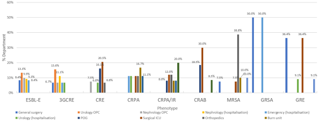
The Orthopedics Department accounted for 38% of MRSA isolates, followed by the Pediatrics and Burn Units, each representing 10.1% of isolates. Additionally, CRPA/IR strains were isolated in 20% of cases within the CVTS department and in 12% of cases distributed between the Surgical ICU and Orthopedics (Figure 1).
MDR isolates were CRAB in 45% of cases in the Cardiovascular and Thoracic Surgery department and in 51% of cases in the Surgical ICU. ESBL-E were isolated across multiple departments, representing 59.6% of MDR isolates in the Emergency Department, 66% in Pediatrics, 61.5% in Nephrology, and 57.7% in Urology.
In the Medical ICU, MDR isolates consisted of CRAB, ESBL-E, and CRE in 77.8%, 16.7%, and 5.6% of cases, respectively (Table 4).
Figure 1. The main hospital departments contributing to each MDR phenotype.
CRAB: Carbapenem resistant Acinetobacter baumannii; CRPA: Pseudomonas aeruginosa resistant to ceftazidime; CRPA/IR: Pseudomonas aeruginosa resistant to both ceftazidime and imipenem; CRE: Carbapenem-resistant Enterobacterales; ESBL-E: Extended-spectrum β-lactamase-producing Enterobacterales; GRE: Enterococcus faecium resistant to glycopeptides; MRSA: Methicillin-resistant Staphylococcus aureus; GRSA: Glycopeptide-resistant Staphylococcus aureus; 3GCRE: Enterobacterales resistant to third-generation cephalosporins (other than ESBL); OPC: Outpatient consultations; POG: General Post-Operative Unit; CVTS: Cardiovascular and Thoracic Surgery.
Table 6. Distribution of MDR phenotypes in the departments most contributing to MDR cases.

Service

ESBL-E

3GCRE

CRE

CRPA

CRPA/IR

CRAB

GRSA

MRSA

GRE

Total

Surgical Intensive Care: n (%)

16(8,4)

3(1,5)

61(32,1)

3(1,5)

5(2,6)

97(51)

0

6(3,1)

4(2,1)

190(100)

POG: n (%)

7(9,6)

0

26(35,6)

1(1,4)

2(2,7)

37(50,7)

0

0

0

73(100)

Surgical Intensive Care Unit: n (%)

9(7,6)

2(1,7)

33(27,7)

2(1,7)

3(2,5)

60(50,4)

0

6(5)

4(3,4)

119(100)

Anesthesia Outpatient Consultations:

n (%)

0

1(33,3)

2(66,7)

0

0

0

0

0

0

3(100)

Urology: n (%)

67(57,7)

10(8,6)

21(18,1)

2(1,7)

0

14(12)

0

1(0,8)

1(0,8)

116(100)

Nephrology Outpatient Consultations: n (%)

43(64,2)

7(10,4)

10(14,9)

0

0

7(10,4)

0

0

0

67(100)

Urology Inpatients: n (%)

24(49)

3(6,1)

11(22,4)

2(4,1)

0

7(14,3)

0

1(2)

1(2)

49(100)

Nephrology: n (%)

65(61,9)

10(9,5)

19(18)

1(0,95)

0

7(6,66)

0

3(2,85)

0

105(100)

Nephrology Outpatient Consultations: n (%)

32(66,7)

3(6,3)

12(25)

0

0

1(2,1)

0

0

0

48(100)

Nephrology Inpatients: n (%)

30(60)

5(10)

7(14)

1(2)

0

6(12)

0

1(2)

0

50(100)

DPC-Unit: n (%)

2(50)

0

0

0

0

0

0

2(50)

0

4(100)

HD- Unit: n (%)

1(100)

0

0

0

0

0

0

0

0

1(100)

CVTS (total): n (%)

14(17,5)

2(2,5)

18(22,5)

0

8(10)

36(45)

0

2(2,5)

0

80(100)

CVTS Inpatients: n (%)

5(13,9)

1(2,8)

6(16,7)

0

5(13,9)

17(47,2)

0

2(5,6)

0

36(100)

Post-Operative CVTS : n (%)

1(7,1)

0

3(21,4)

0

1(7,1)

9(64,3)

0

0

0

14(100)

Post-Pediatric CVTS: n (%)

8(26,7)

1(3,3)

9(30)

0

2(6,7)

10(33,3)

0

0

0

30(100)

Orthopedics: n (%)

17(22,7)

2(2,7)

11(14,7)

2(2,7)

3(4)

9(12)

0

31(41,3)

0

75(100)

General Surgery: n (%)

27(49,1)

3(5,5)

8(14,5)

1(1,8)

1(1,8)

4(7,3)

1(1,8)

6(10,9)

4(7,3)

55(100)

Emergency Department : n (%)

31(59,6)

3(5,8)

7(13,4)

3(5,8)

0

4(7,6)

1(2)

3(5,8)

0

52(100)

Emergency Admissions: n (%)

27(61,4)

3(6,8)

5(11,4)

2(4,5)

0

3(6,8)

1(2,3)

3(6,8)

0

44(100)

Emergency Outpatient Consultations: n (%)

4(50)

0

2(25)

1(12,5)

0

1(12,5)

0

0

0

8(100)

Pediatrics: n (%)

31(66)

3(6,3)

2(4,2)

0

0

1(2,2)

0

9(19,1)

1(2,2)

47(100)

Pediatric Emergency: n (%)

4(66,7)

1(16,7)

0

0

0

0

0

1(16,7)

0

6(100)

Pediatric Inpatients: n (%)

26(65)

2(5)

2(5)

0

0

1(2,5)

0

8(20)

1(2,5)

40(100)

Pediatric Outpatient Consultations: n (%)

1(100)

0

0

0

0

0

0

0

0

1(100)

Neurosurgery: n (%)

9(39,1)

2(8,7)

4(17,4)

0

1(4,3)

4(17,4)

0

3(13)

0

23(100)

Physical Medicine: n (%)

4 (21,1)

2(10,5)

3(15,8)

2(10,5)

2(10,5)

4(21,1)

0

2(10,5)

0

19(100)

Medical Intensive Care Unit: n (%)

3(16,7)

0

1(5,6)

0

0

14(77,8)

0

0

0

18(100)

Others*: n (%)

40(48,7)

5(6)

6(7,3)

4(4,9)

5(6)

6(7,3)

0

15(18,2)

1(1,2)

82(100)

Total

323(37,3)

45(5,2)

161(18,6)

18(2)

25(2,9)

200(23,1)

2(0,2)

80(9,2)

11(1,3)

865(100)

POG: General Post-Operative Unit; CVTS: Cardiovascular and Thoracic Surgery; CRAB: Carbapenem resistant Acinetobacter baumannii; CRPA: Pseudomonas aeruginosa resistant to ceftazidime; CRPA/I: Pseudomonas aeruginosa resistant to ceftazidime and imipenem; CRE: Carbapenem-resistant Enterobacterales; ESBL-E: Extended-spectrum β-lactamase-producing Enterobacterales; GRE: Enterococcus faecium resistant to glycopeptides; MRSA: Methicillin-resistant Staphylococcus aureus; GRSA: Glycopeptide-resistant Staphylococcus aureus; 3GCRE: Enterobacterales resistant to third-generation cephalosporins.
Others: Maxillofacial surgery, cardiology, gastroenterology, neurology, internal medicine, and personalized medicine departments.
3.4. Main Specimens with Positive MDR Cultures
MDR bacteria were isolated from urine, blood cultures, protected specimen brush, and deep pus in 44.2%, 13.1%, 12.1%, and 8.3% of cases, respectively (Table 7).
Table 7. Distribution of MDR isolates according to specimen type.

Sample

N

%

Urine

382

44,2

Blood

113

13,1

protected specimen brush

105

12,1

Deep pus

72

8,3

Catheter

45

5,2

Operative site infection pus (ISO)

24

2,8

Biopsy

25

2,9

Swab pus

20

2,3

Peritoneal pus

10

1,2

Others*

69

7,9

Total

865

100

*Others: cerebrospinal fluid samples, pleural fluids, joint fluids, ascitic fluids, sputum, tracheal aspirates, bronchoalveolar lavages, stool samples, ear swabs, drain tips, urinary catheters, tracheal tubes, osteosynthesis materials, and shunt devices.
ESBL-E were isolated from urine samples in 72.1% of cases. Carbapenem resistant Acinetobacter baumannii strains were isolated from PSB and urine samples in 34.5% and 20.5% of MDR infection cases, respectively. MRSA strains were isolated from deep pus in 29.1% and from blood cultures in 20.3% of MDR infection cases (Table 8).
Table 8. Main specimen sources according to phenotype.

Sample

EBLSE: n (%)

3GCRE: n (%)

CRE: n (%)

CRPA: n (%)

CRPA/I: n (%)

CRAB: n (%)

GRSA: n (%)

MRSA: n (%)

GRE: n (%)

Total

Biopsy

34(2,1)

0

4(2,5)

3(16,6)

2(8)

4(2)

0

4(5,1)

0

PSB

4(1,2)

2(4,4)

17(10,6)

2(11,1)

7(28)

69(34,5)

0

4(5,1)

0

Pleural fluid

1(0,3)

0

0

0

3(12)

2(1)

0

0

0

Deep pus

15(4,6)

2(4,4)

14(8,7)

1(5,6)

4(16)

11(5,5)

1(50)

23(29,1)

1(9,1)

Blood

25(7,7)

3(6,7)

26(16,1)

4(22,2)

1(4)

33(16,5)

1(50)

16(20,3)

4(36,4)

Urine

233(72,1)

31(68,9)

61(37,9)

6(33,3)

2(8)

41(20,5)

0

5(6,3)

3(27,3)

Catheter

3(0,9)

1(2,2)

18(11,2)

0

2(8)

17(8,5)

0

4(5,1)

0

Others

35(10,8)

6(13,3)

21(13)

2(11,1)

4(16)

23(11,5)

0

24(29,1)

3(27,2)

Total

323(100)

45(100)

161(100)

18(100)

25(100)

200(100)

2(100)

80(100)

11(100)

865

PSB: Protected specimen brush
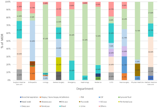
Urine samples represented the main source of MDR bacteria in several departments, reaching over 70% in the urology, nephrology, pediatrics, and physical medicine departments. MDR bacteria were isolated from PSB in 39.5%, 38.5%, and 20% of cases in the surgical ICU, medical ICU, and CVTS departments, respectively (Figure 2).
CSF: Cerebrospinal fluid; CVTS: Cardiovascular and thoracic surgery; PSB: Protected specimen brush; ISO pus: Operative site infection pus

Download: Download full-size image

Figure 2. Distribution of MDR bacteria-positive samples by hospital department.
3.5. Monthly Evolution of MDR Bacteria Cases
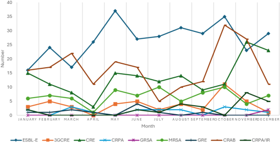
The monthly evolution of MDR bacteria showed fluctuations throughout the year. ESBL-E cases ranged from 16 to 37 per month, with a peak observed in May (37 cases). CRAB cases fluctuated between 5 and 32, reaching a maximum in October (32 cases) and minimum values in July (5 cases) and August (10 cases). MRSA cases varied between 1 and 10 per month, with peaks in July and October (10 cases each) and troughs in April (1 case) and August (5 cases) (Figure 3).
Figure 3. Monthly evolution of MDR bacteria.
CRAB: Carbapenem resistant Acinetobacter baumannii; CRPA: Pseudomonas aeruginosa resistant to ceftazidime; CRPA/IR: Pseudomonas aeruginosa resistant to both ceftazidime and imipenem; CRE: Carbapenem-resistant Enterobacterales; ESBL-E: Extended-spectrum β-lactamase-producing Enterobacterales; GRE: Enterococcus faecium resistant to glycopeptides; MRSA: Methicillin-resistant Staphylococcus aureus; GRSA: Glycopeptide-resistant Staphylococcus aureus; 3GCRE: Enterobacterales resistant to third-generation cephalosporins
3.6. Annual Incidence Rate of Isolated MDR Bacteria
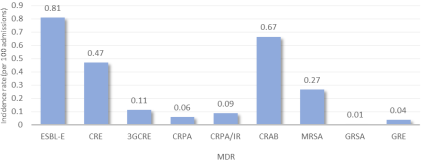
The results of our study revealed an annual incidence rate of 0.67 per 100 admissions for infections caused by CRAB. Similarly, infections due to carbapenem-resistant Enterobacterales had an incidence of 0.47 per 100 admissions, while MRSA infections reached 0.26 per 100 admissions (Figure 4).
Figure 4. Annual incidence rate of MDR bacteria isolated at CHU Sahloul.
CRAB: Carbapenem resistant Acinetobacter baumannii; CRPA: Pseudomonas aeruginosa resistant to ceftazidime; CRPA/IR: Pseudomonas aeruginosa resistant to both ceftazidime and imipenem; CRE: Carbapenem-resistant Enterobacterales; ESBL-E: Extended-spectrum β-lactamase-producing Enterobacterales; GRE: Enterococcus faecium resistant to glycopeptides; MRSA: Methicillin-resistant Staphylococcus aureus; GRSA: Glycopeptide-resistant Staphylococcus aureus; 3GCRE: Enterobacterales resistant to third-generation cephalosporins
3.7. Antibiotic Resistance Profile
3.7.1. Enterobacterales
ESBL-E were resistant to amoxicillin-clavulanic acid, ceftazidime, and ciprofloxacin in 66%, 88%, and 57% of cases, respectively (Table 9).
CRE showed resistance to imipenem, amikacin, ciprofloxacin, and trimethoprim/sulfamethoxazole in 83.1%, 68.1%, 94.3%, and 81.2% of isolates, respectively. Among the 33 strains tested for colistin, three Klebsiella pneumoniae isolates were found to be resistant.
Overall, 82.6% (n=133) of CRE isolates were classified as XDR (Table 10).
For 3GCRE, 100% of the isolates were resistant to amoxicillin/clavulanic acid, piperacillin/tazobactam, while two isolates were resistant to gentamicin, and none were resistant to amikacin (Table 11).
Resistance to third-generation cephalosporins was frequent among 3GCRE isolates. Resistance to cefotaxime and ceftazidime in E. coli was 91.6% and 83.3%, respectively, 93.3% for both antibiotics in K. pneumoniae, and 100% in P.mirabilis. E. cloacae exhibited low resistance rates with only 5.8% resistance to cefotaxime and ceftazidime.
High resistance rates were detected for ofloxacin (85.7%) and nalidixic acid (100%) when tested, with overall resistance to ciprofloxacin observed in 13 out of 42 isolates, predominantly among E. coli (50%) and K. pneumoniae (46.6%), while resistance was low in E. cloacae (5.8%).
Table 9. Antibiotic Resistance Profile of ESBL-E.

Antibiotic

E.coli ESBL n/N (%)

K.pneumoniae ESBL n/N (%)

P.mirabilis ESBL n/N (%)

E.cloacae ESBL n/N (%)

Total ESBL-E n/N (%)

Ampicillin

159/159(100)

-

5/5

-

164/164(100)

Ticarcillin

159/159(100)

-

NT

5/5

164/164(100)

Piperacillin

159/159(100)

-

0/1

5/5

164/165(99)

Amoxicillin/Clavulanic acid

108/155(69,7)

95/153(62,1)

2/2

-

205/310(66)

Amoxicillin/Clavulanic acid (Cystitis)

14/61(22,9)

19/67(28,3)

3/3

-

36/131(27)

Ampicillin/sulbactam

NT

1/5

NT

1/5

Piperacillin/tazobactam

35/158(22,2)

35/154(22,7)

NT

5/5

75/317(23,6)

Cephalexin

159/159(100)

154/154(100)

4/4

-

317/317(100)

Cefoxitin

12/159(7,8)

33/153(21,6)

1/3

-

46/315(14,6)

Cefuroxime

NT

5/5

NT

5/5

Cefixime (Cystitis)

NT

2/5

NT

2/5

Ceftriaxone

NT

0/5

NT

0/5

Cefotaxime

152/156(97,4)

149/153(97,3)

NT

5/5

306/314(97,4)

Ceftazidime

131/159(82,5)

142/152(93,4)

NT

4/4

277/315(88)

Cefepime

NT

7/7(100)

NT

NT

7/7

Ertapenem

0/152(0)

0/150(0)

0/3

0/3

0/305(0)

Imipenem

0/144(0)

0/145(0)

-

0/5

0/294(0)

Meropenem

1/10(10)

0/5(0)

NT

NT

1/15

Aztreonam

9/11(81,8)

1/6(16,7)

NT

1/1

11/18(61)

Nalidixic acid

10/13(76,9)

14/15(93,3)

4/5

1/1

29/34(85,3)

Ofloxacin

18/25(72)

15/21(71,5)

4/4

1/1

38/51(74,5)

Norfloxacin

2/2(100)

0/1(0)

5/5

NT

7/8

Ciprofloxacin

99/150(66)

72/152(47,4)

NT

5/5

176/307(57)

Levofloxacin

3/5(60)

0/1(0)

NT

1/1

4/7

Moxifloxacin

NT

3/3

NT

3/3

Amikacin

7/142(4,9)

4/147(2,7)

0/1

1/5

12/295(4)

Gentamicin

26/54(48,1)

34/144(23,6)

NT

4/4

64/202(31,6)

Tobramycin

5/15(33,3)

4/15(26,7)

NT

1/1

10/31(32,2)

Fosfomycin IV

9/144(6,3)

1/12(8,3)

1/5

0/1

11/162(6,7)

Nitrofurantoin (cystitis)

3/25(12)

13/15(86,7)

-

0/1

16/41(39)

Tigecycline

0/139(0)

0/5(0)

-

NT

0/144(0)

Chloramphenicol

0/6(0)

0/2(0)

NT

NT

0/8

Trimethoprim/sulfamethoxazole

97/156(62,2)

95/151(62,9)

1/5

5/5

198/317(62,4)

Total

159

154

5

5

323

n: number of strains resistant to a given antibiotic, N: number of strains tested for that antibiotic, NT: not tested
Table 10. Antibiotic Resistance Profile of CRE.

Antibiotic

E. coli CRE n/N (%)

K.pneumoniae CRE n/N (%)

P.mirabilis CRE n/N (%)

E. cloacae CRE n/N (%)

Total CRE n/N (%)

Ampicillin

6/6

-

15/15

-

21/21

Ticarcillin

6/6

-

13/13

23/23

42/42(100)

Piperacillin

6/6

-

12/12

25/25

43/43(100)

Amoxicillin/Clavulanic acid

4/6

113/115(98,3)

14/14

-

131/135(97)

Amoxicillin/Clavulanic acid (Cystitis)

2/2

35/35(100)

3/3

-

40/40(100)

Piperacillin/tazobactam

4/5

113/115(98,3)

14/14

25/25

156/159(98,1)

Cephalexin

5/5

100/100(100)

15/15

-

120/120(100)

Cefoxitin

6/6

109/111(98,2)

15/15

-

130/132(98,4)

Cefotaxime

4/6

112/114(98,2)

15/15

25/25

156/160(97,5)

Ceftazidime

4/6

111/112(99,1)

15/15

25/25

155/158(98,1)

Cefepime

NT

9/9

1/1

3/3

13/13

Ertapenem

5/5

106/106(100)

15/15

25/25

151/151

Imipenem

2/6

101/111(91)

11/14

14/23

128/154(83,1)

Meropenem

NT

5/5

NT

NT

5/5

Aztreonam

NT

6/6

0/1

1/1

7/8

Nalidixic acid

1/1

10/10

NT

2/2

13/13

Ofloxacin

1/1

16/16

NT

4/4

21/21

Norfloxacin

NT

1/1

NT

1/1

2/2

Ciprofloxacin

3/6

106/112(94,6)

15/15

22/25

149/158(94,3)

Levofloxacin

NT

4/4

NT

1/1

5/5

Moxifloxacin

NT

NT

NT

1/1

1/1

Amikacin

1/6

90/112(80,4)

14/14

0/22

105/154(68,1)

Gentamicin

1/1

83/111(74,8)

14/14

18/23

116/149(77,8)

Tobramycin

1/1

10/11

NT

0/2

11/14

Colistin

0/1

3/27

-

0/3

3/31

Fosfomycin IV

1/5

2/14

1/1

4/4

8/24

Nitrofurantoin (cystitis)

NT

12/12

-

4/4

16/16

Tigecycline

0/5

2/8

-

NT

2/13

Chloramphenicol

NT

2/3

1/1

1/1

4/5

Trimethoprim/sulfamethoxazole

3/6

93/114(81,6)

15/15

19/25

130/160(81,2)

Total

6

115

15

25

161

n: number of strains resistant to a given antibiotic, N: number of strains tested for that antibiotic, NT: not tested
3.7.2. Non-Fermenting Gram-Negative Bacilli
All CRAB strains were resistant to ciprofloxacin, with resistance rates of 95.2% for gentamicin, 67.9% for amikacin, and 59.4% for trimethoprim/sulfamethoxazole (Table 11).
Regarding CRPA/IR, the strains were resistant to ciprofloxacin in 92% of cases and to amikacin in 84% of cases (Table 12).
Overall, 96% of CRAB and CRPA/IR isolates were classified as extensively drug-resistant, while only two isolates were considered pan-drug resistant.
Table 11. Antibiotic resistance profile of CRAB, CRPA, and CRPA/IR strains.

Antibiotic

CRAB n/N (%)

CRPA n/N (%)

CRPA/IR n/N (%)

Ticarcillin

196/198(99)

18/18

25/25(100)

Piperacillin

195/195(100)

18/18

24/24(100)

Ticarcillin- Clavulanic acid

10/12

6/6

6/6

Piperacillin/tazobactam

200/200(100)

17/18

25/25(100)

Ceftazidime

200/200(100)

18/18

25/25(100)

Cefepime

13/13

5/5

7/7

Ceftazidime/avibactam

NT

NT

1/1

Imipenem

193/193(100)

0/18

24/24(100)

Meropenem

10/11(90,9)

0/11

8/9

Aztreonam

NT

5/6

6/7

Ciprofloxacin

189/189(100)

4/18

23/25(92)

Levofloxacin

NT

5/7

5/6

Amikacin

131/193(67,9)

1/18

21/25(84)

Gentamicin

178/187(95,2)

4/17

23/25(92)

Tobramycin

9/9

0/6

5/7

Colistin

1/58

NT

0/9

Tigecyclin

11/11

-

-

Fosfomycin IV

-

NT

2/4

Trimethoprim/sulfamethoxazole

117/197(59,4)

-

-

Total

200

18

25

n: number of strains resistant to a given antibiotic, N: number of strains tested for that antibiotic, NT: not tested
3.7.3. Staphylococcus Aureus
All MRSA strains were fully susceptible to linezolid, nitrofurantoin, and glycopeptides, and resistant to fusidic acid and erythromycin in 52% and 20.8% of cases, respectively. Three isolates (3.7%) were classified as extensively drug-resistant (XDR) (Table 13).
Two GRSA strains were isolated and confirmed using the broth microdilution method. Both were resistant to erythromycin, ampicillin, and vancomycin and susceptible to trimethoprim/sulfamethoxazole, fluoroquinolones (ofloxacin, ciprofloxacin, moxifloxacin), and aminoglycosides (gentamicin and tobramycin).
Table 12. Antibiotic resistance profile of MRSA.

Antibiotique

SARM n/N (%)

Benzylpenicill

80/80(100)

Oxacillin

80/80(100)

Ampicillin

79/79(100)

Amoxicillin/Clavulanic acid

80/80(100)

Cefotaxime

72/72(100)

Ofloxacin

10/69(14,4)

Levofloxacin

12/78(15,3)

Ciprofloxacin

10/74(13,5)

Moxifloxacin

10/71(14)

Gentamicin

11/78(14,1)

Kanamycin

2/5(40)

Tobramycin

11/77(14,2)

Teicoplanin

0/73(0)

Vancomycin

0/74(0)

Erythromycin

17/78(21,8)

Clindamycin

8/79(10)

Lincomycin

4/23(17,4)

Tetracycline

27/72(37,5)

Doxycyclin

28/70(40)

Tigecyclin

3/79(3,7)

Minocyclin

1/44(2,3)

Fusidic acid

40/76(52)

Fosfomycin IV

6/73(8,2)

Nitrofurantoin

0/72(0)

Rifampicin

3/8(37,5)

Linézolid

0/79(0)

Chloramphenicol

1/5(20)

Trimethoprim/sulfamethoxazole

8/76(10,5)

Total

80

n: number of strains resistant to a given antibiotic, N: number of strains tested for that antibiotic, NT: not tested
3.7.4. Enterococcus Faecium
All GRE isolates were classified as extensively drug-resistant (XDR). They were resistant to β-lactams (amoxicillin, amoxicillin/clavulanic acid, and imipenem), fluoroquinolones (levofloxacin), glycopeptides (teicoplanin and/or vancomycin), chloramphenicol, and had high-level resistance to aminoglycosides (gentamicin and streptomycin). These GRE strains were fully susceptible to quinupristin/dalfopristin and linezolid.
4. Discussion
The alarming proliferation of multidrug-resistant bacteria constitutes a global health challenge, the magnitude of which varies across countries and hospital departments depending on prescribing practices and hygiene protocols, reflecting the quality of care provided . The emergence of these strains raises serious concerns due to their significant pathogenic potential, high dissemination capacity, clonal nature, and the ease of transfer of genetic resistance determinants. This combination of factors leads to therapeutic dead-ends, increased morbidity and mortality, and higher healthcare costs .
During the study period, 3,207 clinical specimens were analyzed, resulting in the identification of 865 MDR isolates, representing an overall prevalence of 27%. Among these isolates, 57.9% were from male patients, yielding a male-to-female ratio of 1.38. These results differ slightly from a Moroccan study, in which the MDR isolation rate was 32% with a male-to-female ratio of 1.84 .
Of the MDR isolates identified, 83% were from hospitalized patients, with a median age of 56 years. Among strains isolated from these patients, 74.1% were of nosocomial origin. A study by Meng Wang et al. on 3,223 patients with nosocomial infections reported that 42.5% of these infections were caused by MDR bacteria. Moreover, patients over 60 years of age were the most affected by MDR bacterial infections .
In our study, the three main species responsible for MDR infections were Klebsiella pneumoniae, Acinetobacter baumannii, and Escherichia coli. These bacteria are part of the ESKAPE group, which includes six highly pathogenic and antibiotic-resistant bacteria: Enterococcus faecium, Staphylococcus aureus, Klebsiella pneumoniae, Acinetobacter baumannii, Pseudomonas aeruginosa, and Enterobacter spp. Occasionally, Escherichia coli is also included, making the acronym ESKAPEE .
The most common MDR bacteria were extended-spectrum β-lactamase-producing Enterobacterales (ESBL-E), CRAB, and carbapenem-resistant Enterobacterales, accounting for 37.3%, 23.1%, and 18.6% of all MDR isolates in 2022, respectively. Our results differ from a Nigerian study, where ESBL-producing Enterobacterales constituted the majority of MDR bacteria (61.8%), followed by multidrug-resistant Pseudomonas aeruginosa (26.5%) and MRSA (11.7%) .
The prevalence of infections caused by multidrug-resistant Gram-negative bacilli is increasing, while that of MRSA is declining. According to J.-C. Lucet and G. Birgand, this is explained by the higher inoculum of ESBL-producing Enterobacterales, such as E. coli, whose resistance spreads easily in the gastrointestinal tract. Antibiotic use further promotes the selection of these bacteria over MRSA, increasing the risk of colonization and cross-transmission. Additionally, patients carrying ESBL-E disseminate more bacteria, notably via excretions. Finally, healthcare workers’ awareness has historically focused more on MRSA than on ESBL-E .
The Surgical Intensive Care Unit was the main contributor of MDR isolates, followed by the Urology, Nephrology, CVTS, and Orthopedics departments, representing 22.5%, 13.4%, 11.9%, 9.3%, and 8.7% of cases, respectively. A Moroccan study documented a significant concentration of MDR isolates in intensive care units (40.34%) and surgical departments (36.02%) . Similarly, a Chinese investigation highlighted a predominance of MDR infections in polyvalent intensive care units (20.7%), neurosurgical units (16.1%), neonatal units (13.6%), and neurology departments (15.0%) .
The high prevalence of MDR bacteria in intensive care and surgical units is attributable to a combination of factors. Frequent use of broad-spectrum antibiotics, immunocompromised patient status, and invasive procedures create a favorable environment for the proliferation of resistant bacteria. Additionally, overcrowding, inadequate hygiene practices, and the movement of people and food facilitate their spread .
A marked prevalence of urinary-origin MDR isolates was observed in the Urology, Nephrology, Pediatrics, and Physical Medicine departments, exceeding 70% of isolates. Deep pus samples accounted for a significant proportion of MDR isolates in intensive care units, particularly in the Surgical ICU (39.5%) and Medical ICU (38.5%). Positive blood cultures for MDR bacteria were especially frequent in the Emergency and CVTS Departments (25% each). Deep pus constituted the main source of MDR isolates in Orthopedics (52.2%), whereas catheter-associated MDR isolates were more frequently found in the Surgical ICU (13.8%).
A study conducted at the Avicenne Military Hospital in Marrakech revealed that in intensive care units, 66% of MDR isolates were obtained from deep pus samples. Surgical departments reported a predominance of MDR isolates in suppurative infections, representing 55% of isolates. In medical departments, urine samples were the main source of MDR isolates, reaching 78% of cases. In the Emergency Department, MDR bacteria were primarily detected in urine samples, followed by suppurations .
The heterogeneous distribution of MDR-positive samples across hospital departments can be explained by unit-specific factors. In Urology and Nephrology, the predominance of urine cultures reflects the nature of urinary pathologies. In intensive care and critical care units, invasive procedures (ventilation, catheters) and patient vulnerability favor positive deep pus and catheter samples. Bacteremia is more frequent in CVTS due to complex surgical procedures and prolonged vascular access. This diversity underscores the need to tailor infection prevention and control strategies to each department and sample type .
ESBL-producing Enterobacterales accounted for 37.3% of all MDR isolates. Among these ESBL-E, Escherichia coli and Klebsiella pneumoniae predominated, representing 49.2% and 47.7% of isolates, respectively. Urine samples were the primary source of ESBL-producing Enterobacterales (72.1%), consistent with the predominance of E. coli and K. pneumoniae as uropathogens . Other relevant samples included blood cultures (7.7%) and deep pus specimens (4.6%).
According to the conclusions of the latest report from the Global Antimicrobial Resistance and Use Surveillance System (GLASS) published by the World Health Organization (WHO) in 2022, resistance of Escherichia coli and Klebsiella pneumoniae to third-generation cephalosporins was on a concerning rise, with median rates of 19% and 23%, respectively. These percentages varied significantly depending on geographical regions, sources of isolates (blood, urine, other), and age groups .
Analysis of antibiotic susceptibility in ESBL-producing Enterobacterales revealed variable resistance profiles depending on the molecule and species. For the amoxicillin-clavulanic acid combination, resistance rates were 69.7% for E. coli and 62.1% for K. pneumoniae. Regarding ceftazidime, 82.5% of ESBL-producing E. coli and 93.4% of ESBL-producing K. pneumoniae were resistant. Resistance to ciprofloxacin was observed in 66% of E. coli and 47.4% of K. pneumoniae. Amikacin retained good activity with low resistance rates: 4.9% for E. coli and 2.7% for K. pneumoniae. Finally, for the trimethoprim-sulfamethoxazole combination, resistance rates were similar between the two species: 62.2% for E. coli and 62.9% for K. pneumoniae.
Although our study did not include molecular analysis of ESBL-producing Enterobacterales, it is useful to refer to previous Tunisian research describing genotypic resistance profiles in hospital settings. The study by Ayari et al. highlighted the predominance of the CTX-M-15 gene among ESBL-producing E. coli, frequently associated with the ISEcp1 insertion element, which could explain its widespread dissemination . Additionally, Dziri et al. studied non-E. coli ESBL-producing Enterobacterales in the hospital environment (inanimate surfaces and hands of staff and patients), identifying several K. pneumoniae strains carrying blaCTX-M-15, blaTEM-1, and various blaSHV variants, illustrating the diversity and complexity of resistance mechanisms present in Tunisian hospitals .
The prevalence of carbapenem-resistant Enterobacterales in our study accounted for 18.6% of all multidrug-resistant bacteria isolated. Among these CRE, Klebsiella pneumoniae predominated, representing 71.4% of isolates, followed by Enterobacter cloacae (15.5%) and Proteus mirabilis (9.3%). The distribution of CRE varied by hospital unit, with the highest proportions found in the surgical intensive care unit (20.5%), the general surgery post-operative unit (16.1%), and the nephrology outpatient department (7.5%). Regarding the source of isolates, CRE were mainly recovered from urine samples (37.9%), followed by blood cultures (16.1%), catheter tips (11.2%), and protected specimen brush (10.6%).
According to the 2022 PARES mission report on the surveillance of antibiotic consumption and bacterial resistance in healthcare facilities, the distribution of carbapenemase-producing Enterobacterales (CPE) revealed a predominance of Klebsiella pneumoniae (36.0%), followed by Escherichia coli (27.6%) and the Enterobacter cloacae complex (17.4%). The analysis of specimen origins showed that urinary samples were the main source of CPE isolation (42.9%), followed by blood cultures (12.4%) and deep pus or serous fluid samples (8.9%) .
The antimicrobial susceptibility evaluation of CRE isolates revealed heterogeneous resistance patterns depending on the tested antimicrobial agents and bacterial species.
For Klebsiella pneumoniae, high resistance rates were observed for imipenem (91%), ciprofloxacin (94.6%), amikacin (80.4%), gentamicin (74.8%), and trimethoprim/sulfamethoxazole (81.6%). Notably, the emergence of colistin resistance—a last-resort antibiotic for MDR infections—was detected in 3 out of 27 tested isolates.
For Enterobacter cloacae, 14 of 23 tested isolates were resistant to imipenem, and 22 of 25 showed resistance to ciprofloxacin. Gentamicin was ineffective against 18 of the 23 isolates, while trimethoprim/sulfamethoxazole demonstrated limited efficacy, with 19 resistant strains out of 25 tested. Conversely, amikacin retained excellent activity, with no resistance observed among the 22 isolates tested.
Among all CRE isolates, 82.6% were classified as extensively drug-resistant. Colistin, considered a last-resort therapeutic option against multidrug-resistant Gram-negative bacilli, has been placed by the World Health Organization in the “Reserve” category of antimicrobials . In our study, the emergence of 3 resistant strains out of 32 tested CRE highlights the urgent need for a strict antimicrobial stewardship policy, particularly regarding colistin use, to prevent the dissemination of resistance and preserve its therapeutic efficacy against CRE infections.
A molecular analysis conducted by Jaidane et al. on 13 colistin-resistant Klebsiella pneumoniae isolates from Sahloul University Hospital characterized the genetic mechanisms underlying colistin resistance. The study demonstrated that resistance was associated with mutations in the mgrB gene, which encodes the transmembrane regulatory protein MgrB. Genetic variations—including deletions, insertions, and substitutions within the mgrB, pmr, and pho operons—were identified, suggesting altered protein function .
The epidemiology of the first Klebsiella pneumoniae carbapenemase-producing (KPC) strains isolated at Sahloul University Hospital between 2012 and 2014 was described by Messaoudi et al. Phenotypic analysis revealed that Klebsiella pneumoniae resistant to temocillin was predominant, suggesting the production of class D carbapenemases (OXA-48 type), along with a smaller proportion of strains producing metallo-β-lactamases (NDM-1 and NDM-4) .
In 2017, a study conducted at the National Burn and Trauma Center in Tunis aimed to characterize the molecular profile of carbapenemase-producing Enterobacterales (CPE). The results showed that the blaNDM and blaOXA-48 genes were the most commonly identified, with prevalences of 59% and 33%, respectively. Furthermore, 7% of CPE co-expressed both genes simultaneously. Among the CPE, Klebsiella pneumoniae and Proteus mirabilis were the most frequent species .
Although our study did not include phenotypic or molecular analyses, recent data from a study conducted in Ben Arous subsequent to the work of Messaoudi et al.suggest a significant epidemiological shift in the distribution of carbapenemase-encoding genes among Enterobacterales in Tunisia.
CRAB emerged as the second most frequently isolated multidrug-resistant pathogen in our study, accounting for 23.1% of all MDR identified. Moreover, CRAB represented 84.7% of all Acinetobacter baumannii isolates, underscoring the high prevalence of imipenem resistance within this species. A meta-analysis conducted in Iran reported an imipenem resistance rate of 74.2% among Acinetobacter baumannii isolates. This study also highlighted the predominance of blaOXA-23-carrying strains among the resistant isolates .
The distribution of imipenem-resistant Acinetobacter baumannii isolates within the hospital revealed a predominance in the surgical intensive care unit (30%), followed by the postoperative general surgery unit (18.5%) and the cardiovascular and thoracic surgery department (8.5%). Regarding the origin of the samples, CRAB were mainly isolated from protected specimen brush (34.5%), urine specimens (20.5%), and blood cultures (16.5%).
The World Health Organization has classified Acinetobacter baumannii among the critically prioritized multidrug-resistant pathogens, emphasizing the urgent need to develop novel therapeutic options to counter this growing threat . CRAB isolates are primarily associated with healthcare-related infections, while their occurrence in community-acquired infections remains rare .
Antimicrobial susceptibility testing of CRAB isolates revealed alarming resistance profiles. Total resistance was observed for piperacillin/tazobactam, ceftazidime, and ciprofloxacin. Gentamicin showed very limited efficacy, with a resistance rate of 95.2%. Amikacin retained moderate activity, with 67.9% of isolates exhibiting resistance. Trimethoprim/sulfamethoxazole demonstrated slightly better performance, with 59.4% of isolates resistant. Notably, colistin remained the most effective antibiotic, with only one resistant isolate among the 58 tested.
The increasing resistance of Acinetobacter baumannii to carbapenems is often attributed to the production of Ambler class D β-lactamases (OXA-type). A. baumannii carries intrinsic chromosomal OXA-51-type carbapenemases, as well as four major groups of acquired carbapenemases: OXA-23, OXA-24 (OXA-40-like), OXA-58-like, and OXA-143-like. Among these, OXA-23 is particularly associated with widespread dissemination and strong carbapenem resistance, whereas OXA-24 exhibits moderate hydrolytic activity against carbapenems .
Our study revealed a relatively low prevalence of multidrug-resistant Pseudomonas aeruginosa among the isolates analyzed. These strains accounted for 14% of the total Pseudomonas aeruginosa isolates.
The distribution of CRPA/IR isolates was predominant in the Cardiovascular and Thoracic Surgery (CVTS) Department, while CRPA isolates were mainly recovered from the Burn Unit. Christian M. Gill et al. highlighted that carbapenem-resistant Pseudomonas aeruginosa (CRPA) represents an increasing challenge, not only in intensive care units but also across other hospital departments. Their study revealed that 59% of CRPA isolates originated from patients hospitalized outside intensive care units .
Understanding the circulating carbapenemase types is essential, as new antimicrobial agents have been developed with well-defined spectra of activity against multidrug-resistant Gram-negative bacilli. Currently, the only available agent in Tunisia is the ceftazidime-avibactam combination. This molecule shows in vitro activity against Enterobacterales producing class A carbapenemases (KPC) and certain class D carbapenemases (OXA-48), against carbapenem-resistant Enterobacterales that do not produce carbapenemases, and against ESBL-producing strains. It is also active against Pseudomonas aeruginosa strains that do not produce metallo-β-lactamases. However, ceftazidime-avibactam remains ineffective against CRAB and Enterobacterales producing metallo-β-lactamases, which currently appear to be the main MDR pathogens circulating in Tunisia .
The increasing emergence of multidrug-resistant Gram-negative bacilli contrasts with the decreasing prevalence of MRSA, GRSA, and GRE reported in recent years . Our findings align with this epidemiological trend, showing relatively low prevalence rates of MRSA (9.2%), GRSA (0.2%), and GRE (1.3%) among all MDR isolates in our study.
The distribution of MRSA isolates within our hospital showed a marked predominance in the Orthopedic Department, accounting for 38.8% of cases, followed by the Burn Unit and Pediatrics, each representing 10% of MRSA cases. Deep pus specimens and blood cultures were the most frequent sources of MRSA isolation, accounting for 29.1% and 20.3% of cases, respectively. Our results are comparable to a Moroccan study, which reported that MRSA represented 19% of isolates in the Burn Unit, 11.5% in Dermatology, and 10% in Traumatology. In that study, S. aureus isolates were mainly recovered from pus, respiratory, and blood samples .
Regarding the two GRSA isolates, they were recovered from the Emergency Department and the General Surgery Department, originating respectively from a blood culture and a deep pus sample.
Antibiotic susceptibility testing of MRSA strains revealed that fusidic acid showed the highest resistance rate, reaching 52% of isolates. Tetracycline also exhibited limited efficacy, with 37.5% of strains being resistant. Erythromycin demonstrated moderate resistance at 21.8%. Fluoroquinolones, represented by levofloxacin and moxifloxacin, showed similar resistance rates of 15.3% (12/78) and 14% (10/71), respectively. Gentamicin and tobramycin displayed comparable resistance profiles, around 14%. Clindamycin maintained better activity, with only 10% of isolates resistant. Tigecycline proved to be the most effective agent, showing a low resistance rate of 3.7%.
GRE A six-year study conducted at the National Bone Marrow Transplant Center (CNGMO) in Tunisia revealed a significant increase in the incidence of GRE, rising sharply from 2% in 2011 to 27% in 2016. All isolates were found to carry the vanA gene. Moreover, molecular characterization of resistance mechanisms to other antimicrobials showed a high prevalence of the ermB, tetM, and aac(6′)-Ie-aph(2′’)-Ia genes .
GRE primarily spread through direct or indirect contact, often via contaminated healthcare workers’ hands or environmental surfaces. Intestinal colonization by GRE —frequently asymptomatic—plays a crucial role in their dissemination, acting as a reservoir for potential pathogens .
Monthly variations in MDR bacteria displayed fluctuations throughout the year. For ESBL-E, the number of cases ranged from 16 to 37 per month, with a notable peak in May. In contrast, CRE isolates varied between 3 and 26 per month, with incidence peaks recorded in November and December, and a minimum observed in April.
The analysis of antibiotic resistance seasonality conducted by Bandy et al. demonstrated significant temporal variations in Escherichia coli and Klebsiella pneumoniae. A marked increase in carbapenem resistance was observed during winter compared to other seasons, with odds ratios of 2.55 (95% CI: 1.21-5.37; p<0.01) for imipenem and 2.18 (95% CI: 1.01-4.67; p<0.04) for meropenem. Conversely, the frequency of ESBL-producing isolates showed a significant decrease during the second, third, and fourth quarters compared with the first quarter of the year .
The temporal distribution of CRAB isolates demonstrated fluctuating monthly incidence rates, ranging from 5 to 32 isolates. The highest peak was observed in October (n=32), whereas the lowest counts were recorded in July (n=5) and August (n=10). Güvenir et al. reported a predominance of A. baumannii infections during the summer, followed by a gradual decline in subsequent seasons. Their findings revealed a peak in carbapenem resistance in October; however, statistical analysis did not demonstrate a significant correlation between resistance and seasonality (p=0.202) .
The temporal distribution of MRSA isolates exhibited monthly fluctuations ranging from 1 to 10 cases. Incidence peaks were recorded in July and October, contrasting with the lowest counts observed in April and August. These trends are consistent with an American study reporting a periodic pattern of MRSA infections characterized by a minimal incidence from February to May, followed by a peak between July and October .
Epidemiological indicators play a crucial role in the surveillance of multidrug-resistant bacteria. The analysis of annual incidence rates per 100 admissions in our institution revealed a distinct hierarchical distribution. ESBL-E infections exhibited the highest incidence (0.81/100 admissions), followed by CRAB (0.67/100 admissions) and CRE (0.47/100 admissions). MRSA infections showed a comparatively moderate incidence (0.27/100 admissions).
Preventive strategies remain central to the control of MDR dissemination. Hand hygiene—performed before any patient contact, between procedures, and after exposure to biological fluids—represents the first line of defense . Active screening, targeting high-risk departments and patients, enables early detection of carriers and contact tracing, complemented by environmental surveillance measures to limit cross-transmission. Rational antibiotic stewardship is essential to prevent the emergence of multidrug-resistant bacteria. It relies on prior microbiological diagnosis to favor targeted antimicrobial therapy. Dose adjustments consider the patient’s characteristics, the site of infection, renal function, and pharmacokinetic properties to avoid under- or overdosing. Treatment duration is regularly reassessed to adjust or discontinue unnecessary antibiotics . Therapeutic de-escalation, which replaces broad-spectrum agents with targeted molecules based on antibiogram results, reduces selective pressure . Finally, patient education and continuous professional training are crucial to ensure adherence, update knowledge, and enforce protocol compliance .
The spread of MDR in the community represents a major public health challenge. Optimizing outpatient prescriptions through accurate diagnostics and rapid testing helps limit unnecessary antibiotic therapy, particularly for respiratory infections. Coordination among healthcare professionals improves prescription appropriateness and therapeutic monitoring . Health education aims to change public perception of antibiotics and promote rational use . Strengthening legislation governing antibiotic dispensation is necessary to reduce self-medication. Additionally, reducing antibiotic use in livestock and agriculture mitigates selective pressure on pathogenic bacteria . The One Health concept emerges as a fundamental framework, recognizing the inseparable interconnection between human, animal, and environmental health in understanding and managing this complex issue.
The One Health concept emerges as a fundamental framework, recognizing the inseparable interconnection between human, animal, and environmental health in understanding and managing this complex issue .
5. Conclusion
The emergence and dissemination of multidrug-resistant bacteria constitute a major public health challenge, exacerbated by the widespread and sometimes inappropriate use of antibiotics. Resistance mechanisms have multiplied, initially affecting hospital settings and now extending into the community, with a concerning prevalence in resource-limited countries such as Tunisia.
Our study at CHU Sahloul in 2022 analyzed 3,207 bacteriological samples, identifying 865 MDR strains (overall prevalence 27%), primarily nosocomial (74.1%). The distribution by hospital unit showed a concentration in the surgical intensive care unit (22.5%), followed by urology (13.4%), nephrology (11.9%), and cardiovascular and thoracic surgery (CVTS, 9.3%).
The main pathogens isolated were Klebsiella pneumoniae (32.8%), Acinetobacter baumannii (23.1%), and Escherichia coli (20.5%). ESBL-E were the most prevalent (37.3%), followed by CRAB (23.1%) and CRE (18.6%). Resistance profiles varied according to the unit: in intensive care, ABRI predominated; in urology and nephrology, ESBL-E were dominant, primarily in urinary samples; and in the CVTS department, ABRI and K. pneumoniae were mainly isolated from blood cultures and respiratory samples.
The observed resistance severely limits therapeutic options, with colistin often remaining the last resort for ABRI, while CRE exhibit extensive multidrug resistance. These findings underscore the necessity of a multidimensional response, including active surveillance, molecular characterization of strains, and a One Health approach integrating human, animal, and environmental health.
Several areas for improvement are apparent: strengthening hygiene measures, optimizing and de-escalating antibiotic protocols, continuous training of healthcare personnel, patient education, development of computerized surveillance systems and regional databases, and research in molecular epidemiology and therapeutic innovation.
Abbreviations

3GCRE

Enterobacterales Resistant to Third-generation Cephalosporins (other than ESBL)

BMD

Broth Microdilution

CA-SFM/EUCAST

Antibiogram Committee of the French Society of Microbiology / European Committee on Antimicrobial Susceptibility Testing

CPE

Carbapenemase-producing Enterobacterales

CRAB

Carbapenem-resistant Acinetobacter Baumannii

CRE

Carbapenem-resistant Enterobacterales

CRPA

Pseudomonas Aeruginosa Resistant to Ceftazidime

CRPA/IR

Pseudomonas Aeruginosa Resistant to Both Ceftazidime and Imipenem

CSF

Cerebrospinal Fluid

CVTS

Cardiovascular and Thoracic Surgery

DPC

Continuous Peritoneal Dialysis Unit

ESBL

Extended-spectrum β-lactamase

ESBL-E

Extended-spectrum β-lactamase-producing Enterobacterales

ESKAPE

Enterococcus Faecium, Staphylococcus Aureus, Klebsiella Pneumoniae, Acinetobacter Baumannii, Pseudomonas Aeruginosa, Enterobacter spp. (+ Escherichia Coli)

ESKAPEE

Enterococcus Faecium, Staphylococcus Aureus, Klebsiella Pneumoniae, Acinetobacter Baumannii, Pseudomonas Aeruginosa, Enterobacter spp. Escherichia Coli

GLASS

Global Antimicrobial Resistance and Use Surveillance System

GRE

Enterococcus Faecium Resistant to Glycopeptides

GRSA

Glycopeptide-resistant Staphylococcus Aureus

HD

Hemodialysis Unit

ICU

Intensive Care Unit

ISO

Operative Site Infection

KPC

Klebsiella Pneumoniae Carbapenemase-producing

MDR

Multidrug-resistant

MIC

Minimum Inhibitory Concentration

MRSA

Methicillin-resistant Staphylococcus Aureus

NDM

New Delhi Metallo-β-lactamase

OPC

Outpatient Consultations

OXA

Oxacillinase (Carbapenemase Family)

PARES

National Mission for Surveillance of Antibiotic Consumption and Bacterial Resistance (Tunisia)

POG

General Post-operative Unit

PSB

Protected Specimen Brush

SHV

Sulfhydryl Variable (β-lactamase family)

SPSS

Statistical Package for the Social Sciences

SSI

Surgical Site Infection

TEM

Temoneira (β-lactamase Family)

WHO

World Health Organization

XDR

Extensively Drug-resistant

Author Contributions
Zied Fehri: Conceptualization, Formal Analysis, Methodology, Writing – original draft
Farah Azouzi: Conceptualization, Formal Analysis, Methodology, Supervision, Validation, Writing – original draft
Hela Ghali: Supervision, Validation
Sameh Boughattas: Supervision, Validation
Lamia Tilouche: Supervision, Validation
Soumaya Ketata: Supervision, Validation
Yomna Ben Lamine: Supervision, Validation
Abdelhalim Trabelsi: Supervision, Validation
Conflicts of Interest
The authors declare no conflicts of interest.
References
[1] Hutchings MI, Truman AW, Wilkinson B. Antibiotics: past, present and future. Current Opinion in Microbiology. 2019, 51, 72?80.
[2] Binkley S, Fish DN. Principles of Antiinfective Therapy. In: Mandell, Douglas, and Bennett's Principles and Practice of Infectious Diseases. 9th ed. Elsevier; 2019.
[3] Muylaert, A., Mainil, J. G. Bacterial resistance to antibiotics: mechanisms and their “contagiousness”. Annales de Médecine Vétérinaire. 2012, 156, 109–123.
[4] Thabet L, Frigui S, Mellouli A, Gargouri M, Maamar B, Harzallal I, et al. Correlation between antibiotic consumption and resistance rates in Pseudomonas aeruginosa in a Tunisian burn ICU: six?year study (2014?2019). Annals of Burns and Fire Disasters. 2021, 34(3), 245?251.
[5] Bouldouyre, M.-A., Mollo, B., Stordeur, F., Nutte, L. Establishment of regional antimicrobial stewardship coordination centres (CRAtb) in French administrative regions: definition and role of these new actors against antimicrobial resistance. 2024, 37, 1-7.
[6] World Health Organization. Global antimicrobial resistance and use surveillance system (GLASS) report: 2022. Geneva: World Health Organization; 2022.
[7] Coignard, B. Antimicrobial resistance: the situation in France and worldwide. Bulletin of the National Academy of Medicine. 2019, 203.
[8] French Society of Microbiology. REMIC: Reference in Medical Microbiology. 7th ed. Paris: French Society of Microbiology; 2022.
[9] WHO. WHO publishes list of bacteria for which new antibiotics are urgently needed. 2017. Available at:
[10] Fumat, C. Medical Bacteriology: Standard Techniques. 3rd ed. Paris: Elsevier Masson; 2016.
[11] French Society of Microbiology (SFM). French Society of Microbiology [Internet]. Paris: French Society of Microbiology. Available from:
[12] bioMérieux France. VITEK® 2 [Internet]. Available from: bioMérieux France – VITEK® 2
[13] Courvalin, P., Leclercq, R. Antibiotic Susceptibility Testing. 3rd ed. Paris: Eska Publishing; 2012.
[14] Correa-Martínez, C. L., Idelevich, E. A., Sparbier, K., Kostrzewa, M., Becker, K. Rapid Detection of Extended-Spectrum β-Lactamases (ESBL) and AmpC β-Lactamases in Enterobacterales: Development of a Screening Panel Using the MALDI-TOF MS-Based Direct-on-Target Microdroplet Growth Assay. Frontiers in Microbiology. 2019, 10, Article 13.
[15] ROSCO Diagnostica. Insert for Kit 98006/98010/98015 [Internet]. [cited 2023 Sep 3]. Available from: ROSCO Diagnostica – Kits.
[16] bioMérieux France. ETEST® [Internet]. [cited 2023 Sep 3]. Available from: bioMérieux France – ETEST®
[17] Atmani, B., Hamidi, M. Study of colistin susceptibility in Klebsiella pneumoniae at EHS Salim Zemirli. Algerian Journal of Pharmacy. 2023, 5(2), 111-118.
[18] Bruker. UMIC [Internet]. [cited 2023 Nov 30]. Available from: Bruker – UMIC.
[19] Sutton, K. F., Ashley, L. W. Antimicrobial resistance in the United States: origins and future directions. Epidemiology and Infection. 2024, 152, e33.
[20] Vivas, R., Barbosa, A. A. T., Dolabela, S. S., Jain, S. Multidrug-Resistant Bacteria and Alternative Methods to Control Them: An Overview. Microbial Drug Resistance. 2019, 25(6), 890-908.
[21] El Harrak, K., Al Faqir, I., El Ghorba, E. M., Amrani Souhli, R., Chahid, N., Arbai, M., El-Hassouni, M., Bousgheiri, F., Najdi, A., Rissoul, K. Epidemiological burden of multidrug-resistant bacteria and carbapenemases in Northern Morocco: a one-year observational study. BMC Infectious Diseases. 2026, 26(1), 805.
[22] Wang, M., Wei, H., Zhao, Y., Shang, L., Di, L., Lyu, C., et al. Analysis of multidrug-resistant bacteria in 3223 patients with hospital-acquired infections (HAI) from a tertiary general hospital in China. Bosnian Journal of Basic Medical Sciences. 2019, 19(1), 86-93.
[23] De Oliveira, D. M. P., Forde, B. M., Kidd, T. J., Harris, P. N. A., Schembri, M. A., Beatson, S. A., et al. Antimicrobial Resistance in ESKAPE Pathogens. Clinical Microbiology Reviews. 2020, 33(3), e00181-19.
[24] Abdoulaye, O., Bacha, B. S. M., Aghali, N. H., Abdoulaye, I., Abdoulaye, M. B., Lo, G., et al. Profile of multidrug-resistant clinical bacterial isolates at the National Hospital of Zinder (NHZ), Niger Republic in 2021. African Journal of Clinical and Experimental Microbiology. 2022, 23(4), 369-377.
[25] Lucet, J. C., Birgand, G. Multidrug-resistant Gram-negative bacilli: where are we heading? Journal of Anti-Infective Agents. 2011, 13(2), 122-132.
[26] Saharman, Y. R., Karuniawati, A., Severin, J. A., Verbrugh, H. A. Infections and antimicrobial resistance in intensive care units in lower-middle income countries: a scoping review. Antimicrobial Resistance and Infection Control. 2021, 10(1), 22.
[27] Bekkaoui, D., Boughalem, Y., Kamouni, Y., Arsalane, L., Zouhair, S. Prevalence of bacteremia and antimicrobial resistance profile in patients in South Morocco. GSC Advanced Research and Reviews. 2026, 26(1), 43-50.
[28] Moglad, E. H. Antibiotics Profile, Prevalence of Extended-Spectrum Beta-Lactamase (ESBL), and Multidrug-Resistant Enterobacteriaceae from Different Clinical Samples in Khartoum State, Sudan. International Journal of Microbiology. 2020, 2020(1), 8898430.
[29] Ayari, K., Bourouis, A., Chihi, H., Mahrouki, S., Naas, T., Belhadj, O. Dissemination and genetic support of broad-spectrum beta-lactam-resistant Escherichia coli strains isolated from two Tunisian hospitals during 2004-2012. African Health Sciences. 2017, 17(2), 346-355.
[30] Dziri, R., Klibi, N., Alonso, C. A., Said, L. B., Bellaaj, R., Slama, K. B., et al. Characterization of extended-spectrum beta-lactamase (ESBL)-producing Klebsiella, Enterobacter, and Citrobacter bacteria recovered from environmental samples of a Tunisian hospital. Diagnostic Microbiology and Infectious Disease. 2016, 86(2), 190-193.
[31] French Public Health Agency. Surveillance of Antibiotic Consumption and Bacterial Resistance in Healthcare Facilities: SPARES Mission. 2022 Results. Saint-Maurice: French Public Health Agency; 2023. Available from: SPARES Mission – French Public Health Agency.
[32] El-Mahallawy, H. A., El Swify, M., Abdul Hak, A., Zafer, M. M. Increasing trends of colistin resistance in patients at high risk of carbapenem-resistant Enterobacteriaceae. Annals of Medicine. 2022, 54(1), 2748-2756.
[33] Hammami, S., Dahdeh, C., Mamlouk, K., et al. Genomic Insights into Colistin-Resistant Klebsiella pneumoniae from a Tunisian Teaching Hospital. PubMed [Internet]. [cited 2024 Oct 28]. Available from: PubMed – Genomic Insights into Colistin-Resistant Klebsiella pneumoniae from a Tunisian Teaching Hospital.
[34] Messaoudi, A., Mansour, W., Jaidane, N., Chaouch, C., Boujaâfar, N., Bouallègue, O. Epidemiology of resistance and phenotypic characterization of carbapenem resistance mechanisms in Klebsiella pneumoniae isolates at Sahloul University Hospital-Sousse, Tunisia. African Health Sciences. 2019, 19(2), 2008-2020.
[35] Maamar, B., Messadi, A. A., Thabet, L. Molecular profile and antibiotic resistance of carbapenemase-producing Enterobacterales in burn patients. Annals of Burns and Fire Disasters. 2019, 32(3), 203-209.
[36] Derakhshan Sefidi, M., Heidary, L., Shams, S. Prevalence of Imipenem-Resistant Acinetobacter baumannii Isolates in Iran: A Meta-Analysis. Infection Epidemiology and Microbiology. 2021, 7(1), 77-99.
[37] Bartal, C., Rolston, K. V. I., Nesher, L. Carbapenem-resistant Acinetobacter baumannii: colonization, infection and current treatment options. Infectious Diseases and Therapy. 2022, 11(2), 683-694.
[38] Vázquez-López, R., Solano-Gálvez, S. G., Juárez Vignon-Whaley, J. J., Abello Vaamonde, J. A., Padró Alonzo, L. A., Rivera Reséndiz, A., et al. Acinetobacter baumannii Resistance: A Real Challenge for Clinicians. Antibiotics. 2020, 9(4), 205.
[39] Gill, C. M., Nicolau, D. P. Carbapenem-resistant Pseudomonas aeruginosa: an assessment of frequency of isolation from ICU versus non-ICU, phenotypic and genotypic profiles in a multinational population of hospitalized patients. Antimicrobial Resistance & Infection Control. 2022, 11, 146.
[40] Paul, M., Carrara, E., Retamar, P., Tängdén, T., Bitterman, R., Bonomo, R. A., et al. European Society of Clinical Microbiology and Infectious Diseases (ESCMID) guidelines for the treatment of infections caused by multidrug-resistant Gram-negative bacilli (endorsed by the European Society of Intensive Care Medicine). Clinical Microbiology and Infection. 2022, 28(4), 521-547.
[41] Lahlou, Y. B., Comlan, A. G., Maleb, A., Lemnouer, A., Elouennass, M. Epidemiology and resistance profile of Gram-positive bacteria at Mohammed V Military Training Hospital in Rabat, Morocco. Journal Marocain des Sciences Médicales. 2020, 22(2). Available from: Journal Marocain des Sciences Médicales – Article.
[42] Raddaoui, A., Chebbi, Y., Frigui, S., Latorre, J., Ammeri, R. W., Abdejlil, N. B., et al. Genetic characterization of vancomycin-resistant Enterococcus faecium isolates from neutropenic patients in Tunisia: spread of the pandemic CC17 clone associated with high genetic diversity in Tn1546-like structures. Journal of Applied Microbiology. 2024, 135(9), lxae225.
[43] Gouliouris, T., Coll, F., Ludden, C., Blane, B., Raven, K. E., Naydenova, P., et al. Quantifying acquisition and transmission of Enterococcus faecium using genomic surveillance. Nature Microbiology. 2021, 6(1), 103-111.
[44] Bandy, A., Tantry, B. ESBL Activity, MDR, and Carbapenem Resistance among Predominant Enterobacterales Isolated in 2019. Antibiotics. 2021, 10(6), 744.
[45] Güvenir, M., Güler, E., Süer, K. Do Seasonal Changes and Climate Affect the Prevalence of Antibiotic Resistance of Acinetobacter calcoaceticus-baumannii Complex? Polish Journal of Environmental Studies. 2021, 30(2), 1155-1159.
[46] Delorme, T., Garcia, A., Nasr, P. A longitudinal analysis of methicillin-resistant and sensitive Staphylococcus aureus incidence in respect to specimen source, patient location, and temperature variation. International Journal of Infectious Diseases. 2017, 54, 50-57.
[47] Bouza, E., Muñoz, P., Burillo, A. Role of the Clinical Microbiology Laboratory in Antimicrobial Stewardship. Medical Clinics. 2018, 102(5), 883-898.
[48] Wisconsin Department of Health Services. Recommendations for Prevention and Control of Multidrug-Resistant Organisms for Health Care Settings. Wisconsin Healthcare-Associated Infections (HAI) Prevention Program. Available from: Wisconsin Department of Health Services – Healthcare-Associated Infections Prevention Program.
[49] Mills, J. P., Marchaim, D. Multidrug-Resistant Gram-Negative Bacteria: Infection Prevention and Control Update. Infectious Disease Clinics of North America. 2021, 35(4), 969-994.
[50] Vink, J., Edgeworth, J., Bailey, S. L. Acquisition of MDR-GNB in hospital settings: a systematic review and meta-analysis focusing on ESBL-E. Journal of Hospital Infection. 2020, 106(3), 419-428.
[51] Majumder, M. A. A., Rahman, S., Cohall, D., Bharatha, A., Singh, K., Haque, M., et al. Antimicrobial Stewardship: Fighting Antimicrobial Resistance and Protecting Global Public Health. Infection and Drug Resistance. 2020, 13, 4713-4738.
[52] Morris, S., Cerceo, E. Trends, Epidemiology, and Management of Multidrug-Resistant Gram-Negative Bacterial Infections in the Hospitalized Setting. Antibiotics. 2020, 9(4), 196.
[53] Salam, M. A., Al-Amin, M. Y., Salam, M. T., Pawar, J. S., Akhter, N., Rabaan, A. A., et al. Antimicrobial Resistance: A Growing Serious Threat for Global Public Health. Healthcare. 2023, 11(13), 1946.
[54] Simon, R. R., Barbara, H., Jakob, Z., editors. Integrated Approaches to Health. Wageningen: Wageningen Academic Publishers; 2018. 256 p.
Cite This Article
  • APA Style

    Fehri, Z., Azouzi, F., Ghali, H., Boughattas, S., Tilouche, L., et al. (2026). Surveillance of Multidrug-resistant Bacteria at a Tunisian University Hospital in 2022. Biomedical Sciences, 12(3), 58-79. https://doi.org/10.11648/j.bs.20261203.12

    Copy | Download

    ACS Style

    Fehri, Z.; Azouzi, F.; Ghali, H.; Boughattas, S.; Tilouche, L., et al. Surveillance of Multidrug-resistant Bacteria at a Tunisian University Hospital in 2022. Biomed. Sci. 2026, 12(3), 58-79. doi: 10.11648/j.bs.20261203.12

    Copy | Download

    AMA Style

    Fehri Z, Azouzi F, Ghali H, Boughattas S, Tilouche L, et al. Surveillance of Multidrug-resistant Bacteria at a Tunisian University Hospital in 2022. Biomed Sci. 2026;12(3):58-79. doi: 10.11648/j.bs.20261203.12

    Copy | Download

  • @article{10.11648/j.bs.20261203.12,
      author = {Zied Fehri and Farah Azouzi and Hela Ghali and Sameh Boughattas and Lamia Tilouche and Soumaya Ketata and Yomna Ben Lamine and Abdelhalim Trabelsi},
      title = {Surveillance of Multidrug-resistant Bacteria at a Tunisian University Hospital in 2022},
      journal = {Biomedical Sciences},
      volume = {12},
      number = {3},
      pages = {58-79},
      doi = {10.11648/j.bs.20261203.12},
      url = {https://doi.org/10.11648/j.bs.20261203.12},
      eprint = {https://article.sciencepublishinggroup.com/pdf/10.11648.j.bs.20261203.12},
      abstract = {The development of antibiotics has significantly reduced mortality from bacterial infections; however, the emergence of antimicrobial-resistant bacteria poses a global threat. In response, an international action plan was launched, and Tunisia has participated by implementing a national program to monitor bacterial resistance to antibiotics. This study aimed to identify multidrug-resistant (MDR) bacteria isolated at Sahloul University Hospital in 2022, determine their prevalence and annual incidence, and describe their epidemiological and microbiological profiles. This cross-sectional study included all bacterial strains isolated from clinical specimens collected from patients at Sahloul University Hospital, Sousse, between January 1 and December 31, 2022. Among 3,207 clinical specimens analyzed, 865 MDR strains were identified, corresponding to an overall prevalence of 27%. Klebsiella pneumoniae and Acinetobacter baumannii accounted for 32.8% and 23.1% of the isolates, respectively. Extended-spectrum β-lactamase-producing Enterobacterales (ESBL-E) represented 37.3% of MDR isolates, followed by carbapenem-resistant A. baumannii (CRAB, 23.1%) and carbapenem-resistant Enterobacterales (CRE, 18.6%). The most affected departments were the surgical intensive care unit (22.5%), urology (13.4%), and nephrology (11.9%). MDR strains were mainly isolated from urine (44.2%), blood cultures (13.1%), and protected specimen brush samples (12.1%). Monthly trends showed fluctuations, with ESBL-E ranging from 16 to 37 cases per month and CRAB from 5 to 32 cases per month. Annual incidence rates were 0.67 per 100 admissions for CRAB, 0.47 per 100 admissions for CRE, and 0.26 per 100 admissions for methicillin-resistant Staphylococcus aureus (MRSA). All CRAB strains were resistant to ciprofloxacin, and 95.2% were resistant to gentamicin. Among the 33 CRE isolates tested for colistin susceptibility, 3 were resistant. In our hospital, the main MDR pathogens were ESBL-E, CRAB, and CRE, with the surgical intensive care unit, urology, and nephrology departments being the most affected. These findings highlight the need for urgent and targeted interventions to control the spread of MDR bacteria and strengthen antimicrobial resistance surveillance and infection prevention and control measures.},
     year = {2026}
    }
    

    Copy | Download

  • TY  - JOUR
    T1  - Surveillance of Multidrug-resistant Bacteria at a Tunisian University Hospital in 2022
    AU  - Zied Fehri
    AU  - Farah Azouzi
    AU  - Hela Ghali
    AU  - Sameh Boughattas
    AU  - Lamia Tilouche
    AU  - Soumaya Ketata
    AU  - Yomna Ben Lamine
    AU  - Abdelhalim Trabelsi
    Y1  - 2026/11/11
    PY  - 2026
    N1  - https://doi.org/10.11648/j.bs.20261203.12
    DO  - 10.11648/j.bs.20261203.12
    T2  - Biomedical Sciences
    JF  - Biomedical Sciences
    JO  - Biomedical Sciences
    SP  - 58
    EP  - 79
    PB  - Science Publishing Group
    SN  - 2575-3932
    UR  - https://doi.org/10.11648/j.bs.20261203.12
    AB  - The development of antibiotics has significantly reduced mortality from bacterial infections; however, the emergence of antimicrobial-resistant bacteria poses a global threat. In response, an international action plan was launched, and Tunisia has participated by implementing a national program to monitor bacterial resistance to antibiotics. This study aimed to identify multidrug-resistant (MDR) bacteria isolated at Sahloul University Hospital in 2022, determine their prevalence and annual incidence, and describe their epidemiological and microbiological profiles. This cross-sectional study included all bacterial strains isolated from clinical specimens collected from patients at Sahloul University Hospital, Sousse, between January 1 and December 31, 2022. Among 3,207 clinical specimens analyzed, 865 MDR strains were identified, corresponding to an overall prevalence of 27%. Klebsiella pneumoniae and Acinetobacter baumannii accounted for 32.8% and 23.1% of the isolates, respectively. Extended-spectrum β-lactamase-producing Enterobacterales (ESBL-E) represented 37.3% of MDR isolates, followed by carbapenem-resistant A. baumannii (CRAB, 23.1%) and carbapenem-resistant Enterobacterales (CRE, 18.6%). The most affected departments were the surgical intensive care unit (22.5%), urology (13.4%), and nephrology (11.9%). MDR strains were mainly isolated from urine (44.2%), blood cultures (13.1%), and protected specimen brush samples (12.1%). Monthly trends showed fluctuations, with ESBL-E ranging from 16 to 37 cases per month and CRAB from 5 to 32 cases per month. Annual incidence rates were 0.67 per 100 admissions for CRAB, 0.47 per 100 admissions for CRE, and 0.26 per 100 admissions for methicillin-resistant Staphylococcus aureus (MRSA). All CRAB strains were resistant to ciprofloxacin, and 95.2% were resistant to gentamicin. Among the 33 CRE isolates tested for colistin susceptibility, 3 were resistant. In our hospital, the main MDR pathogens were ESBL-E, CRAB, and CRE, with the surgical intensive care unit, urology, and nephrology departments being the most affected. These findings highlight the need for urgent and targeted interventions to control the spread of MDR bacteria and strengthen antimicrobial resistance surveillance and infection prevention and control measures.
    VL  - 12
    IS  - 3
    ER  - 

    Copy | Download

Author Information
  • ¹Department of Medical Microbiology, Faculty of Medicine of Sousse, Sousse, Tunisia

  • ¹Department of Medical Microbiology, Faculty of Medicine of Sousse, Sousse, Tunisia

  • Department of Preventive and Community Medicine, Faculty of Medicine of Sousse, Sousse, Tunisia

  • Department of Medical Microbiology, Faculty of Pharmacy of Monastir, Sousse, Tunisia

  • Department of Medical Microbiology, Faculty of Pharmacy of Monastir, Sousse, Tunisia

  • Department of Medical Microbiology, Faculty of Pharmacy of Monastir, Sousse, Tunisia

  • Department of Medical Microbiology, Faculty of Pharmacy of Monastir, Sousse, Tunisia

  • Department of Medical Microbiology, Faculty of Pharmacy of Monastir, Sousse, Tunisia

  • Abstract
  • Keywords
  • Document Sections

    1. 1. Introduction
    2. 2. Methods
    3. 3. Results
    4. 4. Discussion
    5. 5. Conclusion
    Show Full Outline
  • Abbreviations
  • Author Contributions
  • Conflicts of Interest
  • References
  • Cite This Article
  • Author Information