1. Introduction
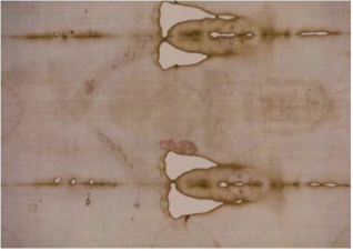
In 1978, a group of scientists was permitted to have approximately five days access to the Shroud of Turin, a 4.4 by 1.1 meters cloth containing the ventral and dorsal images of a man with blood marks corresponding to scourging and crucifixion (
Figure 1). The majority of these scientists were members of the Shroud of Turin Research Project team (STURP), co-organized by the physicists Drs. John Jackson and Eric Jumper
| [1] | Jumper E. J., Adler A. D., et al. A comprehensive examination of the various stains and images on the Shroud of Turin. Archaeological Chemistry III, ACS Advances in Chemistry 205, 22, American Chemical Society, Washington DC; p. 447‒476 (1984). |
| [2] | Schwalbe LA, Rogers RN. Physics and chemistry of the Shroud of Turin, a summary of the 1978 investigation. Anal. Chem Acta. 135: 4‒49 (1982).
https://doi-org.utk.idm.oclc.org/10.1016/S0003-2670(01)85263-6 |
| [3] | Jackson, J. Critical Summary of Shroud Observations, Data and Hypotheses. Colorado Springs, CO: Turin Shroud Center of Colorado Publishing; 2019. |
[1-3]
. Sample collection by STURP was performed by tape lifting of cloth surface fibers, most of which were approximately 10-15 μm in diameter and ranged from 40 μm to several mm in length
. To date, this remains the only detailed scientific investigation of the whole cloth that has been conducted. Studies have established that the bloodstains contain authentic blood components although it is currently unknown if additional substances may be present as well
| [3] | Jackson, J. Critical Summary of Shroud Observations, Data and Hypotheses. Colorado Springs, CO: Turin Shroud Center of Colorado Publishing; 2019. |
| [5] | Heller, J. H. and Adler, A. D. Blood on the Shroud of Turin. Applied Optics, 19: 2742-2744 (1980). |
[3, 5]
. The image does not appear to result from the simple addition of pigments or dyes and is devoid of characteristic brushstrokes like those seen in a typical painting. Understandably, it is unknown how the linen and the image may differ from their original appearance some 700-2,000 years ago. The molecular basis for creation of the image remains enigmatic but has been suggested to involve a wide range of possibilities, including acid treatment, photochemical effects, Maillard-type reactions, coronal discharge, proton emission, ultraviolet radiation, and neutron projection
| [3] | Jackson, J. Critical Summary of Shroud Observations, Data and Hypotheses. Colorado Springs, CO: Turin Shroud Center of Colorado Publishing; 2019. |
[3]
.
Figure 1. Photograph of the Shroud of Turin during the 2015 public exhibition made by the author. This photo was taken at a distance of approximately 15 feet using a zoom lens which shows a close up of the ventral portion of the image, including the facial region.
Biochemical and immunological studies have established that authentic blood components are present, including hemoglobin, albumin, and immunoglobulin, although the blood species of origin remains to be verified
| [5] | Heller, J. H. and Adler, A. D. Blood on the Shroud of Turin. Applied Optics, 19: 2742-2744 (1980). |
| [6] | Kearse, K. P. Unanticipated issues in the serological analysis of blood species: The Shroud of Turin as a case example, Forensic. Sci. Int. 2: 100073 (2020).
https://doi.org/10.1016/j.fsir.2020.100073 |
| [7] | Kearse, K. P. The aggrandizement of scientific data in the media: The Shroud of Turin blood marks as a case example, Forensic Sci. Int. Report 12: 100430 (2025).
https://doi.org/10.1016/j.fsir.2025.100430 |
[5-7]
. In the 1980s, STURP scientist Alan Adler treated blood containing fibers that were removed from an image area with protease digestion enzymes, revealing that no image-type coloration was present underneath. Control image fibers (lacking blood) were unaffected by exposure to protease, establishing that a protein component was not responsible for the image
. These findings also led to the suggestion that blood was added to the cloth first, providing subsequent protection from image creation, which occurred secondarily
| [8] | Adler, A. D. The nature of the body images on the Shroud of Turin, The Orphaned Manuscript, A gathering of Publications on the Shroud of Turin, Shroud Spectrum International Special Issue. 1st Edition, ed. D. Crispino, Effata Editrice, Italy (2002). |
[8]
. This idea has become assimilated as a type of “scientific proof” that the image could not have been created artistically. Certainly, a blood first/image second scenario is in opposition as to how an artist might likely work, i.e. fashioning an image around the blood marks instead of the other way around. However, it must be cautioned that extrapolation is not equivalent to scientific confirmation.
One of the most intriguing properties of the Shroud is that colored and non-colored fibers exist adjacent to one another
| [1] | Jumper E. J., Adler A. D., et al. A comprehensive examination of the various stains and images on the Shroud of Turin. Archaeological Chemistry III, ACS Advances in Chemistry 205, 22, American Chemical Society, Washington DC; p. 447‒476 (1984). |
| [3] | Jackson, J. Critical Summary of Shroud Observations, Data and Hypotheses. Colorado Springs, CO: Turin Shroud Center of Colorado Publishing; 2019. |
[1, 3]
. The issue has been raised that since Adler’s digestion experiments were performed at the fiber level, the possibility of obtaining a colorless “neighbor” within an image area in these types of experiments should be considered
| [9] | Is the Shroud of Turin Authentic or a Medieval Fake? Hugh Farey Reveals the Truth?! Interview on the Joe Brock Show (2024). Accessed June 2026. |
[9]
. As the number of samples available for these original studies was quite limited, it is unknown if the lack of color is a general characteristic of fibers underneath most of the blood marks in image areas of the Shroud.
Historian and artist Nicholas Allen has commented that blood has a “bleaching effect” on fabric
| [10] | Allen, N. P. L. Verification of the Nature and Causes of the Photo-negative Images on the Shroud of Lirey-Chambéry-Turin. De Arte, 30(51), 21–35. (1995).
https://doi.org/10.1080/00043389.1995.11761214 |
| [11] | P. Loysona, P., Vorstera, N. M. and Allen, N. Image formation on the Shroud of Turin: a proposed chemical model. South African Journal of Science 101: 36-42 (2005). |
[10, 11]
, offering an alternative explanation for the absence of colored fibers underneath the bloodstains. Several decades ago, he proposed the use of a camera obscura together with light sensitive silver compounds in the creation of the Shroud image
| [10] | Allen, N. P. L. Verification of the Nature and Causes of the Photo-negative Images on the Shroud of Lirey-Chambéry-Turin. De Arte, 30(51), 21–35. (1995).
https://doi.org/10.1080/00043389.1995.11761214 |
| [11] | P. Loysona, P., Vorstera, N. M. and Allen, N. Image formation on the Shroud of Turin: a proposed chemical model. South African Journal of Science 101: 36-42 (2005). |
| [12] | Allen, N. Turin Shroud: Testament to a Lost Technology. LAP Lambert Academic Publishing, Beau Bssin, Mauritius (2017). |
[10-12]
. Most objections to Allen’s theory center largely around historical arguments that such technology would not have been available for use by a medieval artist, which he disputes
| [12] | Allen, N. Turin Shroud: Testament to a Lost Technology. LAP Lambert Academic Publishing, Beau Bssin, Mauritius (2017). |
| [13] | Schwortz, B. Is the Shroud of Turin a Medieval Photograph (2000) Is the Shroud of Turin a Medieval Photograph? Accessed June 2026. |
[12, 13]
. It should be noted that specifics of Allen’s theory are often misunderstood. He does not hypothesize that silver is directly responsible for the image as in a photographic type system; rather, he proposes that following the quenching of the silver salts with ammonium solution and removal of residual silver by washing, the chemically altered cellulose fibers left behind are responsible for the coloration
| [11] | P. Loysona, P., Vorstera, N. M. and Allen, N. Image formation on the Shroud of Turin: a proposed chemical model. South African Journal of Science 101: 36-42 (2005). |
| [12] | Allen, N. Turin Shroud: Testament to a Lost Technology. LAP Lambert Academic Publishing, Beau Bssin, Mauritius (2017). |
[11, 12]
.
In the initial studies of Shroud image properties, STURP scientist Adler reported that fibrils “identical in all chemical and microscopic characteristics to those found on the Shroud” could be prepared by exposure of linen to concentrated sulfuric acid
. The use of acid-containing gall inks in the Shroud’s creation has been proposed by several investigators
| [14] | Accetta, J. S. Origins of a 14th Century Turin Shroud Image (2014) Origins of a 14th Century Turin Shroud Image Accessed June 2026. |
| [15] | Farey, H. How was it done? (2025) How was it done? – The Medieval Shroud Accessed June 2026. |
[14, 15]
. In 2009, Garlaschelli utilized a frottage technique with a mixture of pigment and sulfuric acid added to a cloth-draped body and bas relief in an attempt to create a full-size Shroud image
. Certain macroscopic features were congruent between the facsimile and the real Shroud, although other characteristics, such as no image in non-contact areas were absent. As the authors acknowledge, the experimental results of their initial attempts can doubtlessly be improved
. The experiments did not address the issue of bloodstains.
More recently, extremely short (nanosecond) pulses of ultraviolet (uv) radiation have been used to reproduce the coloration of the Shroud image on linen
| [17] | Di Lazzaro, P., Murra, D., Nichelatti, E., Santoni, A. and Baldacchini, G. Superficial and Shroud-like coloration of linen by short laser pulses in the vacuum ultraviolet. Appl. Opt. 51: 8567–8578 (2012). https://doi.org/10.1364/AO.51.008567 |
| [18] | Murra, D., Di Lazzaro, P., et al. Shroud-like coloration of linen by nanosecond laser pulses in the vacuum ultraviolet ENEA report (2015)
https://www.researchgate.net/publication/26220133
(Accessed June 2026). |
[17, 18]
. These results have led to the developing idea that energetic projection from a body is responsible for image formation, although obviously, this has not been reproduced in the laboratory. The characteristics of fiber coloration by laser irradiation have been studied at macroscopic, microscopic, and chemical levels and show strong similarities to samples from the Shroud
| [17] | Di Lazzaro, P., Murra, D., Nichelatti, E., Santoni, A. and Baldacchini, G. Superficial and Shroud-like coloration of linen by short laser pulses in the vacuum ultraviolet. Appl. Opt. 51: 8567–8578 (2012). https://doi.org/10.1364/AO.51.008567 |
| [18] | Murra, D., Di Lazzaro, P., et al. Shroud-like coloration of linen by nanosecond laser pulses in the vacuum ultraviolet ENEA report (2015)
https://www.researchgate.net/publication/26220133
(Accessed June 2026). |
[17, 18]
. Two types of coloration by laser treatment have been described, that which is immediately visible after nanosecond pulsing, and latent coloration, which is not visually detectable unless the sample is subsequently heated to simulate artificial aging, or aged naturally
| [17] | Di Lazzaro, P., Murra, D., Nichelatti, E., Santoni, A. and Baldacchini, G. Superficial and Shroud-like coloration of linen by short laser pulses in the vacuum ultraviolet. Appl. Opt. 51: 8567–8578 (2012). https://doi.org/10.1364/AO.51.008567 |
| [18] | Murra, D., Di Lazzaro, P., et al. Shroud-like coloration of linen by nanosecond laser pulses in the vacuum ultraviolet ENEA report (2015)
https://www.researchgate.net/publication/26220133
(Accessed June 2026). |
| [19] | Di Lascio, A. Di Lazzaro, P., Iacomussi, P., Missori, M., and Murra, D. Investigating the color of the blood stains on archaeological cloths: the case of the Shroud of Turin. Applied Optics 57: 6626-6631 (2018). https://doi.org/10.1364/AO.51.008567 |
[17-19]
. Interestingly, the laser dose required for immediate color visualization on linen was found to vaporize bloodstains
| [19] | Di Lascio, A. Di Lazzaro, P., Iacomussi, P., Missori, M., and Murra, D. Investigating the color of the blood stains on archaeological cloths: the case of the Shroud of Turin. Applied Optics 57: 6626-6631 (2018). https://doi.org/10.1364/AO.51.008567 |
[19]
. Only settings corresponding to latent coloration were suitable for no apparent damage of bloodstains by a general macroscopic evaluation
| [19] | Di Lascio, A. Di Lazzaro, P., Iacomussi, P., Missori, M., and Murra, D. Investigating the color of the blood stains on archaeological cloths: the case of the Shroud of Turin. Applied Optics 57: 6626-6631 (2018). https://doi.org/10.1364/AO.51.008567 |
[19]
.
In the current study, various systems were utilized to gain information pertaining to the possible sequential order of blood and image addition to the Shroud of Turin cloth. It is important to emphasize at the onset that the purpose of this study was not to refute or promote authentic or non-authentic viewpoints of the origin of the Shroud of Turin. Rather, the goal of these efforts was to provide empirical data related to a principal feature of the Shroud, which although frequently cited as important evidence, has never progressed scientifically since the idea was first suggested over forty years ago.
2. Materials and Methods
Human blood was obtained from healthy volunteers by the fingerstick method using a Health Lancing device (CVS pharmacy®, USA) fitted with a micro lancet (CVS pharmacy®, USA). Human blood serum was obtained from Innovative Research (Innovative Research Company, Novi, MI), purchased as pooled human serum off the clot.
For acid coloration, 20 microliters of HCl were added, allowed to dry, and samples heated at 110°C for 10 minutes. With experiments involving blood and serum, acid was added, allowed to dry, and then a similar volume of blood or serum was added and allowed to dry before heating. Protease treatment was performed as previously described
| [20] | Kearse K. P. Environmental influence on blood serum detection using ultraviolet 365. J Forensic Sci Res., 5: 030-036 (2021). https//doi.org/ 0.1364/AO.57.006626 |
[20]
.
The UV light system used for photography in these studies was a ultraviolet 365 UV flashlight, 365 nm, LED-UV301-356 nm (Shenzhen Lightfe Light Limited, Shenzhen, China). Photographs were taken using Sony RX100 and Sony alpha 6500 digital cameras. For various treatments, either Whatman® 1 mm filter paper (Cytiva, Marlborough, MA) or natural unprocessed linen (chemical free, unbleached, undyed) made from 100% organically grown flax (Sand Dunes) purchased from Rawganique (Blaine, WA) was used. Laser irradiation was carried out on an Advanced Technology Lasers ATLEX-ILR argon-fluoride (ArF) excimer laser with a wavelength of 193 nm. The laser dimensions were measured with direct thermal paper exposed to the laser and calipers. A Coherent FieldMax II external energy meter with a J-50MUV-193 sensor was used to measure the laser fluence prior to sample exposure. For latent conditions, 7.05 mJ per pulse, 5 ns, N of 486 was used for FT = 16.8 J/cm2; and for low level latent conditions, 9.17 mJ per pulse, 5 ns, N of 250 was used for FT = 10.5 J/cm2. For above threshold conditions, 7.05 mJ per pulse, 5 ns, N of 729 was used for FT = 25.2 J/cm2 7.05 mJ per pulse, 5 ns, N of 1146 was used for FT = 39.6 J/cm2. To visualize latent coloration effects, samples were heated at 190°C for 60 seconds.
For silver treatment, a 0.5% solution of AgNO
3 solution was added to linen, and after drying, exposed to sunlight for 8-16 hours. Samples were then quenched in 6M NH
4OH solution and washed in copious amounts of water as described
| [10] | Allen, N. P. L. Verification of the Nature and Causes of the Photo-negative Images on the Shroud of Lirey-Chambéry-Turin. De Arte, 30(51), 21–35. (1995).
https://doi.org/10.1080/00043389.1995.11761214 |
| [11] | P. Loysona, P., Vorstera, N. M. and Allen, N. Image formation on the Shroud of Turin: a proposed chemical model. South African Journal of Science 101: 36-42 (2005). |
[10, 11]
.
All of the results presented in this manuscript are representative of at least three separate experiments.
3. Results
Blood contains several chemical buffering systems which help to maintain a strict pH balance in the range of 7.35-7.45, without which the body would cease to function properly. These systems exist to offset the influx of H
+ or OH
- ions, thereby preventing a drastic drop or rise in pH
. As mentioned in the introduction, the possibility has been raised that some type of acid treatment may have been used in creation of the Shroud image. If true, then it is reasonable to assume that blood addition may serve to counterbalance the chemical etching effect of H
+ treatment in such areas.
As shown in
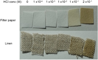
Figure 2, acid treatment followed by mild heating resulted in the rapid coloration of filter paper and linen (
Figure 2). Samples treated at or below a concentration of .01 M (1 x 10
-2) M HCl (pH ~ 2) did not show a detectable change (
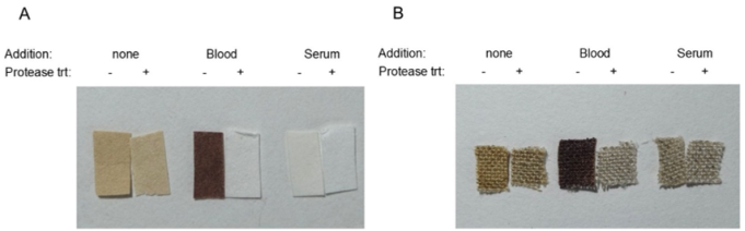
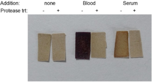
Figure 2). The degree and tone of color was variable depending upon acid concentration and heating conditions; a concentration of 0.2 M was typically used for subsequent experiments as that gave a color that was easily visible and like the Shroud image, did not fluoresce under ultraviolet (uv), (data not shown). To examine the effect of blood on acid coloration of filter paper and linen, samples were acid-treated, and after drying, a similar volume of blood or serum was applied to the same area. After a second round of drying, the samples were briefly heated as before, and one-half of each group was treated with proteolytic enzymes to remove blood and serum. As shown in
Figure 3A, proteolytic digestion was effective at removing blood and serum from filter paper but did not diminish the coloration by acid treatment in control groups without such additions (
Figure 3A).
Figure 2. Acid coloration of filter paper and linen. Samples were treated with the indicated concentration (M = molarity) of HCl, allowed to dry, and heated for 10 minutes at 110°C.
Importantly, these data show that when blood was removed from acid-treated samples, no coloration was observed underneath (
Figure 3A). Because of the relative transparency of serum, the counteracting effect on acid coloration was apparent even without proteolytic digestion (
Figure 3A). Similar results were obtained when linen was used (
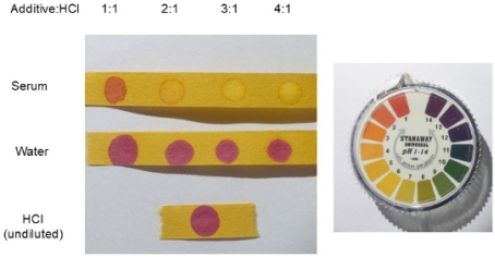
Figure 3B). Taken together, these data demonstrate that both blood and serum were able to efficiently nullify coloration by acid when applied secondarily. The neutralizing effect of blood products on acid is directly demonstrated in
Figure 4. Serum was used for these experiments as the opacity of whole blood obscures a visual readout. Unlike water, serum very effectively raised the pH of acid solution; water addition was necessary to equalize the volumes for comparison (
Figure 4). As noted previously in
Figure 2, an increase to pH ~2 or above is sufficient to nullify the effect of acid on the coloration of cellulose containing material. Taken together, these data indicate that blood addition offsets the effect of acid by increasing the pH by at least 1.3 units, consistent with our results in
Figures 2 and 3.
Figure 3. Blood nullifies acid coloration of filter paper and linen. (A) Samples were treated with acid and allowed to dry; a similar volume of blood or serum was added and allowed to dry, after which samples were heated for 10 minutes at 110°C. Blood and serum were removed by digestion with protease enzymes as indicated. Note that protease treatment had no effect on acid coloration in control groups (no additions). (B) Similar to (A), using linen, see Materials and Methods for details.
Figure 4. Blood serum effectively neutralizes acid. Serum or water was mixed with 0.2 M HCl in the indicated ratio and spotted onto pH indicator paper. A key wheel with the correlation between color and approximate pH value is shown on the right. A pH value of 1-6 is considered acidic, 7 is neutral, and 8-14 represents the basic range.
In the next set of studies, experiments were performed to evaluate the possibility that blood has a “bleaching effect”, an idea suggested by Allen
| [12] | Allen, N. Turin Shroud: Testament to a Lost Technology. LAP Lambert Academic Publishing, Beau Bssin, Mauritius (2017). |
[12]
, although no experimental data was provided to support it. If true, then application of blood to linen that had already undergone a coloration process would be expected to be reversed. The initial studies were conducted using the silver salt method for coloration; in this system, linen was treated with silver salts, exposed to sunlight, quenched with ammonium nitrate solution, and washed as previously described
| [10] | Allen, N. P. L. Verification of the Nature and Causes of the Photo-negative Images on the Shroud of Lirey-Chambéry-Turin. De Arte, 30(51), 21–35. (1995).
https://doi.org/10.1080/00043389.1995.11761214 |
| [12] | Allen, N. Turin Shroud: Testament to a Lost Technology. LAP Lambert Academic Publishing, Beau Bssin, Mauritius (2017). |
[10, 12]
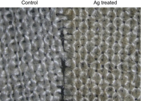
. Treated samples were easily distinguishable from control groups and gave the type of coloration that has been reported
| [10] | Allen, N. P. L. Verification of the Nature and Causes of the Photo-negative Images on the Shroud of Lirey-Chambéry-Turin. De Arte, 30(51), 21–35. (1995).
https://doi.org/10.1080/00043389.1995.11761214 |
| [12] | Allen, N. Turin Shroud: Testament to a Lost Technology. LAP Lambert Academic Publishing, Beau Bssin, Mauritius (2017). |
[10, 12]
, from yellowish to sepia in tone (
Figure 5). The extent and tone of color was somewhat variable depending upon the amount of sun exposure provided; silver-treated linen did not fluoresce under uv
| [10] | Allen, N. P. L. Verification of the Nature and Causes of the Photo-negative Images on the Shroud of Lirey-Chambéry-Turin. De Arte, 30(51), 21–35. (1995).
https://doi.org/10.1080/00043389.1995.11761214 |
| [12] | Allen, N. Turin Shroud: Testament to a Lost Technology. LAP Lambert Academic Publishing, Beau Bssin, Mauritius (2017). |
[10, 12]
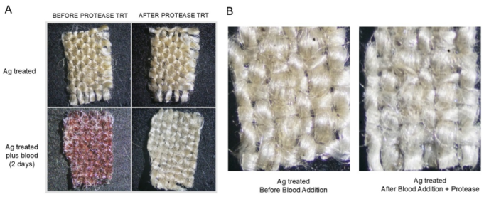
, (data not shown). Silver-treated samples were divided into two groups, with and without blood addition, and subjected to protease treatment as described above. As shown in
Figure 6, coloration of samples without blood addition was similar before and after protease digestion (
Figure 6A). In contrast, blood-containing samples appeared visually lighter afterwards (
Figure 6A, 6B).
It was reasoned that if the observed “bleaching effect” of blood was likely relevant to image formation on the Shroud, then blood would have a similar effect on linen coloration achieved by other means. Two systems were used for these experiments: i) acid coloration as done before and ii) linen exposed to nanosecond pulses with an ultraviolet laser. The laser system has the advantages that it does not rely on a particular chemical treatment for coloration, and the product characteristics match up very well with what has been described for the Shroud
| [17] | Di Lazzaro, P., Murra, D., Nichelatti, E., Santoni, A. and Baldacchini, G. Superficial and Shroud-like coloration of linen by short laser pulses in the vacuum ultraviolet. Appl. Opt. 51: 8567–8578 (2012). https://doi.org/10.1364/AO.51.008567 |
[17]
. If blood can truly “bleach” linen that has undergone oxidation, dehydration, and conjugation, then a decrease/reversal of color would be predicted to occur within other systems as well.
Figure 5. Coloration of linen treated with silver salt solution. A solution of 0.5% AgNO3 was added to linen, allowed to dry, and then exposed to sunlight for 16 hours. Samples were then soaked in 6M NH4OH to quench the reaction, washed with several rinsings of water, and allowed to dry (see Materials and Methods for details).
Figure 6. The “bleaching effect” of linen following blood addition and protease treatment on linen treated with silver salt solution. Blood was added to linen that had been treated with silver (Ag) salt solution and two days later digested with protease for blood removal. The overall color of linen appeared lighter following blood addition and removal.
Figure 7. Blood is unable to reverse filter paper coloration by acid: no “bleaching effect”. Samples were treated with 0.2 M HCl, allowed to dry, and heated for 10 minutes at 110°C. Blood or serum was added and two days later samples were digested with protease as indicated.
Unlike the experiments shown in
Figure 3 where the preventive effect of blood on acid coloration was evaluated (
Figure 3), the “bleaching effect” was examined by adding blood after coloration had already taken place. As shown using protease digestion in
Figure 7, the coloration of acid-treated filter paper was unaffected by blood or serum addition (
Figure 7).
Previous studies have shown that the threshold value for ArF laser coloration of linen occurs when the total fluence (F
T) is greater than 22 J/cm
2, with a yellowish coloring occurring when F
T is ~ 25-27 J/cm
2 | [17] | Di Lazzaro, P., Murra, D., Nichelatti, E., Santoni, A. and Baldacchini, G. Superficial and Shroud-like coloration of linen by short laser pulses in the vacuum ultraviolet. Appl. Opt. 51: 8567–8578 (2012). https://doi.org/10.1364/AO.51.008567 |
| [18] | Murra, D., Di Lazzaro, P., et al. Shroud-like coloration of linen by nanosecond laser pulses in the vacuum ultraviolet ENEA report (2015)
https://www.researchgate.net/publication/26220133
(Accessed June 2026). |
| [19] | Di Lascio, A. Di Lazzaro, P., Iacomussi, P., Missori, M., and Murra, D. Investigating the color of the blood stains on archaeological cloths: the case of the Shroud of Turin. Applied Optics 57: 6626-6631 (2018). https://doi.org/10.1364/AO.51.008567 |
[17-19]
. When linen is treated below the threshold value with F
T = 16 J/cm
2, no coloration is observed but becomes immediately visible following a brief exposure to heat, referred to as latent coloration. Latent coloration also develops over time (without heating) if the sample is allowed to age naturally
| [18] | Murra, D., Di Lazzaro, P., et al. Shroud-like coloration of linen by nanosecond laser pulses in the vacuum ultraviolet ENEA report (2015)
https://www.researchgate.net/publication/26220133
(Accessed June 2026). |
| [19] | Di Lascio, A. Di Lazzaro, P., Iacomussi, P., Missori, M., and Murra, D. Investigating the color of the blood stains on archaeological cloths: the case of the Shroud of Turin. Applied Optics 57: 6626-6631 (2018). https://doi.org/10.1364/AO.51.008567 |
[18, 19]
. Using these parameters as a guideline, two levels of laser treatment were chosen for our studies: a setting below the threshold (latent), F
T = 16.8 J/cm
2 and a setting above the threshold, F
T = 39.6 J/cm
2. As shown in
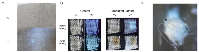
Figure 8, latent level treatment resulted in modification of linen that was essentially imperceptible at the macroscopic level under visible light but was readily detectable under uv (
Figure 8A). When irradiated samples were subjected to brief heating to simulate artificial aging, the affected areas darkened and a shifting from uv fluorescent to non-fluorescent was observed, which did not occur in control groups (
Figure 8B). Cross-sectional analysis indicated that coloration was primarily restricted to the uppermost part of the threads, as expected (
Figure 8C).
Figure 8. Irradiation of linen with levels corresponding to latent coloration. (A) Linen was irradiated with FT = 16.8 J/cm2 and evaluated under visible light (VL) and ultraviolet light (UV). (B) Control and irradiated linen (FT = 16.8 J/cm2) were evaluated before and after 60 sec of heating at 190°C to visualize latent coloration. Samples were evaluated under visible light (VL) and ultraviolet light (UV). (C) Magnified cross-section of a thread from irradiated samples described in (B). Only the uppermost part of the thread is colored.
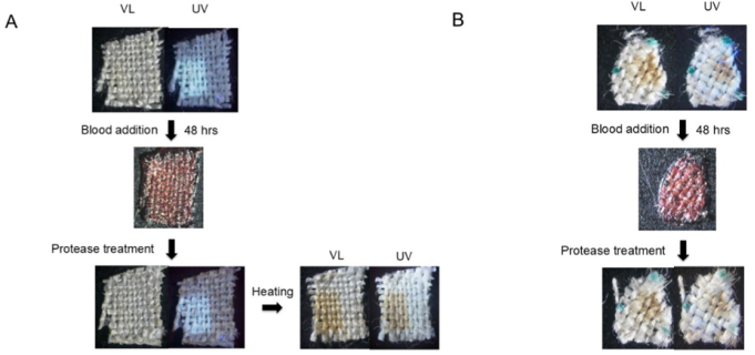
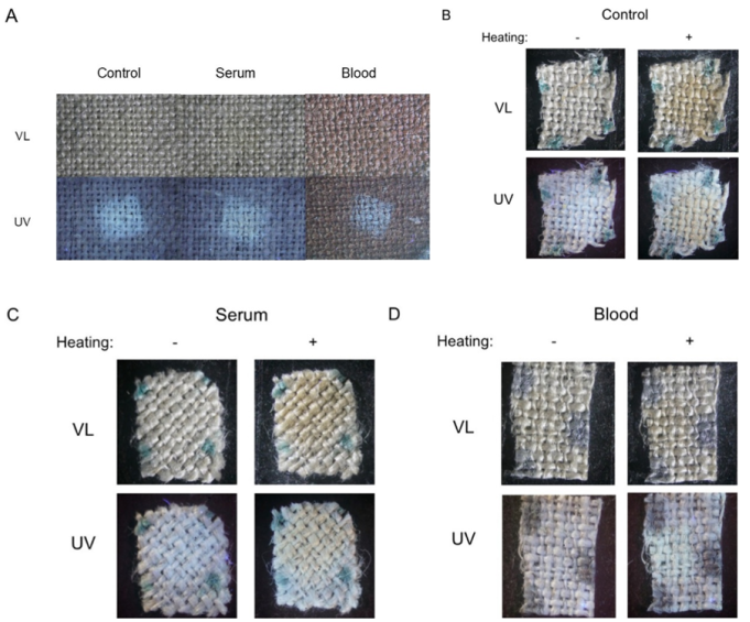
To evaluate the potential blood “bleaching effect” on irradiated linen, blood was added to linen that had been laser-treated both below (latent), and above the threshold for immediate color visualization, and several days later removed by protease treatment (
Figure 9). Samples in (A) were heated following protease digestion to visualize the color effectively. As is evident, a “bleaching effect” was not observed in either case (
Figure 9). Collectively, these results indicate that the “bleaching effect” of blood was limited to the system involving silver salt treatment (see Discussion).
Figure 9. Blood does not reverse the effects of radiation on linen: no “bleaching effect”. Samples that had been irradiated with FT = 16.8 J/cm2 (latent), (A) or FT = 39.6 J/cm2 (above the threshold for immediate coloration), (B) were layered with blood and two days later digested with protease. Samples were examined under visible light (VL) and ultraviolet light (UV). Following removal of blood by protease, the samples in (A) were heated to visualize coloration; heating was not necessary for samples in (B) as they were irradiated above the threshold for immediate coloration. See text for details. The green spots result from a Sharpie marking pen which was used for orientation during removal of samples from a larger piece of linen. Similar results were obtained when blood was digested after a 7 week period of incubation (data not shown).
The central feature of the blood first/image second premise is that no image coloration will exist under bloodstains within image areas because blood performs a protective/blocking function for underlying linen as image creation occurs. The laser irradiation system offers the unique opportunity to investigate if bloodstains are truly protective of underlying linen under conditions that reproduce many of the Shroud image characteristics. As noted above, the threshold value for linen coloration reported in previous studies was F
T > 22 J/cm
2 | [17] | Di Lazzaro, P., Murra, D., Nichelatti, E., Santoni, A. and Baldacchini, G. Superficial and Shroud-like coloration of linen by short laser pulses in the vacuum ultraviolet. Appl. Opt. 51: 8567–8578 (2012). https://doi.org/10.1364/AO.51.008567 |
| [18] | Murra, D., Di Lazzaro, P., et al. Shroud-like coloration of linen by nanosecond laser pulses in the vacuum ultraviolet ENEA report (2015)
https://www.researchgate.net/publication/26220133
(Accessed June 2026). |
[17, 18]
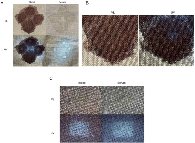
. No apparent damage to bloodstains or serum stains was visible by macroscopic analysis when a setting below the threshold (latent, F
T = 16.8 J/cm
2) was used (
Figure 10A, 10B). When examined microscopically, however, a slight darkening in the target areas was noticeable (
Figure 10C). Experiments using settings above the coloration threshold (F
T = 25 J/cm
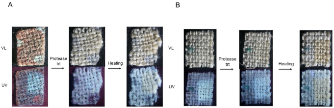
2) were found to be disruptive to bloodstains (data not shown). Thus, it was decided that for this type of experiment, latent settings would be the upper limit. Protease digestion showed that areas underneath both blood and serum were affected by irradiation, which was verified by subsequent heating to enhance visualization (
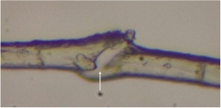
Figure 11A, 11B). Previous studies have shown that microscopic examination of individual fibers that may have become damaged during the procedure (or through just general manipulation) can provide information regarding the extent of radiation penetration through the primary cell wall
| [17] | Di Lazzaro, P., Murra, D., Nichelatti, E., Santoni, A. and Baldacchini, G. Superficial and Shroud-like coloration of linen by short laser pulses in the vacuum ultraviolet. Appl. Opt. 51: 8567–8578 (2012). https://doi.org/10.1364/AO.51.008567 |
| [18] | Murra, D., Di Lazzaro, P., et al. Shroud-like coloration of linen by nanosecond laser pulses in the vacuum ultraviolet ENEA report (2015)
https://www.researchgate.net/publication/26220133
(Accessed June 2026). |
| [19] | Di Lascio, A. Di Lazzaro, P., Iacomussi, P., Missori, M., and Murra, D. Investigating the color of the blood stains on archaeological cloths: the case of the Shroud of Turin. Applied Optics 57: 6626-6631 (2018). https://doi.org/10.1364/AO.51.008567 |
[17-19]
. It should be noted that the vast majority of fibers appear undamaged following treatment, but with thorough examination, certain fibers may be found that are physically damaged, exposing the central area. Threads were removed from bloodstained linen which had been irradiated, digested with protease, and heated (necessary for visualization of color) as in
Figure 11A; colored threads were teased with forceps to separate into individual fibers. Similar to what has been described for linen irradiated above the threshold for color visualization
, microscopic images of isolated single fibers damaged in the central part showed that the inner part of the fibers were colorless (
Figure 12). These data indicate that the observed effects on linen underneath blood did not result from undue penetration/strength of the radiation treatment.
Figure 10. Latent level irradiation of bloodstains on linen. (A) Linen containing dried blood or blood serum was irradiated with FT = 16.8 J/cm2 and evaluated under visible light (VL) and ultraviolet light (UV). (B) Close up of an individual bloodstain in (A) that was targeted for laser treatment. (C) Microscopic evaluation of blood and serum areas following latent level irradiation treatment. Samples were evaluated under visible light (VL) and ultraviolet light (UV).
Figure 11. Blood and serum do not protect underlying linen from the effects of latent level irradiation. (A) Linen containing dried blood was irradiated with FT = 16.8 J/cm2, blood was removed by protease treatment and evaluated under visible light (VL) and ultraviolet light (UV). Samples were subsequently heated to effectively visualize latent coloration. (B) Similar to (A), using serum. The silver and green spots result from a Sharpie marking pen which was used for orientation during removal of samples from a larger piece of linen.
Finally, experiments were performed using a latent laser setting much less than shown in
Figure 11 to try to identify a point at which blood might protect underlying linen from the full effect of radiant energy, referred to as low latent, F
T = 10.5 J/cm
2, (see Materials and Methods for details). As shown in
Figure 13A, little to no darkening was observed in the targeted area (
Figure 13A). When samples were digested with protease and examined, it was clear that serum was not protective of underlying linen as the results were similar to control groups with no additive (
Figure 13B, 13C). In contrast, the radiation effect was much less pronounced in blood-containing samples, with little to no color change apparent under visible light following heating (
Figure 13D). Additionally, uv analysis showed that in heated samples, fluorescence only increased to a level similar to that of unheated control and serum-containing samples (
Figure 13C, 13D). Taken together, these results suggest that latent irradiation conditions may be identified in which blood, but not serum, protects underlying linen from the full effect of energy transfer.
Figure 13. Protective effect of blood under low level latent irradiation conditions. Linen samples were irradiated with low level latent settings FT = 10.5 J/cm2 (see Materials and Methods for details). (A) Microscopic analysis of irradiated areas under visible light (VL) and ultraviolet light (UV). (B). Control samples were digested with protease treatment and evaluated before and after heating. (C) and (D) are similar to (B), except that serum- or blood-containing linens were used, respectively. The green (B, C) and silver (D) spots result from a Sharpie marking pen which was used for orientation during removal of samples from a larger piece of linen.
4. Discussion
The current report has examined various systems for linen coloration to gather potential information regarding the vertical juxtaposition of blood and image on the Shroud of Turin. Obviously, it is impossible to say with scientific certainty which came first on the Shroud, the blood or the image, although certain insight may be gained through experimental investigation. In extension of Adler’s original findings, these studies represent an important principal step in the further evaluation of this interesting characteristic of the Shroud.
The data in the current report show that acid coloration of linen may be effectively prevented by blood (or serum) addition, findings which are logical given the known buffering properties of blood that exist as part of its natural function. These results provide evidence that if blood is added to linen after a primary application of acid, chemical etching will be nullified within those areas. A brief and gentle heating was required for immediate visualization of acid coloration in this study, which used relatively low acid concentrations. Adler’s findings indicate that heating is not requisite if stronger concentrations are used and that direct application may be bypassed if used in the form of a vapor
. In the current system, color preclusion only required a shift from pH ~ 0.7 to ~ 2.0, which is well within the buffering capacities of blood or serum. These data provide a potential route by which no coloration would exist underneath if blood were added in an artistic-type scenario. Clearly, there are additional microscopic characteristics that would need to be fulfilled, which were not explored in this study. More detailed investigation using varying conditions may aid in the future development of model systems that increasingly correspond to the features of colored fibers found on the Shroud.
The suggested “bleaching effect” of blood was examined in multiple systems and found to be operative only in the case of linen coloration by silver salt treatment. The observed effect in linen treated with silver salts could potentially be explained by removal of residual, trace amounts of silver which could be responsible for the coloration. Although Allen has provided evidence that remaining silver is lessened after quenching and washing in this system, it is unclear if it is completely absent
| [11] | P. Loysona, P., Vorstera, N. M. and Allen, N. Image formation on the Shroud of Turin: a proposed chemical model. South African Journal of Science 101: 36-42 (2005). |
| [12] | Allen, N. Turin Shroud: Testament to a Lost Technology. LAP Lambert Academic Publishing, Beau Bssin, Mauritius (2017). |
[11, 12]
. In modern antimicrobial manufacturing, microscopic amounts of silver are added to certain fabrics which release charged silver ions that disrupt bacterial cellular functions and thereby help to reduce odor. Release of silver ions from solid silver is catalyzed in a humid environment (sweat)
| [22] | Wagener, S., Dommershausen, N. et al. Textile Functionalization and Its Effects on the Release of Silver Nanoparticles into Artificial Sweat. Environ. Sci. Technol. 50: 5927–5934 (2016). https//doi.org/10.1021/acs.est.5b06137 |
[22]
; thus, it is reasonable that a similar process may occur following blood addition as blood is 75-80% water. Moreover, as blood proteins are composed of sulfur-containing amino acids (like cysteine and methionine) which have a strong chemical affinity for binding with silver ions, it is possible that the observed blood “bleaching effect” in this system results from the removal of trace amounts of silver. Importantly, this effect was not operative in the other coloration systems that were examined, leading to the general conclusion that blood added after coloration is unable to reverse it. It is possible that extensively aged blood may yield a different outcome; our findings, of course, were limited to short-term observations. Relatedly, the current results do not rule out the possibility of blood plus an unknown additive having an effect.
The present study provides the first analysis of linen underneath blood and serum stains under irradiation conditions. These studies document that blood and serum were not (fully) protective of underlying linen when energy transfer corresponding to latent conditions were used. Differences were noted between blood and serum, with serum not able to provide a shielding effect, even under low latent settings. Our experiments were restricted to levels below the threshold for immediate visualization of coloration as results indicated settings above the threshold resulted in bloodstain damage. Relatedly, previous studies using a different type of laser system (Lambda Physik LPX-305, 308 nm, 0.4 J/pulse, 30 ns) have shown that settings required for immediate visualization of linen coloration resulted in bloodstain vaporization
| [19] | Di Lascio, A. Di Lazzaro, P., Iacomussi, P., Missori, M., and Murra, D. Investigating the color of the blood stains on archaeological cloths: the case of the Shroud of Turin. Applied Optics 57: 6626-6631 (2018). https://doi.org/10.1364/AO.51.008567 |
[19]
. The ArF 193 nm laser system used in our studies is most similar to the ArF 193 nm lasers used to establish the parameters relevant to Shroud-like coloration of linen
| [17] | Di Lazzaro, P., Murra, D., Nichelatti, E., Santoni, A. and Baldacchini, G. Superficial and Shroud-like coloration of linen by short laser pulses in the vacuum ultraviolet. Appl. Opt. 51: 8567–8578 (2012). https://doi.org/10.1364/AO.51.008567 |
| [18] | Murra, D., Di Lazzaro, P., et al. Shroud-like coloration of linen by nanosecond laser pulses in the vacuum ultraviolet ENEA report (2015)
https://www.researchgate.net/publication/26220133
(Accessed June 2026). |
[17, 18]
, although the laser in our system had shorter pulse duration and lower pulse energy than those in previous reports (5 ns vs 12 ns and 7.05 mJ vs 80 mJ, respectively). Linen coloration is proportional to the total fluence and is not related to the intensity or fluence of each pulse
. As each pulse affects to some extent the surface modified by the previous pulse
, this could potentially influence the final outcome. Importantly, it was observed that irradiated linen in our study showed similar properties as have been described for treatment with longer pulse durations and higher energy, especially those related to latent coloration. While artificial aging through brief heating is useful to visualize areas involved in latent coloration, it is not equivalent to aging by natural means. Indeed, it has been documented in previous studies that susceptibility of blood to protease treatment is affected by increasing heat exposure whereas aged blood was readily digestible
| [20] | Kearse K. P. Environmental influence on blood serum detection using ultraviolet 365. J Forensic Sci Res., 5: 030-036 (2021). https//doi.org/ 0.1364/AO.57.006626 |
[20]
. During the current study, it was noted that brief heating of irradiated blood samples hindered subsequent protease digestion (K. P. Kearse, unpublished observations), making it difficult to compare samples that had been artificially aged prior to blood removal.
The previous observation that irradiation conditions necessary for immediate visualization of colored linen is not compatible with the subsistence of bloodstains
| [19] | Di Lascio, A. Di Lazzaro, P., Iacomussi, P., Missori, M., and Murra, D. Investigating the color of the blood stains on archaeological cloths: the case of the Shroud of Turin. Applied Optics 57: 6626-6631 (2018). https://doi.org/10.1364/AO.51.008567 |
[19]
, together with the data in the current report raise the interesting possibility that the Shroud image may have developed in a delayed manner. Alternatively, some might debate that such results argue against analogous energy transfer type mechanisms being involved in image creation. Regarding Adler’s original protease experiments, the best fit with a radiation-type model would require levels below the threshold for immediate visualization of coloration; it should be noted, however, that serum was not protective, even when very low latent levels were used. Recognizably, there are limitations in trying to approximate the specifics of unknown events that may have been important in the creation of the Shroud. As previous investigators have written, such experiments may be “difficult to control and characterize. We are not the conclusion, we are composing pieces of a fascinating and complex scientific puzzle.”
| [17] | Di Lazzaro, P., Murra, D., Nichelatti, E., Santoni, A. and Baldacchini, G. Superficial and Shroud-like coloration of linen by short laser pulses in the vacuum ultraviolet. Appl. Opt. 51: 8567–8578 (2012). https://doi.org/10.1364/AO.51.008567 |
[17]
. Clearly, there is much more work to be done to increase our understanding of the enigmatic properties associated with this cloth. Like many studies pertaining to the Shroud, the current investigation is limited by the lack of new exploration of the cloth’s features. Together with previous analyses, the current results may provide stimulation for consideration of future scientific data gathering from this historically important textile.
Lastly, it is in the hands of the reader to decide how the current results might affect their opinion of the blood first/image second hypothesis related to the Shroud. As demonstrated here, pathways exist to circumvent the difficulty of no image underneath the bloodstains using artistic methods, although this by no means demonstrates that the Shroud was created in such a fashion. In a corresponding manner, the significance of the observation that blood was not (fully) protective of underlying linen from even low latent levels of radiation will vary, depending on one’s perspective. For some, an interest in the Shroud’s blood marks may stimulate a reevaluation of the details of image formation and vice versa.
In summary, the current report has investigated various circumstances related to the sequential order of blood and image addition to the Shroud of Turin. These studies show that addition of blood to acid-treated linen nullifies subsequent coloration, offering a potential example of no image underneath blood involving artistic creation. Additionally, the suggested “bleaching effect” of blood was addressed and shown to be restricted to only one out of three coloration systems that were examined. Hence, the general conclusion is that blood added after coloration is unable to reverse it. Lastly, these studies document that linen underneath blood (or serum) was affected by energy transfer from radiation, even under conditions corresponding to latent coloration.





 Share This Article
 Twitter
 Linked In
 Facebook
 Copy
Copy Download