Research Article | | Peer-Reviewed

Isolation, Phenotypic Characterization, and Distribution of Normal Bacterial Flora from the Hand Skin of Healthy Female Students at Wolkite University, Ethiopia

Received: 27 May 2026     Accepted: 8 June 2026     Published: 23 June 2026
Views:       Downloads:
Abstract

The cutaneous microbiota serves as a critical first line of defense through bacterial interference, but can transition into opportunistic pathogens if introduced into deep tissues via mechanical trauma. This study aimed to isolate, phenotypically characterize, and assess the distribution profile of normal hand skin bacterial flora among healthy female students at Wolkite University, Ethiopia. A laboratory-based cross-sectional study was conducted from December 2024 to June 2025 at the Department of Biotechnology Laboratory. Hand skin swab samples were collected from 60 healthy female students using simple random sampling. Isolated colonies were purified and classified to the genus level based on macroscopic morphology, Gram reaction, cellular shapes, and standard biochemical verification arrays. A total of 27 distinct bacterial isolates were recovered from the 60 samples. Phenotypic and biochemical profiling identified seven distinct bacterial groups. The family Enterobacteriaceae was the most prevalent group (10 isolates, 37.07%), followed by Staphylococci (6 isolates, 22.22%) and Lactobacilli (4 isolates, 14.81%). The findings demonstrate that while female hand skin maintains protective resident commensals (Staphylococci and Lactobacilli), it frequently harbors transient enteric and environmental bacteria (Enterobacteriaceae) due to continuous exposure to shared institutional touch points. This underscores the critical importance of implementing consistent personal hygiene, systematic hand-sanitation protocols, and enhanced public health awareness within the university campus ecosystem to minimize hand-borne opportunistic infections.

Published in International Journal of Pharmacy and Chemistry (Volume 12, Issue 1)
DOI 10.11648/j.ijpc.20261201.12
Page(s) 11-18
Creative Commons

This is an Open Access article, distributed under the terms of the Creative Commons Attribution 4.0 International License (http://creativecommons.org/licenses/by/4.0/), which permits unrestricted use, distribution and reproduction in any medium or format, provided the original work is properly cited.

Copyright

Copyright © The Author(s), 2026. Published by Science Publishing Group

Keywords

Enterobacteriaceae, Hand Skin, Normal Flora, Phenotypic Characterization, Staphylococci

1. Introduction
The mixture of microorganisms regularly found at any anatomical site of humans is referred to as normal flora, and those which reside on the human skin are specifically known as skin flora or skin microbiota . Usually skin flora are non-pathogenic and they are either of commensal (harm full to their host) or mutualistic (without causing detectable harm) . In a healthy individual, this normal microflora is relatively stable, with specific genera populating particular body regions during predictable periods of life . This normal flora is established in an orderly succession after birth, eventually developing into the complex adult ecosystem observed across different skin boundaries.
The skin acts as an intricate microbial ecosystem driven by interactions between diverse microbial populations and host immune responses . Culturable bacteria widely isolated from normal skin surfaces include Staphylococci, Micrococci, Corynebacteria, Brevibacteria, Propionibacteria, and Acinetobacter species . The primary factors determining both the absolute quantity and species quality of the local microbiota depend directly on the anatomical site and its regional microenvironment specifically fluctuating micro-gradients of pH, moisture, temperature, oxygen availability, and surface lipid concentrations . Consequently, the composition of the cutaneous flora varies drastically between sebaceous (oily), moist, and dry skin areas . Human hands function primarily as dry environments with intermittent moisture, representing a unique ecologic zone heavily exposed to external environments .
The skin barrier provides essential selective advantages for survival, concurrently preventing the escape of physiological moisture and halting the deep invasion of toxic chemical substances or infectious agents. Because of this, normal skin flora contributes significantly as a critical component of the first line of innate host defense. On outer epithelial cells and mucous membranes, the established resident normal flora blocks the colonization of incoming pathogenic invaders, successfully preventing clinical disease through a process known as bacterial interference. The functional mechanisms driving bacterial interference are multi-faceted; they involve direct competition for cellular receptors or binding sites on host tissue, competitive consumption of limited local nutrients, mutual metabolic inhibition via acidic or toxic by-products, and the targeted secretion of specialized antimicrobial proteins or bacteriocins .
Despite their protective evolutionary roles, members of the normal flora can shift behaviors to become pathogenic under specific micro environmental or systemic circumstances. Cutaneous microbes are highly adapted to non-invasive lifestyles defined by the strict structural limitations of the outer skin layers. However, if these organisms are forcefully displaced from their natural boundaries and introduced into deep tissues or the bloodstream via mechanical trauma, compromised skin barriers, burns, or wounds, they rapidly transition into highly aggressive opportunistic pathogens . This risk is severely amplified in hospitalized populations or individuals experiencing profound immunosuppression. In clinical settings where opportunistic infections emerge directly from the patient’s own commensal community, immediate and precise phenotypic isolation is required to differentiate normal flora from true contaminants and guide effective antibiotic selection.
Furthermore, external environmental and behavioral pressures can rapidly alter the homeostatic equilibrium of the skin, causing abrupt disruptions in community structures. In female populations, the frequent and varied use of topical cosmetics, moisturizing agents, and chemical cleansers alters local pH and lipid barriers, often reducing or shifting the baseline distribution of protective normal flora. While international programs have deeply characterized human internal mucosal populations, our fundamental understanding of localized hand microflora dynamics across specific institutional environments remains surprisingly limited . Cutaneous flora can be operationally categorized into transient (highly fluctuating, non-reproducing contaminants), temporary resident, and true resident (actively replicating and growing) flora . On human hands, transient floras are constantly deposited through contact with shared public touch points. At present, the structural composition and changing dynamics of these bacterial communities have not been established under localized laboratory baselines at Wolkite University. To address this gap, this study focuses strictly on evaluating the hands of healthy female university students. This targeted phenotypic evaluation maps out exactly what commensal and transient bacterial genera are distributed across the hand skin under standard institutional conditions, utilizing classical Gram staining alongside detailed biochemical validation arrays.
2. Materials and Methods
2.1. Description of Study Area
The study was conducted at the Biotechnology Laboratory, Department of Biotechnology, Wolkite University, Ethiopia. Wolkite University is located in the Gurage Zone, Central Ethiopian Region. Wolkite, the capital town, is located 155 km southwest of Addis Ababa. The Gurage Zone comprises altitudes ranging from 1001 to 3500 meters above sea level. Based on the local agro-climatic classification, the zone is divided into three agro-climatic zones. Dega (high altitude) covers 28.3% of the area and ranges between 2500 and 3662 masl, Weinadega (mid-altitude) at 1500–2500 masl encompasses about 64.9% of the area, and Kola (lowland) at 1000–1500 masl covers 6.8% of the area. The mean annual temperature ranges from 14 to 24°C, with an average of 20.5°C. The rainfall pattern in the Gurage Zone is bimodal, with 80% occurring during the Belg period from February to May.
2.2. Sampling Design and Period
The design of this research was a laboratory-based cross-sectional study to conduct isolates and phenotypically characterize normal bacterial flora from the hand skin of healthy female regular students at Wolkite University with complete random design and each experiment had three replications and each experiments were repeated twice. The study was carried out from December 2024 to June 2025.
2.3. Target Population and Sample Size
The target population included all healthy female regular students residing on the main campus of Wolkite University during the study period. Students who were apparently healthy and volunteered to participate were included in the study. Those with visible skin infections, open wounds on their hands, or those who had used topical antibiotics within the previous two weeks were excluded from the study. A total of 60 healthy female students were selected using a simple random sampling technique. Swab samples were systematically collected from the hand skin surface of each selected participant.
2.4. Experimental Reagents, Material and Equipment
The major equipment used in this study included an autoclave, a hot plate with a magnetic stirrer, a compound light microscope, a bacteriological incubator, a digital weighing balance, a laminar airflow hood, and a refrigerator. The primary culture media and biochemical reagents utilized for isolation and characterization consisted of Nutrient Agar, MacConkey Agar, Mannitol Salt Agar, Triple Sugar Iron (TSI) Agar, Simmon's Citrate Agar, Urea Agar Base, and Hydrogen Peroxide (H2O2) for the catalase test, Crystal Violet, Gram's Iodine, 95% Acetone-Alcohol decolorizer, and Safranin counterstain. Additional laboratory consumables included sterile cotton swabs, normal saline (0.9% NaCl), microscopic glass slides, immersion oil, inoculation loops, and Petri dishes.
2.5. Measurement and Data Collection Procedure
Before sample collection, the purpose of the study was explained to each participant. Swab samples were obtained by rubbing a sterile cotton swab, pre-moistened with sterile normal saline, firmly over the palm and dorsal surfaces of the participant's hands. After collection, the swabs were immediately transported to the biotechnology laboratory for processing.
2.6. Isolation of Bacteria
The collected swab samples from the selected anatomical sites of healthy female student’s hand were directly inoculated onto Nutrient Agar plates by using the streak-plate method to obtain isolated colonies. To select for specific groups, samples were also streaked onto MacConkey Agar (selective for Gram-negative bacteria) and Mannitol Salt Agar (selective for Staphylococci species). The inoculated Petri dishes were then inverted and incubated at 37°C for 24 to 48 hours under aerobic conditions. After the incubation period, distinct, isolated colonies were selected based on their macroscopic appearance for further purification and phenotypic identification.
2.7. Morphological, Cultural, and Biochemical Characterization
The isolated bacterial colonies were systematically classified based on their morphological, cultural, and biochemical characteristics. The macroscopic features of the isolated colonies, such as shape, size, color, margin, elevation, and opacity, were recorded. Microscopic examination was conducted using Gram staining techniques to determine the Gram reaction (Gram-positive or Gram-negative), cellular shape (cocci or bacilli), and structural arrangement of the bacterial cells under the oil immersion objective (100 times) of a compound light microscope.
2.7.1. Gram Staining Identification
Gram staining is a method that differentiates bacteria in two large group gram positive and gram negative. This method differentiates bacteria by the chemical and physical properties of their cell walls by detecting peptidoglycan, in gram positive it is present as a thick layer. A Gram positive results in a purple/blue color while a Gram negative results in a pink/red color . For each bacterial isolate a heat fixed smear from 24 hours- old cultures were prepared, stained with crystal violet solution for 1 minutes, rinsed rapidly with water and iodine solution was added and the smear was blot dried. The slides were washed with 95% ethanol 1 min, rinsed with tap water and stained with safranin for 30 seconds. The slide was air dried and examined under compound light microscope.
2.7.2. Catalase Test
The catalase test was performed to detect the presence of catalase enzyme by transferring a loopful of pure colony to clean microscope slide and addition of one drop of 3% of hydrogen peroxide solution. Positive test was indicated by formation of effervescence or appearance of bubbles, due to the breaking down of hydrogen peroxide to O2 and H2O .
2.7.3. Citrate Utilization Test
This test determines the ability of bacteria to convert citrate into oxaloacetate. Citrate is the only carbon source available to the bacteria in this media. If bacteria cannot use citrate, it was not grow. Positive result was seen if the bacterium grows and the media turns into bright blue color as a result of an increase in the pH of the media .
2.7.4. Triple Sugar Iron Agar Test
Triple Sugar Iron agar slant tubes are used for this test, this multi-test medium contains glucose (at a concentration that is 1/10th that of the other sugars), sucrose and lactose. The inoculation was performed by using a straight wire; the test organism was inoculated deep into the butt of the medium and streak up the slant. These were incubated at 37°C for 24 hr .
2.7.5. Simmon's Citrate Test
Pure isolated colonies were streaked onto the surface of sterile Simmons Citrate Agar slants using a sterile inoculating loop according to Varney et al., . Care was taken to inoculate only the surface of the slant without stabbing the butt of the medium. The inoculated tubes were incubated aerobically at 35–37°C for 24–48 hours. After incubation, the tubes were examined for bacterial growth and color change of the medium. Utilization of citrate as the sole carbon source resulted in alkaline by-products that changed the bromothymol blue indicator from green to deep blue, indicating a positive result. Absence of growth and no color change (medium remaining green) were considered a negative result.
2.7.6. Urease Test
Pure bacterial isolates were inoculated into sterile Urea Agar slants using a sterile inoculating loop by streaking the surface of the medium. The inoculated media were incubated at 35–37°C for 24–48 hours and observed for color development. Production of the urease enzyme hydrolyzed urea into ammonia and carbon dioxide, resulting in an alkaline reaction in the medium. A positive urease test was indicated by a change in color of the medium from yellowish-orange to bright pink due to the phenol red indicator, whereas no color change indicated a negative result .
2.8. Data Analysis and Presentation
The collected laboratory data regarding colony features, staining results, and biochemical reaction profiles were compiled systematically. The distribution, absolute frequencies, and percentage isolation rates of the identified bacterial genera among the participants were analyzed and presented using descriptive statistical summaries, including frequency tables and graphical charts.
3. Results and Discussion
3.1. Socio-Demographic Characteristics of Participants
A total of 60 healthy female regular students from the main campus of Wolkite University were enrolled in this study. Hand skin swab samples were successfully collected from all 60 participants (100% response rate) and processed at the Department of Biotechnology Laboratory for the isolation and phenotypic characterization of normal bacterial flora. The baseline results demonstrate that human hand skin surfaces maintain a highly diverse and complex microenvironment capable of supporting both permanent resident commensals and transient opportunistic microbial communities .
3.2. Isolation and Distribution Rate of Bacterial Genera
From the 60 hand swab samples analyzed, a total of 27 distinct bacterial isolates were isolated (Figure 1; Table 1). Phenotypic characterization based on colony morphology, Gram reaction, and an array of standard biochemical tests identified seven distinct bacterial groups distributing on the hand skin of the study cohort. The distribution of the isolated bacteria showed that the family Enterobacteriaceae was the most frequently isolated group, accounting for 10 out of the 27 total isolates (37.04%). This was followed closely by the genus Staphylococci, which represented 6 isolates (22.22%) and the genus Lactobacilli, which accounted for 4 isolates (14.81%). The remaining bacterial isolates were distributed among the genera Bacillus (2 isolates, 7.4%), Micrococci (2 isolates, 7.4%), Pseudomonas (2 isolates, 7.4%), and Enterococci (1 isolate, 3.70%).
This broad distribution pattern is structurally consistent with established cutaneous ecological principles, which state that the dry, acidic, and nutrient-limiting nature of human skin naturally selects for resilient Gram-positive organisms . The cell wall composition of Gram-positive bacteria allows them to better withstand high osmotic pressures, dehydration, and desiccation compared to thin-walled Gram-negative bacilli .
Table 1. Frequency and percentage distribution of bacterial genera isolated from the hand skin of participants.

No.

Identified Bacterial Genus

Frequency (Number of Isolates)

Percentage Distribution (100%)

1

Enterobacteriaceae

10

37.04

2

Staphylococci

6

22.22

3

Lactobacilli

4

14.81

4

Bacillus

2

7.41

5

Micrococci

2

7.41

6

Pseudomonas

2

7.41

7

Enterococci

1

3.70

Total

All Genera Combined

27

100%

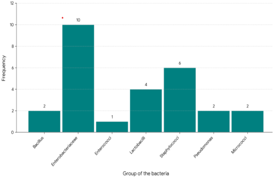
Figure 1. The frequency distribution profile of isolated bacterial groups.
3.3. Morphological and Biochemical Characteristics of Isolates
Out of the 60 hand swab samples processed in the laboratory, a total of 27 distinct bacterial isolates were successfully recovered and classified to the genus level based on their cultural, morphological, and biochemical profiles (Figure 2; Table 2). Gram-positive isolates predominated the sample (n = 15, 55.56%), distributed across five distinct genera/groups, while Gram-negative isolates accounted for the remaining 44.44% (n = 12). Among the Gram-negative isolates, Enterobacteriaceae represented the largest group (n = 10), characterized as motile, medium-sized rods capable of fermenting both lactose and glucose with associated gas production. These isolates tested positive for catalase, citrate utilization, and the Methyl Red (MR) test, but were negative for starch hydrolysis and the Voges-Proskauer (VP) reaction. The remaining Gram-negative isolates were identified as Pseudomonas (n = 2), which exhibited a motile, rod-shaped morphology, tested positive for catalase, citrate, and glucose utilization, but failed to ferment lactose or produce gas. The Gram-positive cohorts were predominantly cocci, led by Staphylococci (n = 6) and followed by Micrococci (n = 2) and Enterococci (n = 1). Both Staphylococci and Micrococci exhibited positive reactions for catalase, glucose, citrate, and Methyl Red, while remaining negative for motility and starch hydrolysis. However, Staphylococci were differentiated by their ability to ferment lactose and a positive VP reaction. Conversely, the single Enterococci isolate was catalase-negative, VP-negative, and non-motile, but positive for lactose, glucose, and MR testing. Rod-shaped Gram-positive isolates comprised Lactobacilli (n = 4) and Bacillus (n = 2). The Lactobacilli isolates were non-motile, catalase-negative, and exhibited variable sizes, showing positive fermentation profiles for lactose, glucose, and MR. Bacillus isolates were characterized as large, motile rods that unique among all tested groups, demonstrated robust starch hydrolysis alongside positive reactions for catalase, glucose, citrate, and Methyl red.
Among the recovered organisms, Enterobacteriaceae was the single most frequently isolated bacterial group on the hands of the participants, representing 10 out of the 27 total isolates (37.04%). The frequent isolation of Enterobacteriaceae which are traditionally classified as enteric or environmental rods presents an interesting ecological finding. Because human hands serve as primary tools for physical interaction, they are highly dynamic zones continuously exposed to external touch points .
In an institutional university setting, students frequently contact shared public surfaces such as cafeteria door handles, lecture hall desks, library books, and restroom fixtures. This dense interaction network facilitates the rapid mechanical acquisition of transient fecal-oral or environmental contaminants, explaining the elevated distribution of Enterobacteriaceae on the participants' hand surfaces .
The genus Staphylococci was the second most prevalent bacterial group identified, accounting for 6 out of the 27 isolates (22.22%). This high isolation rate directly aligns with foundational dermatological studies identifying Staphylococci (particularly coagulase-negative species like Staphylococcus epidermidis) as universal, core components of the human resident skin micro biota . These resident Staphylococci play a critical evolutionary role in the first line of innate host defense. They engage in bacterial interference by physically blocking epithelial binding receptors, competing for limited surface nutrients, and actively secreting specialized antimicrobial peptides or bacteriocins that inhibit the colonization of highly virulent, incoming pathogenic invaders . The remaining microbial distribution on the hand skin included Lactobacilli, Bacillus, Micrococci, Pseudomonas, and Enterococci. The presence of Bacillus and Micrococci represents typical, dry-adapted environmental or soil-borne contaminants that temporarily adhere to the skin as transient flora .
However, the presence of genera such as Pseudomonas and Enterobacteriaceae highlights a critical clinical duality. Under homeostatic baseline conditions, these organisms live completely harmlessly as commensal or transient surface residents on an intact stratum corneum. Yet, if these bacteria are mechanically displaced from their natural cutaneous boundaries and introduced into deep tissues or the systemic bloodstream via physical trauma, accidental punctures, severe burns, or open surgical wounds, they can rapidly transition into highly aggressive opportunistic pathogens . This structural finding underscores the profound public health importance of introducing consistent personal hygiene, systematic hand-sanitation protocols, and enhanced public awareness within dense campus student ecosystems .
Table 2. Morphological and biochemical characterization of bacteria species isolated from female student’s hands.

Bacterial species

Biochemical test

Enterobacteriaceae

Staphylococci

Lactobacilli

Bacillus

Micrococci

Pseudomonas

Enterococci

Number of Isolates

10

6

4

2

2

2

1

Shapes

Rods

Cocci

Rods

Rods

Cocci

Rods

Cocci

Gram rxn.

-

+

+

+

+

-

+

Size

Medium

Small-Medium

Variable

Large

Small-Medium

Medium

Small

Catalase

+

+

-

+

+

+

-

Lactose

+

+

+

-

-

-

+

Glucose

+

+

+

+

+

+

+

Gas prod.

+

-

-

-)

-

-

-

Citrate

+

+

-

+

+

+

-

Starch

-

-

-

+

-

-

-

Methyl red

+

+

+

+

+

-

+

VP

-

+

-

-

-

-

-

Motility

+

-

-

+

-

+

-

+=positive, and - =negative
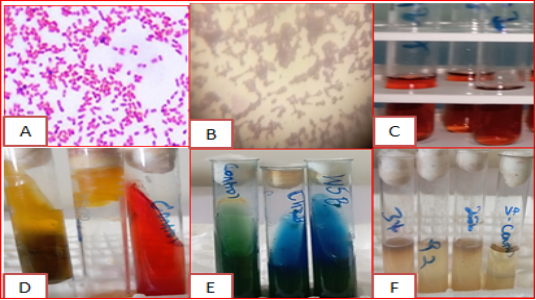
Figure 2. Morphological and biochemical characterization of bacterial isolates: (A): gram-positive isolate; (B): gram-negative; (C): positive methyl red test; (D): TSI positive test; (E): citrate positive; and (F): VP-negative test.
4. Conclusion
This study was successfully conducted to isolate and phenotypically characterize the normal bacterial flora from the hand skin of healthy female regular students at Wolkite University. Out of the 60 hand swab samples processed, a total of 27 bacterial isolates were recovered and identified to the genus level using morphological, staining, and standard biochemical verification arrays. The laboratory findings reveal that Enterobacteriaceae was the most frequently distributed bacterial group on the hands of the participants, accounting for 10 out of the 27 total isolates (37.04%). This was followed closely by the genus Staphylococci, which represented 6 isolates (22.22%). The remaining bacterial distribution consisted of Lactobacilli, Bacillus, Micrococci, Pseudomonas, and Enterococci. These results demonstrate that while the human hand skin maintains a rich community of protective, non-pathogenic commensal resident flora (such as Staphylococci and Lactobacilli), it also frequently harbors transient environmental and enteric bacteria like Enterobacteriaceae. While these organisms exist harmlessly on intact skin surfaces, they possess significant clinical potential to act as opportunistic pathogens if they are forcefully introduced into deep tissues through compromised skin barriers, mechanical trauma, or open wounds. Further research should be carried out with a larger sample size and extended study periods at Wolkite University. Future studies should incorporate advanced molecular identification techniques, such as 16S rRNA gene sequencing, and include antimicrobial susceptibility testing (AST) to map out the resistance profiles of these commensal and transient skin isolates.
Abbreviations

AST

Antimicrobial Susceptibility Testing

MR

Methyl Red Test

VP

Voges-Proskauer Reaction

TSI

Triple Sugar Iron Agar

Acknowledgments
We extend our gratitude to Wolkite University for their invaluable support for this research paper.
Author Contributions
Debebe Landina Lata: Conceptualization, Methodology, Software, Writing – original draft, Writing – review & editing
Abera Kenea: Conceptualization, Data curation, Visualization
Tesfaye Giza: Funding acquisition, Investigation, Supervision
Eyasu Milkias: Formal Analysis, Project administration, Resources
Kaleb Kamayla: Data curation, Resources, Validation
Dawit Regasa: Conceptualization, Data curation
Conflicts of Interest
There are no conflicts of interest to disclose regarding this current paper.
References
[1] Baron, S. Normal flora. In S. Baron (Ed.). Medical microbiology (4th ed., Chapter 6). University of Texas Medical Branch at Galveston. 1996.
[2] Leyden, J. J., McGinley, K. J., Nordstrom, K. M., & Webster, G. F. Skin microflora. Journal of Investigative Dermatology. 1987, 88(3), s65–s72.
[3] Mackowiak, P. A. The normal microbial flora. New England Journal of Medicine. 1982, 307(2), 83–93.
[4] Marples, M. The ecology of the human skin. Charles C. Thomas, Banner stone House, Springfield, II. 1965, 33-78.
[5] Grice, E. A. Skin microbiota: Genomics-based insights into the diversity and role of skin microbes. Trends in Microbiology. 2014, 22(9), 527–537.
[6] Byrd, A. L., Belkaid, Y., & Segre, J. A. The human skin micro biota, Nature Reviews Microbiology. 2018, 16(3), 143–155.
[7] Roth, R. R., & James, W. D. Microbial ecology of the skin. Annual Review of Microbiology. 1988, 42(1), 441–464.
[8] Schlefer W. C. Microbiology of Human Skin. London: Lloyd-Luke. Sears CL. A dynamic partnership: celebrating our gut flora. 2005, 1-7.
[9] Grice, E. A., & Segre, J. A. The skin microbiome. Nature Reviews Microbiology. 2011, 9(4), 244–253.
[10] Sears, C. L. A dynamic partnership: Celebrating our gut flora. Anaerobe. 2005, 11(5), 247–251.
[11] Shaw, C. M., Smith, J. A., McBride, M. E. & Duncan, W. C. An evaluation technique for sampling skin flora. Journal of Investigative Dermatology. 1970, 54, 160-166.
[12] Tripathi, N., Zubair, M., & Sapra, A. Gram staining. Stat Pearls Publishing. 2025, 12-34.
[13] Sophie S Arbefeville, Tristan T Timbrook, Cherilyn D Garner, Evolving strategies in microbe identification—a comprehensive review of biochemical, MALDI-TOF MS and molecular testing methods, Journal of Antimicrobial Chemotherapy. 2024, 79 (1), 2–i8,
[14] Varney, A. M., Mannix-Fisher, E., Thomas, J. C., & McLean, S. Evaluation of phenotypic and genotypic methods for the identification and characterization of bacterial isolates recovered from catheter-associated urinary tract infections. Journal of Applied Microbiology. 2024, 135(7), lxae155.
[15] Rosario Medina, I., Suárez Benítez, M. A., Ojeda-Vargas, M. d. M., Gallo, K., Padilla Castillo, D., Batista-Arteaga, M., Déniz Suárez, S., Díaz Rodríguez, E. L. & Acosta Hernández, B. Investigation of Carriers of Salmonella and Other Hydrogen Sulphide-Positive Bacteria in the Digestive Content of Fish from the Atlantic Area of Macaronesia: A Comparative Study of Identification by API Gallery and MALDI-TOF MS. Animals. 2024, 14(22), 3247.
[16] Timm, K. H., Shayan, M., & Grice, E. A. The skin microbiome in health and disease: Principles, methods, and future directions. Journal of Investigative Dermatology. 2024, 144(4), 692–702.
[17] Amare, A., Eshetie, S., Kasew, D., Amare, A., Abebe, W., & Moges, F. Prevalence of Salmonella spp., Shigella spp., and intestinal parasites among food handlers working in University of Gondar student’s cafeteria, Northwest Ethiopia, Frontiers in Public Health. 2024, 12, 1370338.
Cite This Article
  • APA Style

    Lata, D. L., Kenea, A., Giza, T., Milkias, E., Kamayla, K., et al. (2026). Isolation, Phenotypic Characterization, and Distribution of Normal Bacterial Flora from the Hand Skin of Healthy Female Students at Wolkite University, Ethiopia. International Journal of Pharmacy and Chemistry, 12(1), 11-18. https://doi.org/10.11648/j.ijpc.20261201.12

    Copy | Download

    ACS Style

    Lata, D. L.; Kenea, A.; Giza, T.; Milkias, E.; Kamayla, K., et al. Isolation, Phenotypic Characterization, and Distribution of Normal Bacterial Flora from the Hand Skin of Healthy Female Students at Wolkite University, Ethiopia. Int. J. Pharm. Chem. 2026, 12(1), 11-18. doi: 10.11648/j.ijpc.20261201.12

    Copy | Download

    AMA Style

    Lata DL, Kenea A, Giza T, Milkias E, Kamayla K, et al. Isolation, Phenotypic Characterization, and Distribution of Normal Bacterial Flora from the Hand Skin of Healthy Female Students at Wolkite University, Ethiopia. Int J Pharm Chem. 2026;12(1):11-18. doi: 10.11648/j.ijpc.20261201.12

    Copy | Download

  • @article{10.11648/j.ijpc.20261201.12,
      author = {Debebe Landina Lata and Abera Kenea and Tesfaye Giza and Eyasu Milkias and Kaleb Kamayla and Dawit Regasa},
      title = {Isolation, Phenotypic Characterization, and Distribution of Normal Bacterial Flora from the Hand Skin of Healthy Female Students at Wolkite University, Ethiopia},
      journal = {International Journal of Pharmacy and Chemistry},
      volume = {12},
      number = {1},
      pages = {11-18},
      doi = {10.11648/j.ijpc.20261201.12},
      url = {https://doi.org/10.11648/j.ijpc.20261201.12},
      eprint = {https://article.sciencepublishinggroup.com/pdf/10.11648.j.ijpc.20261201.12},
      abstract = {The cutaneous microbiota serves as a critical first line of defense through bacterial interference, but can transition into opportunistic pathogens if introduced into deep tissues via mechanical trauma. This study aimed to isolate, phenotypically characterize, and assess the distribution profile of normal hand skin bacterial flora among healthy female students at Wolkite University, Ethiopia. A laboratory-based cross-sectional study was conducted from December 2024 to June 2025 at the Department of Biotechnology Laboratory. Hand skin swab samples were collected from 60 healthy female students using simple random sampling. Isolated colonies were purified and classified to the genus level based on macroscopic morphology, Gram reaction, cellular shapes, and standard biochemical verification arrays. A total of 27 distinct bacterial isolates were recovered from the 60 samples. Phenotypic and biochemical profiling identified seven distinct bacterial groups. The family Enterobacteriaceae was the most prevalent group (10 isolates, 37.07%), followed by Staphylococci (6 isolates, 22.22%) and Lactobacilli (4 isolates, 14.81%). The findings demonstrate that while female hand skin maintains protective resident commensals (Staphylococci and Lactobacilli), it frequently harbors transient enteric and environmental bacteria (Enterobacteriaceae) due to continuous exposure to shared institutional touch points. This underscores the critical importance of implementing consistent personal hygiene, systematic hand-sanitation protocols, and enhanced public health awareness within the university campus ecosystem to minimize hand-borne opportunistic infections.},
     year = {2026}
    }
    

    Copy | Download

  • TY  - JOUR
    T1  - Isolation, Phenotypic Characterization, and Distribution of Normal Bacterial Flora from the Hand Skin of Healthy Female Students at Wolkite University, Ethiopia
    AU  - Debebe Landina Lata
    AU  - Abera Kenea
    AU  - Tesfaye Giza
    AU  - Eyasu Milkias
    AU  - Kaleb Kamayla
    AU  - Dawit Regasa
    Y1  - 2026/06/23
    PY  - 2026
    N1  - https://doi.org/10.11648/j.ijpc.20261201.12
    DO  - 10.11648/j.ijpc.20261201.12
    T2  - International Journal of Pharmacy and Chemistry
    JF  - International Journal of Pharmacy and Chemistry
    JO  - International Journal of Pharmacy and Chemistry
    SP  - 11
    EP  - 18
    PB  - Science Publishing Group
    SN  - 2575-5749
    UR  - https://doi.org/10.11648/j.ijpc.20261201.12
    AB  - The cutaneous microbiota serves as a critical first line of defense through bacterial interference, but can transition into opportunistic pathogens if introduced into deep tissues via mechanical trauma. This study aimed to isolate, phenotypically characterize, and assess the distribution profile of normal hand skin bacterial flora among healthy female students at Wolkite University, Ethiopia. A laboratory-based cross-sectional study was conducted from December 2024 to June 2025 at the Department of Biotechnology Laboratory. Hand skin swab samples were collected from 60 healthy female students using simple random sampling. Isolated colonies were purified and classified to the genus level based on macroscopic morphology, Gram reaction, cellular shapes, and standard biochemical verification arrays. A total of 27 distinct bacterial isolates were recovered from the 60 samples. Phenotypic and biochemical profiling identified seven distinct bacterial groups. The family Enterobacteriaceae was the most prevalent group (10 isolates, 37.07%), followed by Staphylococci (6 isolates, 22.22%) and Lactobacilli (4 isolates, 14.81%). The findings demonstrate that while female hand skin maintains protective resident commensals (Staphylococci and Lactobacilli), it frequently harbors transient enteric and environmental bacteria (Enterobacteriaceae) due to continuous exposure to shared institutional touch points. This underscores the critical importance of implementing consistent personal hygiene, systematic hand-sanitation protocols, and enhanced public health awareness within the university campus ecosystem to minimize hand-borne opportunistic infections.
    VL  - 12
    IS  - 1
    ER  - 

    Copy | Download

Author Information
  • Department of Biotechnology, Wolkite University, Wolkite, Ethiopia

    Research Fields: biotechnology, plant biotechnology, plant microbe interactions, plant molecular biology, microbiology and microbial biotechnology, nanotechnology, and bioinformatics

  • Department of Biotechnology, Wolkite University, Wolkite, Ethiopia

    Research Fields: Biotechnology, bioinformatics, molecular biology, nanotechnology, and biochemistry

  • Department of Plant Science, Wolkite University, Wolkite, Ethiopia

    Research Fields: plant science, coffee science, agronomy, plant breeding, and plant pathology

  • Department of Chemistry, Wolkite University, Wolkite, Ethiopia

    Research Fields: chemistry, biochemistry, medical chemistry, analytical and organic chemistry

  • Department of Biology, Wolkite University, Wolkite, Ethiopia

    Research Fields: Biology, medical microbiology, microbiology, entomology, and biotechnology

  • Department of Biotechnology, Wolkite University, Wolkite, Ethiopia

    Research Fields: Biotechnology, molecular biology, microbiology, bioinformatics and biochemistry

  • Abstract
  • Keywords
  • Document Sections

    1. 1. Introduction
    2. 2. Materials and Methods
    3. 3. Results and Discussion
    4. 4. Conclusion
    Show Full Outline
  • Abbreviations
  • Acknowledgments
  • Author Contributions
  • Conflicts of Interest
  • References
  • Cite This Article
  • Author Information