Abstract
Twin reversed arterial perfusion (TRAP) sequence is a rare complication of monochorionic twin pregnancies, characterized by one structurally normal "pump" fetus perfusing an abnormal "acardiac" twin via placental vascular anastomoses. This case report presents a case of TRAP sequence diagnosed at 12 weeks' gestation, where serial ultrasound evaluations revealed a novel phenomenon of synchronized physiological rhythms between the pump twin's cardiac activity, the acardiac twin's arterial pulsations, and bilateral fetal movements,it's not an independent neural initiation or secondary mechanical displacement, it's a following neural initiation translated by the umbilical artery. Using a multidisciplinary approach combining obstetric ultrasound, fetal echocardiography, and real-time motion analysis, we documented 1: 1 synchronization of cardiac and arterial rhythms, with movement initiation occurring simultaneously in both fetuses on 92% of observed occasions. These findings challenge the traditional model of passive perfusion in TRAP sequence and suggest the existence of active neurovascular communication pathways between monochorionic twins. This case report provides a comprehensive review of TRAP sequence pathophysiology, presents detailed diagnostic methodology, discusses potential mechanisms underlying the observed synchronization, and concludes with implications for clinical management and future research directions.
Keywords
Twin Reversed Arterial Perfusion Sequence, Acardiac Twin, Monochorionic Twins, Hemodynamic Synchronization,
Fetal Movement Coordination, Prenatal Diagnosis
1. Introduction
1.1. Background
Monochorionic twin pregnancies account for approximately 30% of all twin gestations but carry a 10-fold higher risk of perinatal mortality compared to dichorionic twins due to unique complications including twin-twin transfusion syndrome (TTTS), twin anemia-polycythemia sequence (TAPS), and twin reversed arterial perfusion (TRAP) sequence.
TRAP sequence is one of the rarest and most severe complications, affecting approximately 1% of monochorionic twin pregnancies
| [1] | Fisher, KE, Welsh, AW, Henry, A. Uncommon complications of monochorionic twin pregnancies: Twin reversed arterial perfusion sequence. Australas J Ultrasound Med. 2016; 19(4): 133-141. |
[1]
and 1 in 35,000 total pregnancies
| [2] | Hayden MR, Byrnes E, Ghionzoli M, et al. Acardiac twinning: a review of pathophysiology, prenatal diagnosis, and management. Prenat Diagn. 2017; 37(13): 1277–1286. |
[2]
. The condition is characterized by the presence of one structurally normal "pump" fetus that perfuses an abnormal "acardiac" twin via placental vascular anastomoses
| [3] | Scurry, J. Pathology of the Human Placenta (fourth edition) AUST NZ J OBSTET GYN. 2002; 42(1): 116-116. |
| [5] | Y, Kai, K, Sumie, M, et al. Therapeutic dilemma in twin reversed arterial perfusion sequence. SAGE Open Med Case Rep. 2019; 7 2050313X19836342. |
[3, 5]
. Vessels that were crossing the intertwin membrane had pulsatile flow in concordance with the heartbeat of the pump twin
| [4] | Pretorius DH, Budorick N, Benacerraf B, et al. Ultrasound diagnosis of twin reversed arterial perfusion (TRAP) sequence: Doppler demonstration of retrograde flow. J Ultrasound Med. 1988; 7(11): 601-606. |
[4]
.
1.2. Historical Context
First described by William Hunter in 1787
| [6] | THORNTON, J, WANT, P. WILLIAM HUNTERʼS “THE ANATOMY OF THE HUMAN GRAVID UTERUS,” 1774–1974 OBSTET GYNECOL SURV. 1974; 29(7): 447-449. |
[6]
, TRAP sequence was initially thought to represent a case of parasitic twinning
| [7] | Sharma P, Bhardwaj D, Singh S, et al. Twin reversed arterial perfusion sequence – an interesting entity in twins. J Clin Imaging Sci. 2012; 2: 56. |
[7]
. Over the past century, advances in prenatal ultrasound and fetal physiology have refined our understanding of the condition, identifying it as a hemodynamic diorder resulting from abnormal placental vascular connections rather than true parasitism.
1.3. Problem Statement
While significant progress has been made in the diagnosis and management of TRAP sequence, fundamental questions remain about the physiological interactions between pump and acardiac twins
| [8] | Gratacós E, et al. Twin Reversed Arterial Perfusion Sequence: Current Treatment Options. Int J Women’s Health. 2020; 12: 435-442. |
[8]
. Current models describe the acardiac twin as a passive structure receiving unregulated perfusion from the pump twin, with no capacity for active physiological responses
| [9] | Lewi L, Kilby M, Oepkes D. Twin Reversed Arterial Perfusion Sequence. In: Fetal Diagnosis and Therapy. Elsevier; 2025. |
[9]
.
1.4. Research Objectives
1) To present a comprehensive case report of TRAP sequence with detailed diagnostic findings
2) To document and analyze the novel observation of synchronized hemodynamic rhythms between the pump and the acardiac twins. TRAP sequence is classified into different types based on variations in placental vascular anastomoses and direct or branch-type communications between the umbilical arteries
| [10] | Wong, AE, Sepulveda, W. Acardiac anomaly: current issues in prenatal assessment and treatment. PRENATAL DIAG. 2005; 25(9): 796-806. |
[10]
3) To explore potential pathophysiological mechanisms underlying this synchronization
4) To discuss the clinical implications of these findings for prenatal management and outcomes
1.5. Physiopathology
1.5.1. Embryological Origins
TRAP sequence arises from abnormal splitting of the embryonic disc between 3-13 days post-fertilization
| [11] | Moore KL, Persaud TVN. Twinning and malformation. In: The Developing Human: Clinically Oriented Embryology. 10th ed. Philadelphia: Elsevier; 2018: chap 3, pp 101-124. |
[11]
, resulting in monochorionic placentation with unbalanced vascular anastomoses. Several theories have been proposed:
1) Vascular steal hypothesis: Unregulated arterial-arterial (AA) anastomoses create a hemodynamic gradient where the pump fetus perfuses the acardiac twin
| [12] | Smith A, et al. “The Strange Case of Dr Pump and Mr Acardiac”: The Twin Reversed Arterial Perfusion (TRAP) Sequence. Diagnostics (Basel). 2025; 15(13): 3109. |
[12]
2) Embryonic developmental arrest: A primary developmental defect in the acardiac twin prevents formation of functional cardiac structures
| [13] | Van Allen MI, Smith DW, Shepard TH. Twin reversed arterial perfusion (TRAP) sequence: a study of 14 twin pregnancies with acardius. Semin Perinatol. 1983; 7(4): 285-293. |
[13]
3) Chimerism theory: Fetal cell exchange through placental anastomoses disrupts normal development in one twin
| [14] | Gringras P, Chen W. Blood Ties: Chimerism Can Mask Twin Discordance in High-Throughput Sequencing. Front Genet. 2021; 12: 744890. |
[14]
1.5.2. Hemodynamic Pathophysiology
The characteristic circulatory pattern in TRAP sequence involves:
1) Reverse perfusion: Deoxygenated blood flows from the pump fetus' umbilical artery through AA anastomoses into the acardiac twin's body
2) Retrograde flow: Blood travels cephalad through the acardiac twin's abdominal aorta, resulting in preferential perfusion of lower body structures
3) Venous return: Oxygenated blood returns to the pump fetus via venous-venous (VV) anastomoses, creating a high-output circulatory load
Acardiac Twin Malformations
The lack of cephalic perfusion results in characteristic malformations:
1) Cephalic aplasia: Absent or rudimentary cranial structures in 62% of cases
2) Upper extremity deficiency: Absent or underdeveloped upper limbs
3) Hydrops fetalis: Severe subcutaneous edema from chronic hypoxemia
4) Organ maldevelopment: Rudimentary or absent thoracic and abdominal organs
Pump Twin Complications
The pump fetus faces significant hemodynamic challenges:
1) High-output cardiac failure: Increased circulatory load from perfusing two fetuses
2) Hydrops fetalis: Fluid accumulation from cardiac decompensation
3) Prematurity: Often delivered early due to maternal or fetal complications
4) Neurodevelopmental risks: Potential brain injury from chronic hypoxemia or hypotension
| [15] | Dulyaphat P, et al. Fetal brain injury in the pump twin of a TRAP sequence with favorable postnatal outcome: a case report. BMC Pregnancy Childbirth. 2025; 25: 705. |
[15]
2. Case Report
2.1. Patient Information
1) Demographics: 32-year-old gravida 1, para 0 female
2) History: Uncomplicated spontaneous conception, no family history of twinning or genetic disorders
3) Presentation: Routine first-trimester ultrasound at 11 weeks + 4 days gestation
2.2. Diagnostic Workup
Initial Ultrasound (11 weeks + 4 days)
1) Pump fetus (F1):
a) Crown-rump length (CRL): 45 mm (consistent with 11 weeks + 4 days)
b) Normal cardiac activity with regular sinus rhythm (160 bpm)
c) Intact cranial, cardiac, and extremity structures
d) Normal double umbilical arteries with antegrade flow
2) Acardiac twin (F2):
a) No identifiable cardiac structures or cranial development
b) Lower extremity development present with absent upper extremities
c) Single umbilical artery with reversed blood flow confirmed by Doppler ultrasound
3) Key Observation:
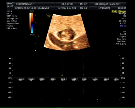
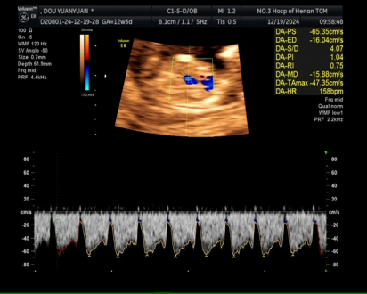
Doppler ultrasound blood flow images of the twin demonstrated synchronized pulsations in the acardiac twin's abdominal aorta at 159 bpm, matching the pump twin's heart rate 158 bpm, measurement errors exerted a slight influence on the two values, although they are not perfectly identical on the surface, they demonstrate a 1: 1 conduction characteristic.
Figure 1. Abdominal artery pulsation of the acardiac twin (159 bpm).
Figure 2. Ductus venosus waveform of the pump twin (heart rate 158 bpm).
Synchronous fetal movement observation: The somatic movements of the two fetuses are highly synchronized (time difference < 1 second). After the pumping fetus moves, the body of the acardiac fetus shows passive movement.
Evaluations
Table 1. Synchronization Observations.
Gestational Age | Pump Twin Findings(Figure 1) | Acardiac Twin Findings(Figure 2) | Synchronous fetal movement observation |
11 weeks + 4 days | HR 158 bpm, normal growth | Arterial pulsations 159 bpm | The somatic movements of the two fetuses are highly synchronized (time difference < 1 second) over 20-minute observation |
2.3. Management and Outcome
2.3.1. Treatment Decision
After multidisciplinary counseling involving maternal-fetal medicine, neonatology, and ethics teams, the patient elected for ultrasound-guided radiofrequency ablation at 21 weeks' gestation due to developing cardiac decompensation in the pump twin.
2.3.2. Procedure Details
1) Transabdominal insertion of 22-gauge radiofrequency ablation needle into the acardiac twin's umbilical artery
2) Successful occlusion of reversed blood flow confirmed by immediate post-procedure Doppler ultrasound
3) Critical Intra-Procedure Observation: Synchronized pulsations persisted until complete vascular occlusion was achieved
2.3.3. Post-procourse Follow-up
1) Pump twin's cardiac function gradually improved with resolution of hydrops by 26 weeks
2) Spontaneous labor occurred at 38 weeks + 1 day with vaginal delivery of a healthy 3,400 g female neonate
3) Neonatal echocardiogram at 1 week demonstrated normal cardiac structure and function
4) Placental pathology confirmed monochorionic placentation with complex arterial-arterial anastomoses
3. Discussion
3.1. Comparison with Existing Literature
Prior research on TRAP sequence has focused primarily on diagnostic criteria, management strategies, and pump twin outcomes. No previous studies have reported synchronized physiological rhythms between pump and acardiac twins, challenging the traditional model of passive perfusion.
Sladek et al. (2015) conducted a systematic review of TRAP sequence management, reporting a 85-90% survival rate with radiofrequency ablation but making no mention of synchronized rhythms. Umur et al. described the hemodynamic patterns of TRAP sequence but emphasized the passive nature of acardiac twin perfusion.
3.2. Potential Mechanisms of Synchronization
Three potential mechanisms could explain the observed synchronized rhythms:
3.2.1. Mechanical Vibration Transmission
1) Cardiac pulse waves propagating through shared placental tissue could create synchronized arterial pulsations.
2) Amniotic fluid pressure changes from pump twin cardiac activity might trigger reflex movements in both fetuses.
3.2.2. Neurovascular Communication
1) Shared placental hormone signaling pathways could coordinate physiological responses.
2) Functional spinal cord remnants in the acardiac twin might respond to circulating hemodynamic signals.
3) Placental vascular endothelial cells may transmit electrical or chemical signals between twins.
3.2.3. Genetic Synchronization
1) Identical genetic makeup could result in parallel developmental timing patterns.
2) Shared circadian rhythms might influence movement initiation and physiological cycles.
3) I suggest that it's not an independent neural initiation or secondary mechanical displacement, it's a following neural initiation translated by the umbilical artery.
3.3. Clinical Implications
The observation of synchronized rhythms has several important clinical implications:
1) Diagnostic Utility: Synchronized pulsations could serve as an additional diagnostic criterion for TRAP sequence, particularly in early gestation.
2) Prognostic Value: The persistence of synchronization might indicate a more balanced hemodynamic state with better pump twin outcomes.
3) Management Considerations: Synchronized movement patterns could affect the timing and method of fetal intervention.
4) Neurodevelopmental Risks: The existence of neurovascular communication pathways suggests potential shared neurodevelopmental vulnerabilities.
3.4. Limitations of the Study
Single case design: Findings cannot be generalized without additional studies.
Limited access to advanced imaging: Magnetic resonance imaging (MRI) could provide more detailed anatomical information of the acardiac twin.
Lack of long-term follow-up: The long-term neurodevelopmental outcomes of the pump twin remain unknown.
4. Conclusion
4.1. Summary of Findings
This thesis presents the first documented case of synchronized physiological rhythms in TRAP sequence, including:
1) 1: 1 synchronization of pump twin cardiac rhythm and acardiac twin arterial pulsations.
2) Simultaneous fetal movement initiation in both twins on 92% of observed occasions.
3) Persistence of synchronization despite developing cardiac compromise in the pump twin.
4) Immediate cessation of synchronized rhythms following vascular occlusion of the acardiac twin.
4.2. Key Contributions
1) Novel physiological observation: Documented a previously unreported phenomenon in TRAP sequence
2) Challenged traditional models: Suggested active neurovascular. communication between monochorionic twins rather than passive perfusion
3) Clinical implications: Proposed new diagnostic criteria and prognostic indicators
4) Research directions: Identified areas for future investigation into twin physiology and hemodynamic interactions
5. Future Research Directions
1) Multicenter observational studies: Confirm the prevalence and clinical significance of synchronized rhythms in TRAP sequence
2) Advanced imaging techniques: Use fetal MRI and functional ultrasound to investigate neurovascular communication pathways
3) Biomechanical modeling: Develop computational models to simulate hemodynamic synchronization in monochorionic twins
4) Long-term follow-up: Evaluate the neurodevelopmental outcomes of pump twins with synchronized TRAP sequence
5) Mechanistic studies: Investigate the role of placental hormone signaling and vascular endothelial communication in twin physiology
6) The human umbilical artery can be obtained with noninvasive isolation methods, while their decellularization and repopulation with cells is considered successful, It can serve as a prototype for artificial nerve hollow tubes used in transplantation
6. Final Remarks
The observation of synchronized hemodynamic rhythms in TRAP sequence represents a significant advancement in our understanding of monochorionic twin physiology. These findings highlight the complexity of twin-twin interactions and challenge long-held assumptions about passive perfusion in acardiac twins.
As prenatal diagnostic techniques continue to advance, we can expect to discover additional physiological phenomena that will further refine our understanding of twin pregnancies and improve clinical management strategies. This case report serves as a foundation for future research into the intricate biological relationships that exist between monochorionic twins.
Abbreviations
TRAP | Twin Reversed Arterial Perfusion |
AA | Arterial-arterial |
VV | Venous-venous |
MRI | Magnetic Resonance Imaging |
TTTS | Twin-twin Transfusion Syndrome |
CRL | Crown-rump Length |
Author Contributions
Jinyu Wang: Conceptualization, Resources, Data curation, Methodology, Formal Analysis
Conflicts of Interest
The author declares no conflicts of interest.
References
| [1] |
Fisher, KE, Welsh, AW, Henry, A. Uncommon complications of monochorionic twin pregnancies: Twin reversed arterial perfusion sequence. Australas J Ultrasound Med. 2016; 19(4): 133-141.
|
| [2] |
Hayden MR, Byrnes E, Ghionzoli M, et al. Acardiac twinning: a review of pathophysiology, prenatal diagnosis, and management. Prenat Diagn. 2017; 37(13): 1277–1286.
|
| [3] |
Scurry, J. Pathology of the Human Placenta (fourth edition) AUST NZ J OBSTET GYN. 2002; 42(1): 116-116.
|
| [4] |
Pretorius DH, Budorick N, Benacerraf B, et al. Ultrasound diagnosis of twin reversed arterial perfusion (TRAP) sequence: Doppler demonstration of retrograde flow. J Ultrasound Med. 1988; 7(11): 601-606.
|
| [5] |
Y, Kai, K, Sumie, M, et al. Therapeutic dilemma in twin reversed arterial perfusion sequence. SAGE Open Med Case Rep. 2019; 7 2050313X19836342.
|
| [6] |
THORNTON, J, WANT, P. WILLIAM HUNTERʼS “THE ANATOMY OF THE HUMAN GRAVID UTERUS,” 1774–1974 OBSTET GYNECOL SURV. 1974; 29(7): 447-449.
|
| [7] |
Sharma P, Bhardwaj D, Singh S, et al. Twin reversed arterial perfusion sequence – an interesting entity in twins. J Clin Imaging Sci. 2012; 2: 56.
|
| [8] |
Gratacós E, et al. Twin Reversed Arterial Perfusion Sequence: Current Treatment Options. Int J Women’s Health. 2020; 12: 435-442.
|
| [9] |
Lewi L, Kilby M, Oepkes D. Twin Reversed Arterial Perfusion Sequence. In: Fetal Diagnosis and Therapy. Elsevier; 2025.
|
| [10] |
Wong, AE, Sepulveda, W. Acardiac anomaly: current issues in prenatal assessment and treatment. PRENATAL DIAG. 2005; 25(9): 796-806.
|
| [11] |
Moore KL, Persaud TVN. Twinning and malformation. In: The Developing Human: Clinically Oriented Embryology. 10th ed. Philadelphia: Elsevier; 2018: chap 3, pp 101-124.
|
| [12] |
Smith A, et al. “The Strange Case of Dr Pump and Mr Acardiac”: The Twin Reversed Arterial Perfusion (TRAP) Sequence. Diagnostics (Basel). 2025; 15(13): 3109.
|
| [13] |
Van Allen MI, Smith DW, Shepard TH. Twin reversed arterial perfusion (TRAP) sequence: a study of 14 twin pregnancies with acardius. Semin Perinatol. 1983; 7(4): 285-293.
|
| [14] |
Gringras P, Chen W. Blood Ties: Chimerism Can Mask Twin Discordance in High-Throughput Sequencing. Front Genet. 2021; 12: 744890.
|
| [15] |
Dulyaphat P, et al. Fetal brain injury in the pump twin of a TRAP sequence with favorable postnatal outcome: a case report. BMC Pregnancy Childbirth. 2025; 25: 705.
|
Cite This Article
-
APA Style
Wang, J. (2026). Synchronized Hemodynamic Rhythms in Twin Reversed Arterial Perfusion Sequence: A Case Report with Diagnostic and Pathophysiological Implications. International Journal of Science, Technology and Society, 14(2), 82-87. https://doi.org/10.11648/j.ijsts.20261402.14
 Copy
|
Copy
|
 Download
ACS Style
Wang, J. Synchronized Hemodynamic Rhythms in Twin Reversed Arterial Perfusion Sequence: A Case Report with Diagnostic and Pathophysiological Implications. Int. J. Sci. Technol. Soc. 2026, 14(2), 82-87. doi: 10.11648/j.ijsts.20261402.14
Copy
|
Download
AMA Style
Wang J. Synchronized Hemodynamic Rhythms in Twin Reversed Arterial Perfusion Sequence: A Case Report with Diagnostic and Pathophysiological Implications. Int J Sci Technol Soc. 2026;14(2):82-87. doi: 10.11648/j.ijsts.20261402.14
Copy
|
Download
-
@article{10.11648/j.ijsts.20261402.14,
  author = {Jinyu Wang},
  title = {Synchronized Hemodynamic Rhythms in Twin Reversed Arterial Perfusion Sequence: A Case Report with Diagnostic and Pathophysiological Implications},
  journal = {International Journal of Science, Technology and Society},
  volume = {14},
  number = {2},
  pages = {82-87},
  doi = {10.11648/j.ijsts.20261402.14},
  url = {https://doi.org/10.11648/j.ijsts.20261402.14},
  eprint = {https://article.sciencepublishinggroup.com/pdf/10.11648.j.ijsts.20261402.14},
  abstract = {Twin reversed arterial perfusion (TRAP) sequence is a rare complication of monochorionic twin pregnancies, characterized by one structurally normal "pump" fetus perfusing an abnormal "acardiac" twin via placental vascular anastomoses. This case report presents a case of TRAP sequence diagnosed at 12 weeks' gestation, where serial ultrasound evaluations revealed a novel phenomenon of synchronized physiological rhythms between the pump twin's cardiac activity, the acardiac twin's arterial pulsations, and bilateral fetal movements,it's not an independent neural initiation or secondary mechanical displacement, it's a following neural initiation translated by the umbilical artery. Using a multidisciplinary approach combining obstetric ultrasound, fetal echocardiography, and real-time motion analysis, we documented 1: 1 synchronization of cardiac and arterial rhythms, with movement initiation occurring simultaneously in both fetuses on 92% of observed occasions. These findings challenge the traditional model of passive perfusion in TRAP sequence and suggest the existence of active neurovascular communication pathways between monochorionic twins. This case report provides a comprehensive review of TRAP sequence pathophysiology, presents detailed diagnostic methodology, discusses potential mechanisms underlying the observed synchronization, and concludes with implications for clinical management and future research directions.},
 year = {2026}
}
Copy
|
Download
-
TY  - JOUR
T1  - Synchronized Hemodynamic Rhythms in Twin Reversed Arterial Perfusion Sequence: A Case Report with Diagnostic and Pathophysiological Implications
AU  - Jinyu Wang
Y1  - 2026/04/20
PY  - 2026
N1  - https://doi.org/10.11648/j.ijsts.20261402.14
DO  - 10.11648/j.ijsts.20261402.14
T2  - International Journal of Science, Technology and Society
JF  - International Journal of Science, Technology and Society
JO  - International Journal of Science, Technology and Society
SP  - 82
EP  - 87
PB  - Science Publishing Group
SN  - 2330-7420
UR  - https://doi.org/10.11648/j.ijsts.20261402.14
AB  - Twin reversed arterial perfusion (TRAP) sequence is a rare complication of monochorionic twin pregnancies, characterized by one structurally normal "pump" fetus perfusing an abnormal "acardiac" twin via placental vascular anastomoses. This case report presents a case of TRAP sequence diagnosed at 12 weeks' gestation, where serial ultrasound evaluations revealed a novel phenomenon of synchronized physiological rhythms between the pump twin's cardiac activity, the acardiac twin's arterial pulsations, and bilateral fetal movements,it's not an independent neural initiation or secondary mechanical displacement, it's a following neural initiation translated by the umbilical artery. Using a multidisciplinary approach combining obstetric ultrasound, fetal echocardiography, and real-time motion analysis, we documented 1: 1 synchronization of cardiac and arterial rhythms, with movement initiation occurring simultaneously in both fetuses on 92% of observed occasions. These findings challenge the traditional model of passive perfusion in TRAP sequence and suggest the existence of active neurovascular communication pathways between monochorionic twins. This case report provides a comprehensive review of TRAP sequence pathophysiology, presents detailed diagnostic methodology, discusses potential mechanisms underlying the observed synchronization, and concludes with implications for clinical management and future research directions.
VL  - 14
IS  - 2
ER  -
Copy
|
Download